576
Carvedilol Inhibits Expressions of Vascular Cell Adhesion Molecule-1, Intercellular Adhesion Molecule-1, Monocyte Chemoattractant-1,
and Interleukin-8 via NF-κB Inhibition in Human Endothelial Cells
Yong Sook Kim, PhD, Youngkeun Ahn, MD, PhD, FACC, FSCAI, Moon Hwa Hong, BS, Soo Yeon Joo, BS, Myung Ho Jeong, MD, Kye Hun Kim, MD, Il Suk Sohn, MD, Hyung Wook Park, MD,
Young Joon Hong, MD, Ju Han Kim, MD, Weon Kim, MD,
Jeong Gwan Cho, MD, Jong Chun Park, MD and Jung Chaee Kang, MD The Heart Center of Chonnam National University Hospital, Gwangju, Korea ABSTRACT
Background and Objectives:Carvedilol is an anti-oxidative, the cardioprotective effects of which are mediated by the inhibition of NF-κB activation. The present study was designed to examine the effects of carvedilol, an α1- and β-blocker, on tumor necrosis factor (TNF)-α stimulated human umbilical vein endothelial cells (HUVEC). Materials and Methods:HUVEC were treated with TNF-α (10 ng/mL) in either the absence or presence of carvedilol. The levels of intracellular reactive oxygen species (ROS) were examined using a fluorescent dye DCFH-DA, with the adhesion of U-937 monocyte to the HUVEC. Nuclear factor kappa B (NF-κB) acti- vation was determined by NF-κB p65 translocation to the nucleus using Western blotting and immunocyto- chemistry. The expressions of NF-κB dependent pro-inflammatory molecules, i.e., vascular cell adhesion molecule (VCAM)-1, intercellular adhesion molecule (ICAM)-1, monocyte chemoattractant protein (MCP)-1 and interleukin (IL)-8, were measured by RT-PCR and ELISA. Bcl-2 and phosphorylation of c-Jun N-terminal protein kinase (JNK) were measured using Western blotting. Results:TNF-α treatment increased the activation of NF- κB, suppressed Bcl-2, and increased the phosphorylation of JNK, the ROS level and the adhesion of U-937.
The levels of mRNA and protein expressions of VCAM-1, ICAM-1, MCP-1 and IL-8 were up-regulated by TNF- α. Carvedilol inhibited the phosphorylation of JNK, ROS formation and the adhesion of U-937 monocyte. In addition, carvedilol reduced the production of VCAM-1, ICAM-1, MCP-1 and IL-8 at the mRNA and protein levels, via the suppression of NF-κB activation. Conclusion:These results suggested that the anti-inflammatory effects of carvedilol on TNF-α stimulated endothelial cells could be explained by its ROS-scavenging and NF-κ B inactivation properties. (Korean Circulation J 2005;35:576-582)
KEY WORDS:Carvedilol;Tumor necrosis factor-α, nuclear factor-κB, endothelial cell.
Introduction
Inflammation has been demonstrated to play an important role in the formation and progression of atherosclerosis.1)2) TNF-α is one of the major inflam- matory mediators that promotes the pathogenesis of
atherosclerosis, and is present in all stages of athero- sclerosis.3) TNF-α enhanced the expressions of the adhesion molecules on the membrane of endothelial cells,4) transcription factors related with inflammation, such as NF-κB, and caused modulation of the acti- vities of various enzymes, such as mitogen-activated protein kinases(MAPK).5)6) In various pathological con- ditions, such as ischemia and acute inflammation, ex- cessively accumulated reactive oxygen species(ROS) promote the activations of MAPKs or apoptosis cas- cades.5)
Carvedilol, a vasodilating and non-selective α1- and β-adrenoreceptor antagonist, is currently used for the treatment of hypertension and heart failure. Besides its anti-hypertensive effects, many reports have demon-
Received:June 22, 2005 Accepted:July 21, 2005
Correspondence : Youngkeun Ahn, MD, PhD, FACC, FSCAI, Interven- tional Cardiologist, Director of Program in Gene and Cell Therapy, Department of Cardiovascular Medicine, Heart Center of Chonnam National University Hospital, 8 Hak-dong, Dong-gu, Gwangju 501-757, Korea
Tel: 82-62-220-4764, Fax: 82-62-223-3105 E-mail: [email protected]
strated that carvedilol reduced infarct sizes and prevented restenosis after angioplasty and coronary atherectomy.7-9) Carvedilol has also been reported to act as an anti- oxidative in vitro, with potency 30 to 80 times higher than vitamin E or probucol.10)
The aim of the present study was to investigate the effects of carvedilol on TNF-α-induced cytokine ex- pressions and NF-κB activation. The involvement of NF-κB activation was confirmed using BAY 11-7082, a specific inhibitor of NF-κB.
Materials and Methods
MaterialsThe carvedilol was kindly provided by Chong Kun Dang Pharm. The BAY 11-7082 was purchased from Biomol(Plymouth Meeting, PA) and the TNF-α from R & D Systems(Minneapolis, MN). The BCA protein assay kit was from Bio-Rad Laboratories(Hercules, CA).
The VCAM-1, human α-actin, Bcl-2 and NF-κB p65 antibodies were from Santa Cruz Biotechnologies(Santa Cruz, CA). The phosphorylated JNK antibody was from Cell Signaling(Beverly, MA). The reverse transcriptase, Taq DNA polymerase, NuPAGE Pre-Cast Gels and We- stern Breeze kits were from Invitrogen(Carlsbad, CA).
The DuoSet ELISA development kit for human IL-8 was from R & D System. The NE-PER nuclear and cy- toplasmic extraction reagents were from Pierce Biotec- hnology(Rockford, IL), the HUVEC from Clonetech (San Diego, CA), and the RBMI1640 and fetal bovine serum(FBS) were from Gibco BRL(Grand Island, NY).
The endothelial basal medium(EBM-2) and Single Quot kit were from Clonetech, and the 2’, 7’-bis-(2- carboxyethyl)-5-(and-6)-carboxyfluorescein acetoxym- ethyl ester(BCECF-AM) and 2’, 7-dichlorofluorescein diacetate(DCFH-DA) were from Sigma(St. Louis, MO).
Apparatus for the cell cultures were from Nunc(Roc- hester, NY).
Cell culture
HUVEC(passage 4-6) were maintained in EBM-2, supplemented with Single Quot kit. Cells were cultured in a culture flask or culture plate until confluence, and then incubated in serum-free medium for a further 12 hours before the addition of the carvedilol (10 μM).
After incubation with the carvedilol for 1 hour, the cells were stimulated with TNF-α(10 ng/mL).
NF-κB p65 activation
Cytoplasmic and nuclear protein fractions from HUVEC were separated using an NE-PER kit, as ac- cording to the manufacture’s instruction. After prepa- ration, Western blot analysis was performed to detect the protein level of NF-κB p65 in the cytoplasmic and nuclear fractions.
Immunocytochemistry was performed to confirm NF-κB activation. HUVEC were pretreated with cur- cumin for 1 hour, and stimulated with TNF-α for 1 hour. Cells were the stained by an indirect immuno- fluorescence method, fixed for 10 minutes with 2%
paraformaldehyde at room temperature and washed three times with PBS. They were then permeabilized for 10 minutes with 0.5% Triton X-100 in PBS, washed three times with PBS and incubated for 10 minutes in 1% BSA in PBS, to block the non-specific binding sites, prior to labeling with the NF-κB p65 antibody. Pri- mary antibodies were applied for 1 hour at room tem- perature in 1% BSA-PBS, and the cells then washed three times with PBS, followed by incubation of the Alexa Fluor 568 goat anti-rabbit antibody. The cardio- myocyte nuclei were counterstained with Hoechst 33342.
The fluorescence signals were recorded and analyzed using the ImagePro software.
Measurement of intracellular ROS
DCFH-DA is a non-polar compound, which enters the cell and is cleaved to form DCFH. Trapped DCFH is oxidized by oxygen free radicals to produce fluore- scent DCF. Cells were cultured on 96-well microplate, preloaded with 10 μM DCFH-DA, for 30 minutes at 37℃, followed by incubation with TNF-α(10 ng/mL) in the presence (10 μM) or absence of carvedilol dis- solved in DMSO. The fluorescence intensity was an- alyzed using a fluorescence reader(Fluoroscan Ascent FL, Labsystmes, Finland), with excitation and emission at 485 and 538 nm, respectively.
U937 Adhesion assay
U937 cells were labeled with BCECF-AM(10 μg/
mL), for 30 minutes at 37℃, washed and then resus- pended in serum-free media. HUVEC were cultured and incubated with reagents on 24-well culture plate, then co-cultured with BCECF-AM-labeled U937 cells for 30 minutes at 37℃. Non-adhering U937 cells were removed, and the 24-well plates washed with PBS. Cells were lysed in 0.1% Triton X-100, in 0.1 M Tris-HCl, at pH 7.4. The fluorescence was measured using a fluo- rescence reader, with excitation and emission at 510 and 531 nm, respectively.
RNA isolation and reverse transcription polymerase chain reaction(RT-PCR)
Total RNA was isolated from the HUVEC, using Trizol, as according to the manufacture’s instruction, and then quantified. 1 μg of total RNA was used as the template for cDNA synthesis at 65℃ for 15 minutes, 25℃ for 10 minutes, 42℃ for 60 minutes and 95℃
for 10minutes, using SuperScrip reverse transcriptase, followed by storage of the products at 4℃. The cDNA was amplified by PCR, with specific primers, and the
products visualized by agarose gel electrophoresis.
Western blot analysis
The HUVEC were pretreated with carvedilol(10 μM) for 1 hour, and stimulated with TNF-α. The cells were washed with ice-cold PBS, resuspended in lysis buffer (20 mM Tris-HCl, pH 7.4, 0.1 mM EDTA, 150 mM NaCl, 1 mM PMSF, 1 μg/mL leupeptin, 1 mM Na3VO4),
and briefly sonicated. After centrifugation(10,000 g, 4℃, 10 minutes), the supernatant was prepared as a protein extract, and the protein concentrations mea- sured with BCA reagents. Whole cell extracts were frac- tionated by electrophoresis on 4-12% gradient gel and transferred onto a PVDF. Non-specific binding was blocked by soaking the PVDF in TTBS(100 mM Tris- HCl, pH 7.5, 150 mM NaCl, and 0.1% Tween 20) con-
칼라
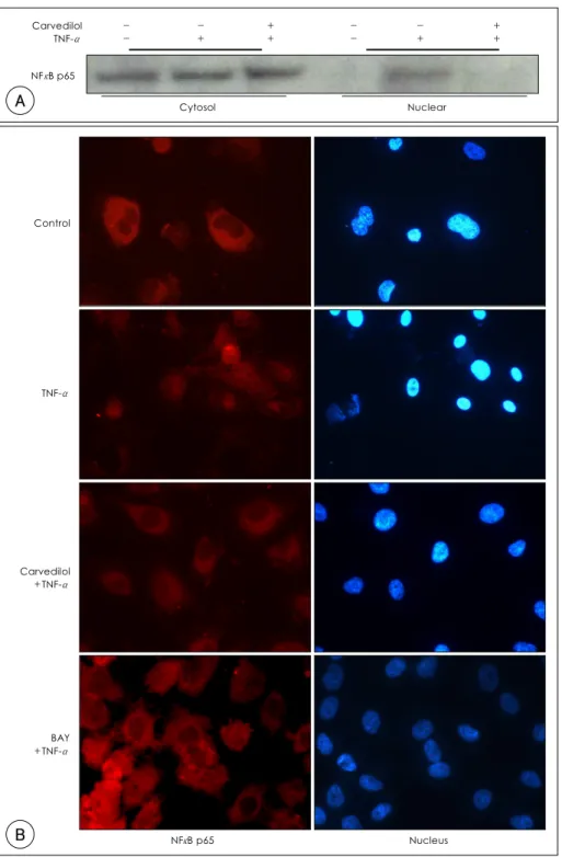
Fig. 1. A: nuclear translocation of NF-κB p65 due to TNF-α was inhibited by carvedilol. Cells were pretreated with carvedilol (10 μM) for 1 hour prior to the addition of TNF-α (10 ng/mL). After 30 minutes, the cytosol and nuclear proteins were fractionized and subjected to western blot. B: intracellular localization of NF-κB p65 was demonstrated by immunocytochemistry, as described in materials and methods. Carvedilol and BAY 11-7082 significantly blocked the NF-κB p65 nuclear translocation. TNF-α: tumor necrosis factor-α, NF-κB: nuclear factor kappa B.
Carvedilol TNF-α
- -
- +
+ +
- -
- +
+ +
Cytosol Nuclear
NFκB p65
Control
TNF-α
Carvedilol +TNF-α
BAY +TNF-α
NFκB p65 Nucleus
A
B
taining 5% skim milk. The polyvinylidene fluoride was blotted against the indicated antibodies. The protein levels were determined using Western Breeze reagents and an Image Reader(LAS-3000 Imaging System, Fuji Photo Film).
ICAM-1, MCP-1, and IL-8 analysis by enzyme-linked immunosorbent assay(ELISA)
Cells were seeded onto a 96-well plate, pretreated with carvedilol (10 μM) for 1 hour, followed by the addition of TNF-α. After incubation for 16 h, in CO2
incubator, the supernatants were collected to measure ICAM-I, MCP-I, and IL-8. The concentrations of cyto- kines were quantified using a commercially available ELISA development system, according to the manufac- turer’s protocol.
Results
Carvedilol inhibited NF-κB p65 nuclear translo- cation induced by TNF-α
To investigate the effect of carvedilol on the NF-κB activation, the protein expression of NF-κB p65 in the cytoplasmic and nuclear fractions were examined. As shown by Western blot and immunocytochemistry, the transcription factor NF-κB p65 is translocated from the cytosol to the nucleus when activated by TNF-α.
NF-κB p65 activation; however, was inhibited by car- vedilol, as well as BAY11-7082, a specific inhibitor of NF-κB(Fig. 1).
Carvedilol inhibited ROS generation and U937 mo- nocyte adhesion induced by TNF-α
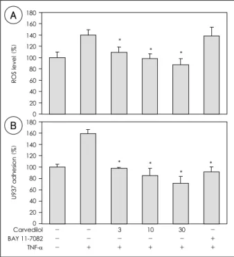
To determine the anti-oxidative effect of carvedilol on TNF-α stimulated HUVEC, the intracellular ROS level was measured. TNF-α increased the intracellular
ROS level at 1 hour(141±5.3% vs. control, p<0.05, n=4). Pretreatment with carvedilol for 1 hour signi- ficantly reduced the TNF-α induced ROS formation,
*
* *
0 20 40 60 80 100 120 140 160 180
ROS level (%)
*
*
* *
U937 adhesion (%)
0 20 40 60 80 100 120 140 160 180
Carvedilol BAY 11-7082 TNF-α
- - -
- - +
3 - +
10 - +
30 - +
- + +
A
B
Fig. 2. A: The effect of carvedilol on the intracellular ROS level in TNF-α-activated HUVEC. HUVEC were treated with carvedilol (3, 10, 30 μM) or BAY 11-7092 (10 μM) 1 hour prior to the addition of TNF-α (10 ng/mL). The DCFH-DA in the cells was labeled, with fluorescence released due to oxidative reduction. A representative of three experiments is shown. Data are presented as the mean±SD (n=4), *: p<0.05 vs. TNF-α alone. B: the effect of car-vedilol on the U937 adhesion to TNF-α-activated HUVEC. HUVEC were pretre- ated with carvedilol, activated with TNF-α (10 ng/mL), and cocul- tured with BCECF-labeled U937. A representative of three experi- ments is shown. Data are the means±SD(n=3), and represent the quantitative fluorescent results obtained from a microplate reader. *:
p<0.05 vs. TNF-α alone. ROS: reactive oxygen species, HUVEC:
human umbilical vein endothelial cells, DCFH-DA: dichlorofluore- scin diacetate, TNF-α: tumor necrosis factor-α, BCECF: 2’,7’-bis (carboxyethyl)-5(6)-carboxyfluorescein.
Carvedilol BAY 11-7082 TNF-α VCAM-1
ICAM-1
MCP-1
IL-B
β-actin - - -
3 - +
10 - +
30 - +
- - +
- - -
- - +
- + +
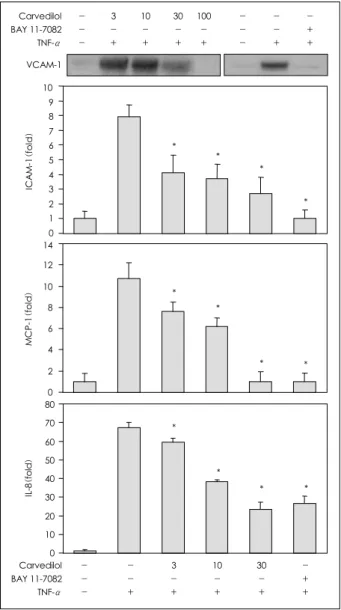
Fig. 3. The effects of carvedilol on the TNF-α induced expressions of VCAM-1, ICAM-1, MCP-1 and IL-8. A, HUVEC were pretreated with carvedilol (3, 10, 30 μM) or BAY 11-7092 (10 μM) for 1 hour. Three hours after TNF-α stimulation, total RNA was extracted and used for RT- PCR to determine the mRNA levels (5 separate experiments). TNF-α: tumor necrosis factor-α, VCAM-1: vascular cell adhesion molecule-1, ICAM-1: intercellular adhesion molecule-1, MCP-1: monocyte chemoattractant protein-1, IL-8: interleukin-8, HUVEC: human umbilical vein endothelial cells, RNA: ribonucleic acid, RT-PCR: reverse transcription polymerase chain reaction.
in a dose-dependent manner, and 10 μM carvedilol almost completely blocked the TNF-α in-duced ROS formation. Conversely, pretreatment with BAY 11-7082 had no effect on the elevated ROS level caused by TNF-α(Fig. 2A).
To examine the effect of carvedilol on the inflam- mation induced by TNF-α, an adhesion assay, using the human premonocytic cell line, U937, was carried out. Adhesion of the U937 cells to the HUVEC was enhanced by TNF-α(161±7% vs. control, p<0.05, n=
3). Pretreatment with carvedilol for 1 hour signifi- cantly inhibited the TNF-α induced adhesion, in a
dose-dependent manner. The BAY 11-7082 also almost completely blocked the U937 adhesion induced by TNF-α(Fig. 2B).
Carvedilol reduced VCAM-1, ICAM-1, MCP-1, and IL-8 expression induced by TNF-α
The expressions of the NF-κB dependent pro-in- flammatory molecules, i.e., VCAM-1, ICAM-1, MCP- 1 and IL-8, were determined at the mRNA and protein levels. TNF-α induced both transcriptions and tran- slations of the VCAM-1, ICAM-1, MCP-1 and IL-8.
Carvedilol reduced the inductions of the VCAM-1, ICAM-1, MCP-1 and IL-8 mRNAs in the HUVEC treated with TNF-α, in a dose dependent manner(Fig.
3). The BAY 11-7082 also suppressed the transcrip- tional expressions of VCAM-1, ICAM-1, MCP-1 and IL-8.
The expressions of the VCAM-1, ICAM-1, MCP-1 and IL-8 proteins were markedly increased in the TNF- α stimulated cells. The TNF-α stimulated cells treated with 10 μM carvedilol demonstrated full inhibition of the VCAM-1 protein, as well as dose-dependent in- hibition of the ICAM-1, MCP-1 and IL-8(Fig. 4).
TNF-α-activated expressions of the VCAM-1, ICAM- 1, MCP-1 and IL-8 proteins were inhibited when the HUVEC were pretreated with BAY 11-7082.
Carvedilol inhibited JNK activation and Bcl-2 re- duction induced by TNF-α
The stress-activated signaling molecules, such as JNK and Bcl-2, were examined to see if they were modu- lated by carvedilol. TNF-α increased the JNK phos- phorylation within 15 minutes, but reduced the Bcl-2 expression within 24 hours. Carvedilol inhibited the
Fig. 4. The whole cell lysates were prepared 16 hours after stimu- lation, separated on SDS-PAGE, transferred to a PVDF membrane, and immunoblotted with an anti-VCAM-1 antibody. Cells were treated, as indicated, for 16 hours, culture media collected and the ICAM-1, MCP-1 and IL-8 levels measured using an ELISA kit.
Data are expressed as the mean±SD of triplicate wells. Results are representative of three independent experiments. SDS-PAGE: so- dium dodecyl sulfate-polyacrylamide gel electrophoresis, PVDF: poly- vinylidene flouride, VCAM-1: vascular cell adhesion molecule-1, ICAM-1: intercellular adhesion molecule-1, MCP-1: monocyte che- moattractant protein-1, IL-8: interleukin-8.
Carvedilol BAY 11-7082 TNF-α VCAM-1
- - -
3 - +
10 - +
- - -
- - +
- + + 30
- +
100 - +
0 1 2 3 4 5 6 7 8 9 10
ICAM-1(fold)
* *
*
*
0 2 4 6 8 10 12 14
MCP-1(fold) *
*
* *
Carvedilol BAY 11-7082 TNF-α
- - -
- - +
3 - +
10 - +
30 - +
- + +
IL-8(fold)
0 10 20 30 40 50 60 70 80
* *
*
*
Carvedilol BAY 11-7082 TNF-α p-JNK
JNK
Bcl-2
anctin - - -
- - +
+ - +
- - -
- - +
- + +
Fig. 5. The effects of carvedilol on JNK phosphorylation and Bcl-2.
HUVEC were pretreated with carvedilol (10 μM) or BAY 11-7082 (10 μM) and then cultured with TNF-α, as indicated, for 15 mi- nutes or 24 hours, to detect JNK phosphorylation and the Bcl-2 level, respectively. The whole cell lysates were prepared, separated on SDS- PAGE, transferred to a PVDF membrane, and immuno-blotted with an anti-phosphorylated JNK or Bcl-2 antibody. Results are represen- tative of three independent experiments. JNK: c-Jun N-terminal pro- tein kinase, HUVEC: human umbilical vein endothelial cells, TNF- α: tumor necrosis faction-α, SDS-PAGE: sodium dodecyl sulfate- polyacrylamide gel electrophoresis, PVDF: polyvinylidene flouride.
TNF-α induced JNK phosphorylation and Bcl-2 re- duction, while with BAY 11-7082 the phosphorylation of JNK and Bcl-2 protein expression patterns rema- ined unchanged(Fig. 5).
Discussion
Inflammatory reactions, such as the expressions of cell adhesion molecules and cytokines with monocyte adhesion, have been observed in atherosclerotic lesions.
Increased pro-inflammatory cytokines and the expres- sions of adhesion molecules, such as VCAM and ICAM, on endothelial cells participate in atherogenesis.11)12) In this study, VCAM-1, ICAM-1, MCP-1 and IL-8, which have been reported to be regulated in a strictly NF-κB dependent fashion, were chosen as parameters of in- flammation.13)
Many studies have revealed carvedilol to be an anti- oxidative, the cardioprotective effects of which are me- diated by the inhibition of NF-κB activation.14)15) ROS are thought to be involved in the development of a number of pathological conditions and diseases, es- pecially of the cardiovascular system.16) The admini- stration of anti-oxidative drugs to treat or prevent such conditions is of great interest. Singh et al.17) reported that carvedilol significantly attenuated lipid peroxida- tion and protected against severe depletion of the an- tioxidant enzyme pool, such as reduced glutathione, glutathione reductase, catalase and SOD in glycerol- treated rats.
NF-κB has been suggested as the major transcrip- tional factor in chronic inflammatory diseases related to endothelial adhesion molecules, such as E-selectin, ICAM-1 and VCAM-1, as well as of chemokines, such as monocyte-chemoattractant protein-1(MCP-1) and IL-8.3) In this study, carvedilol blocked the nuclear translocation of NF-κB p65 stimulated by TNF-α, which in turn reduced the expressions of VCAM-1, ICAM-1, MCP-1, and IL-8 at the transcriptional level.
Carvedilol was also found to attenuate the increase in the intracellular ROS level, in a dose-dependent manner, in TNF-α treated HUVEC.
Carvedilol may inhibit early events in the atheros- clerotic process, possibly due to its interference with NF-κB-dependent transcription. The effects of BAY 11-7082 on the action of TNF-α were tested and com- pare with those of carvedilol. BAY 11-7082, an irre- versible inhibitor of IκBα phosphorylation, also in- hibited the NF-κB p65 translocation induced by TNF- α. BAY 11-7082 inhibited the U937 adhesion, and the expressions of VCAM-1, ICAM-1, MCP-1 and IL- 8; however, unlike carvedilol, the elevated ROS level induced by TNF-α was unchanged.
JNK, a member of MAPKs, plays important roles in signaling pathways for apoptosis, transformation, de-
velopment, immune activation, inflammation and adaptation to environmental changes.5) This enzyme is important in the pathways regulating cell death, which are usually activated by cytokines, such as TNF-α and IL-1, and genotoxic stresses, such as UV. In this report, JNK phosphorylation due to TNFα increased within 15 minutes, but decreased to basal levels within 1 hour (data not shown). Pretreatment of carvedilol for 1 hour significantly inhibited JNK phosphorylation. In addi- tion, the level of Bcl-2, one of the regulatory proteins related to cell survival, was also examined.18) TNF-α has been reported to specifically suppress the expres- sion of Bcl-2 and induce a rise in the concentration of cytoplasmic Ca2+ due to the release of Ca2+ from in- tracellular Ca2+ stores in hepatoma cells.19) Carvedilol rescued the Bcl-2 protein, which was significantly down-regulated by TNF-α. BAY11-7082; however, did not alter the ROS production, JNK phosphorylation or Bcl-2 down-regulation in TNF-α stimulated HUVEC.
Although carvedilol has additional anti-oxidative activity, which is independent of its anti-hypertensive effects, it is not clear whether ROS might enhance the action of TNF-α. To address this question, the effect of antioxidants on TNF-α-induced events should be exa- mined.
In our data, carvedilol almost completely inhibited the U937 monocyte adhesion to TNF-α-activated en- dothelial cells, at least in part, by the inhibition of the expressions of VCAM-1 and ICAM-1. Accordingly, the Western blot data for VCAM-1 supported the in vitro adhesion results. This study has provided insights to the role exerted by carvedilol, and the beneficial effects on TNF-α-stimulated HUVEC through its antioxi- dant activity, NF-κB inhibition and JNK inhibition, as well as α-adrenoreceptor blockade. From the present data, it is suggested that carvedilol could significantly reduce the expressions of inflammatory molecules in- duced by TNF-α. Therefore, the effects of carvedilol might be suggested to be related with its antioxidant activity, contributing significantly to its clinical benefit.
Carvedilol blocked the expression of cytokines by direct modulation at the gene transcription level.
In conclusion, carvedilol has efficacy in inhibiting TNF-α-induced inflammatory responses, as well as apoptosis. These observations suggest that carvedilol plays a significant role in the attenuation of inflam- matory disease and apoptosis in the cardiovascular system.
This study was financially supported by a research fund of Chon- nam National University in 2004.
REFERENCES
1) Ross R. Atherosclerosis: an inflammatory disease. N Eng J Med 1999;340:115-26.
2) Hansson GK. Immune mechanisms in atherosclerosis. Arterio- scler Thromb Vasc Biol 2001;21:1876-90.
3) Barnes PJ, Karin M. Nuclear factor-kappa B: a pivotal transc- ription factor in chronic inflammatory diseases. N Engl J Med 1997;336:1066-71.
4) Molet S, Gosset P, Vanhee D, et al. Modulation of cell adhesion molecules on human endothelial cells by eosinophil-derived me- diators. J Leukoc Biol 1998;63:351-8.
5) Davis RJ. Signal transduction by the JNK group of MAP kinases.
Cell 2000;103:239-52.
6) Adler V, Yin Z, Tew KD, Ronai Z. Role of redox potential and reactive oxygen species in stress signaling. Oncogene 1999;18:
6104-11.
7) Cha DH, Cha YS, Kook JH, et al. Clinical efficacy of carvedilol in patients with moderate to severe congestive heart failure.
Korean Circ J 1998;28:523-31.
8) Lee JM, Kim CM, Youn HJ, et al. The effects of carvedilol on ventricular remodeling after myocardial infarction in rats. Korean Circ J 2001;31:1171-84.
9) Cha KS, Kim MH, Kim JW, et al. The effectiveness of carvedilol, a new antioxidant and antiproliferative beta-blocker, on pre- vention of restenosis after coronary stent implantationa pros- pective, randomized, multicenter study. Korean Circ J 2004;34:
35-40.
10) Yue TL, Mckenns PJ, Lysko PG, et al. SB 211475, a metabolite of carvedilol, a novel antihypertensive agent, is a potent antioxi- dant. Eur J Pharmacol 1994;251:237-43.
11) Schmidt AM, Crandall J, Hori O, Lakatta E. Elevated plasma levels of vascular cell adhesion molecule-1(VCAM-1) in diabetic
patients with microalbuminurea: a marker of vascular dysfunc- tion and progressive vascular disease. Br J Haematol 1996;92:
747-50.
12) Eppihimer MJ. The role of leukocyte-endothelial cell adhesion in cardiovascular disease. Pathophysiology 1998;5:167-84.
13) Hsu MH, Wang M, Browning DD, Mukaida N, Ye RD. NF-κB activation is required for C5a-induced interleukin-8 gene expres- sion in mononuclear cells. Blood 1999;93:3241-9.
14) Oettl K, Greilberger J, Zangger K, Haslinger E, Reibnegger G, Jurgens G. Radical-scavenging and iron-chelating properties of carvedilol, an antihypertensive drug with antioxidative activity.
Biochem Pharmacol 2001;62:241-8.
15) Fahlbusch SA, Tsikas D, Mehls C, et al. Effects of carveilol on oxidative stress in human endothelial cells and healthy volunteers.
Eur J Clin Pharmacol 2004;60:83-8.
16) Griendling KK, Sorescu D, Ushio-Fukai M. NAD(P)H oxidase:
role in cardiovascular biology and disease. Circ Res 2000;86:
494-501.
17) Singh D, Chander V, Chopra K. Carvedilol, an antihypertensive drug with antioxidant properties, protects against glycerol-in- duced acute renal failure. Am J Nephrol 2003;23:415-21.
18) Luo T, Xia Z, Ansley DM, et al. Propofol dose-dependently reduces tumor necrosis factor-alpha-induced human umbilical vein endothelial cell apoptosis: effects on Bcl-2 and Bax expres- sion and nitric oxide generation. Anesth Analg 2005;100:1653-9.
19) Kim BC, Kim HT, Mamura M, Ambudkar IS, Choi KS, Kim SJ.
Tumor necrosis factor induces apoptosis in hepatoma cells by increasing Ca2+ release from the endoplasmic reticulum and suppressing Bcl-2 expression. J Biol Chem 2002;277:31381-9.