Received: 2013.5.21. Revised: 2013.9.3. Accepted: 2013.9.16.
Corresponding author: Byoung-Gie Kim
Department of Obstetrics and Gynecology, Samsung Medical Center, Sungkyunkwan University School of Medicine, 81 Irwon- ro, Gangnam-gu, Seoul 135-710, Korea
Tel: +82-2-3410-3513 Fax: +82-2-3410-0630 E-mail: [email protected]
Articles published in Obstet Gynecol Sci are open-access, distributed under the terms of the Creative Commons Attribution Non-Commercial License (http://creativecommons.
org/licenses/by-nc/3.0/) which permits unrestricted non-commercial use, distribution, and reproduction in any medium, provided the original work is properly cited.
Copyright © 2014 Korean Society of Obstetrics and Gynecology
Introduction
Cervical cancer is still the one of the most common malignan- cies in female, which is the third common cancer in women worldwide, even though the incidence has been steadily decreased in recent years [1,2]. The most of deaths from cervical cancer come from the recurrence of the disease [3].
As a result, predicting recurrence after primary treatment is important not only for counseling the patients about the dis- ease prognosis but also for applying additional treatment to prevent recurrence.
In predicting recurrence of the cervical cancer, clinical stag- ing system has limited value because of inter-observer vari- ability of physical examination and not considering of other important factors such as lymph node (LN) metastasis [4]. As
a result many clinicopathological factors are used to stratify the risk for recurrence of the disease [5-7]. However there is
Prognostic value of pretreatment hemoglobin level in patients with early cervical cancer
Na-Ri Shin, Yoo-Young Lee, Seung-Hyun Kim, Chel Hun Choi, Tae-Joong Kim, Jeong-Won Lee, Duk-Soo Bae, Byoung-Gie Kim
Department of Obstetrics and Gynecology, Samsung Medical Center, Sungkyunkwan University School of Medicine, Seoul, Korea
Objective
The purpose of this study is to investigate the prognostic role of pretreatment anemia in patients with early cervical cancer who underwent radical hysterectomy.
Methods
In this study, we retrospectively enrolled patients with early cervical cancer (International Federation of Obstetrics and Gynecology stage IB to IIA) who were treated at Samsung Medical Center, Seoul, Korea, from 1996 to 2007.
Results
We retrospectively enrolled 805 patients. Median pretreatment hemoglobin (Hb) level was 12.8 g/dL (4.0–16.9) in all patients. Ninety-ninth out of 805 patients had pretreatment anemia (12.3%). Pretreatment anemia was significantly associated with large tumor size, advanced clinical stage, and parametrial invasion. In multivariate analysis, higher pretreatment Hb entailed better prognostic significance in disease free survival (hazard ratio [HR], 0.88; 95%
confidence interval [CI], 0.078–0.99) but not in overall survival (HR, 0.94; 95% CI, 0.80–1.10).
Conclusion
In conclusion, we found that the negative association between pretreatment Hb level and tumor size and the impact of anemia before treatment on disease free survival adjusted for other factors including clinical stage and pathological findings in early stage cervical cancer.
Keywords: Anemia; Hemoglobins; Prognosis; Survival; Uterine cervical neoplasms
http://dx.doi.org/10.5468/ogs.2014.57.1.28pISSN 2287-8572 · eISSN 2287-8580
still a lack of strong predictor for recurrence up to date and many investigation searching for risk factors for recurrence in cervical cancer is ongoing.
Anemia is a common condition in cancer patients [8]. Es- pecially, cervical cancer is among the tumors characterized by higher prevalence of anemia at diagnosis [9]. And, interesting- ly, lower pretreatment hemoglobin (Hb) level or anemia before treatment has been reported as an independent prognostic factor for poor prognosis in locally advanced cervical cancer (LACC) [10,11]. For this reason Hb level before treatment was considered as one of the parameters used in prognostic model predicting recurrence in LACC [12]. However, the prognostic role of pretreatment anemia or lower Hb level in patients with early stage cervical cancer (ECC) is still unclear.
As a result, this study was designed to investigate the role of pretreatment anemia as a prognostic factor in ECC and association between pretreatment Hb level and clinicopatho- logical factors in these patients.
Materials and methods
1. Patients
With institutional review board approval, patients with ECC (International Federation of Obstetrics and Gynecology stage IB to IIA) who were treated at Samsung Medical Cen- ter, Seoul, Korea from 1996 to 2007 were retrospectively enrolled in this study. The patients’ clinical data and patho- logical findings after surgery as well as laboratory results were collected. We excluded patients with IA1 and IA2;
atypical histological subtypes including clear cell, melanoma, metastatic carcinoma, etc.; patients who underwent fertility- saving surgery; patients with concurrent hematologic dis- eases; patients with para-aortic LN metastasis; patients who did not have the results of Hb level within two weeks before starting initial treatment; patients who received transfusion before blood sampling; and patients who had radiation therapy (RT) oriented therapy as a primary treatment. At our institution, anemia is diagnosed when Hb level is less than 11.2 g/dL for adult female. As a result we use the Hb level of 11.2 g/dL as a cut-off value for the analysis.
2. Treatment
We usually performed surgery as a primary treatment in patients with ECC (IB1 to IIA). However, the choice for pri-
mary treatment was dependent on the attending physician’s preference. Since 2000, platinum based concurrent chemo- radiation therapy (CCRT) has been recommended as adju- vant treatment in cases with more than one high-risk patho- logical factor for recurrence after surgery, which is described below.
As we prescribed previously [13], standard surgery con- sisted of type III radical hysterectomy with bilateral pelvic LN dissection. Additional procedures such as bilateral salpingo- oophorectomy and para-arotic LN sampling or dissections were not routinely performed. Adjuvant therapy after surgery was considered based on pathological risk factors. Patients who had more than one of the three high-risk factors (posi- tive pelvic LN, microscopic parametrial invasion, and positive resection margins with tumor) received adjuvant platinum based CCRT. Patients with at least two of the three interme- diate risk factors (stromal invasion of more than half of the cervix or stromal invasion more than 1 cm, lympho-vascular space invasion [LVSI], and the largest pathological diameter of 4 cm or greater) received adjuvant RT alone.
RT protocols were also as previously described [13]. In brief, each patient received external beam RT therapy using 10 to 15-MV photons to the whole pelvis for a total dose of 50.4 Gy. The daily fraction size was 1.8 Gy, administered five times per week. Patients were irradiated with a four-field box tech- nique (anterior, posterior, and bilaterals) to spare some of the small bowel anterior to the iliac nodes.
We follow-up the patients with examinations approximately every three months for the first two years, every six months for the next three years, and every year thereafter. During the rou- tine follow-up, computed tomography or magnetic resonance imaging, and chest X-ray was performed annually. We defined disease free survival as the time from the initial treatment to relapse or to the final follow-up visit, and overall survival was defined as the time from the initial treatment to death or to the final follow-up visit.
3. Statistical analysis
The Wilcoxon rank sum test or two-sample t-test was used to
compare the median or mean values, respectively, after check-
ing whether the data had non-normal or normal distributions
according to the Shapiro-Wilks test. Comparisons of means
or medians among three groups were performed using the
one way ANOVA as a parametric test or Kruskal-Wallis test
as a non-parametric test. Spearman correlation analysis was
used to investigate the association between tumor diameter and pretreatment Hb level. Frequency distributions between categorical variables were compared using the χ
2test. The Fisher’s exact test was used if the expected frequency was
<5. The overall and progression-free survival curves were cal- culated according to the Kaplan-Meier method with the log- rank test. The Cox proportional-hazards model was used for the multivariate analyses. Statistical analyses were performed using PASW ver. 18.0 (SPSS Inc., Chicago, IL, USA). A P-value
<0.05 was considered statistically significant and all P-values were two-sided.
Results
We could enroll 805 patients with ECC (IB1 to IIA) who were treated with surgery with or without adjuvant thera- pies. The characteristics of patients are described in Table 1.
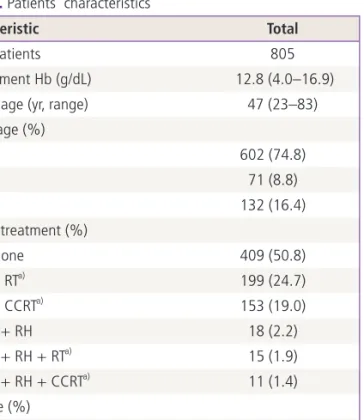
The median age of the patients was 47 years (range, 2–83 years). Median pretreatment Hb level was 12.8 g/dL (range, 4.0–16.9 g/dL) in this study population. Stage IB1 was most common (602, 74.8%) and about a half of patients (409/805, 50.8%) underwent surgery alone, 199 (24.7%) patient underwent surgery with RT, 153 (19.0%) patients underwent surgery with CCRT and the rest of the patients (44/805, 5.5%) had neoadjuvant chemotherapy before sur- gery. Histologically, squamous cell was the most frequent histologic subtype (611/805, 75.9).
The median follow-up for the patient group was 58.3 months with a range of 2 to 181 months and the five-year survival rate was 92.4%. There were 96 (11.9%) recurrences and 56 (7.0%) deaths during the study period.
When we divided the patients into two groups accord- ing to whether the patients had pretreatment anemia or not, there was no statistical significant difference between groups but trends were shown that the patients with pre- treatment anemia may have worse survival outcomes when compared with the patients without anemia (Fig. 1). Then we compared the association between pretreatment anemia and clinico-pathological findings which have been known as risk factors for recurrence. First, patients with IB2 had lower pretreatment Hb level compared with the patients with IB1, which was statistically significant ( P = 0.016), and negative correlation was observed between tumor size and pretreatment Hb level with statistical significance (R
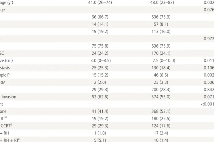
2=- 0.089, P = 0.011) as shown in Fig. 2. Second, among the high risk factors for recurrence, microscopic parametrial invasion was significantly associated with pretreatment ane- mia (P = 0.002) (Table 2). However, pretreatment Hb level was not associated with lymphatic invasion of tumor cells including lympho-vascular space invasion or LN metastasis as well as histologic subtypes and vaginal resection margin with tumor.
In all patients, the univariate analysis revealed that pretreat- ment anemia and other clinicopathological findings such as stage, type of treatment, histological subtype, and intermedi- ate/high risk factors for recurrence had prognostic significance for both disease free and overall survival (Tables 3, 4). In multivariate analyses, lower pretreatment Hb remained as a significant independent poor prognostic factor in disease free survival (P = 0.039) even after adjusting for other factors in- cluding tumor size but this finding was not observed in overall survival (P = 0.404) (Tables 3, 4).
Table 1. Patients’ characteristics
Characteristic Total
No. of patients 805
Pretreatment Hb (g/dL) 12.8 (4.0–16.9)
Median age (yr, range) 47 (23–83)
FIGO stage (%)
IB1 602 (74.8)
IB2 71 (8.8)
IIA 132 (16.4)
Primary treatment (%)
RH alone 409 (50.8)
RH + RTa) 199 (24.7)
RH + CCRTa) 153 (19.0)
NAC + RH 18 (2.2)
NAC + RH + RTa) 15 (1.9)
NAC + RH + CCRTa) 11 (1.4)
Cell type (%)
SCC 611 (75.9)
ASC 51 (6.3)
AC 143 (17.8)
Hb, hemoglobin; FIGO, International Federation of Obstetrics and Gynecology; RH, type III radical hysterectomy; RT, radiation therapy;
CCRT, concurrent chemoradiation therapy; NAC, neoadjuvant che- motherapy; SCC, squamous cell carcinoma; ASC, adenosquamous cell carcinoma; AC, adenocarcinoma.
a)Adjuvant setting.
Discussion
In this study, we found that the negative association between pretreatment Hb level and tumor size and the impact of lower Hb before treatment on disease free survival adjusted for
other factors including clinical stage and pathological findings in ECC.
Anemia is known to be a common finding in cancer patients and up to 30% of cancer patients suffer from anemia [14].
Decreased Hb from the normal range is reported to be associ-
Fig. 1. (A) Disease free survival and (B) overall survival according to the pretreatment hemoglobin (Hb, with anemia vs.without anemia) in patients with early cervical cancer.
P=0.060 P=0.148
0 50 100 150 200 Hb ≥11.2 g/dL
Hb <11.2 g/dL
Hb ≥11.2 g/dL Hb <11.2 g/dL
0 50 100 150 200 1.0
0.8
0.6
0.4
0.2
0.0
1.0
0.8
0.6
0.4
0.2
0.0
Month Month
Cumulative survival Cumulative survival
A B
Fig. 2. The association between pretreatment hemoglobin (Hb) level and International Federation of Obstetrics and Gynecology (FIGO) clinical stage (A) and tumor size (B).
IB1 IB2 IIA P=0.016
R2=-0.089, P=0.011 17.5
15.0 12.5 10.0 7.5 5.0
17.5 15.0 12.5 10.0 7.5 5.0
0.0 2.0 4.0 6.0 8.0 10.0 Tumor size (cm)
FIGO stage
Pretreatment Hb level (g/dL) Pretreatment Hb level (g/dL)
A B
ated with shorter survival times for patients with solid ma- lignant tumors including lung, head and neck, prostate, and hematologic malignancies such as lymphoma and multiple myeloma and the overall estimate increase in risk of death was 65% (range, 54%–77%) in a meta-analysis [8]. Among gynecologic malignancies, anemia was also found to have an independent relationship to poor survival in epithelial ovarian cancer [15], endometrial cancer [16] and LACC [11,17-21].
However, there is no evidence of association between pre- treatment Hb and prognosis in ECC and we found its signifi- cant independent prognostic role in this study.
In our study, the proportion of patients with pretreatment anemia was 12.3% which is relatively lower incidence from that of LACC reported in previous studies, which was about 25% [11,22]. It suggest that bulky tumors might have more
anemic condition compared with small volume tumors and the finding of negative correlation between tumor size and pretreatment Hb level in this study is supporting this idea.
A review article addressing the anemia and cervical cancer also reported that Hb levels prior to and during radiotherapy are strongly correlated with tumor size (R
2=-0.46, P < 0.001) measured based on imaging studies in LACC [9], which is cor- responding well with the result shown in our ECC patients.
Overall, pretreatment Hb may be a surrogate marker for dis- ease burden itself and may partially explain the prognostic impact on survival in patients with ECC in this respect.
Apart from the association between pretreatment anemia and tumor size, we could also find that pretreatment anemia have independent prognostic power despite the impact of tumor size on anemia prior to treatment. Kapp et al. [23]
Table 2. Clinico-pathological findings based on the pretreatment Hb level
Characteristic Hb <11.2 g/dL
(n = 99, 12.3%) Hb ≥11.2 g/dL
(n = 706, 87.7%) P-value
Median age (yr) 44.0 (26–74) 48.0 (23–83) 0.002
FIGO stage 0.076
IB1 66 (66.7) 536 (75.9)
IB2 14 (14.1) 57 (8.1)
IIA 19 (19.2) 113 (16.0)
Cell type 0.972
SCC 75 (75.8) 536 (75.9)
AC/ASC 24 (24.2) 170 (24.1)
Tumor size (cm) 3.0 (0–8.5) 2.5 (0–10.0) 0.011
LN metastasis 25 (25.3) 130 (18.4) 0.106
Microscopic PI 15 (15.2) 46 (6.5) 0.002
Vaginal RM 2 (2.0) 23 (3.3) 0.506
LVSI 29 (29.3) 200 (28.3) 0.842
Depth of invasion 62 (62.6) 374 (53.0) 0.071
Treatment <0.001
RH alone 41 (41.4) 368 (52.1)
RH + RTa) 19 (19.2) 180 (25.5)
RH + CCRTa) 29 (29.3) 124 (17.6)
NAC + RH 1 (1.0) 17 (2.4)
NAC + RH + RTa) 5 (5.1) 10 (1.4)
NAC + RH + CCRTa) 4 (4.0) 7 (1.0)
Values are presented as number (range) or (%).
Hb, hemoglobin; FIGO, International Federation of Obstetrics and Gynecology; SCC, squamous cell carcinoma; AC, adenocarcinoma; ASC, ad- enosquamous cell carcinoma; LN, lymph node; PI, parametrial invasion; RM, resection margin with tumor; LVSI, lympho-vascular space inva- sion; RH, type III radical hysterectomy; RT, radiation therapy; CCRT, concurrent chemoradiation therapy; NAC, neoadjuvant chemotherapy.
a)Adjuvant setting.
reported that not only tumor size but also Hb was one of the significant prognostic factors in a multivariate analysis for survival which is similar with the result of our study. And some authors reported that lower Hb level is related with infiltrative phenotypes of tumors such as corpus invasion and nodal metastases in LACC [24]. Another possible explanation about poor survival and anemia in LACC is that anemia might be a surrogate marker of tumor hypoxia which is known to mediate resistance to radiotherapy [9,25]. So it is difficult to consider anemia as a direct or indirect marker for poor sur- vival, however, it is clear that patients with anemia have lower survival.
Generally anemic conditions can be reversed by transfusion
or administration of erythropoiesis stimulating agents (ESA).
And some authors have suggested that the maintenance of Hb at normal levels would improve not only for quality of life [26,27] but also survival [21,22,28]. However, it is still unknown if transfusion ameliorate hypoxia of tumors and im- prove the outcome of anemic patients. A report showed that only 50% of patients with cervical cancer demonstrated an increase in tumor oxygenation following transfusion [29] and did not improved survival [9]. Furthermore, although transfu- sion may increase tumor oxygenation, does not necessarily improve treatment results as anemia is associated with tumor sizes which has an adverse prognosis independent of oxygen- ation. And in the treatment with ESA, there are still concerns
Table 3. Univariate and multivariate analysis for disease free survivalCharacteristic Univariate Multivariate
HR (95% CI) P-value HR (95% CI) P-value
Pretreatment Hb (continuous value) 0.84 (0.75–0.95) 0.003 0.88 (0.78–0.99) 0.039
Age 1.00 (0.98–1.02) 0.692 1.00 (0.98–1.02) 0.805
FIGO stage <0.001 0.013
IB1 1 1
IB2 4.48 (2.70–7.43) 2.35 (1.29–4.28)
IIA 2.87 (1.79–4.61) 1.72 (1.01–2.93)
Treatment <0.001 0.014
RH alone 1 1
RH+RTa) 2.22 (1.18–4.16) 0.69 (0.32–1.48)
RH+CCRTa) 7.18 (4.21–12.3) 0.75 (0.33–1.74)
NAC+RH 3.83 (1.13–12.9) 4.48 (1.23–16.4)
NAC+RH+RT a) 8.75 (3.28–23.5) 4.00 (1.26–12.4)
NAC+RH+CCRTa) 7.00 (2.07–23.7) 0.79 (0.19–3.29)
Cell type <0.001 <0.001
SCC 1 1
AC/ASC 2.71 (1.81–4.05) 5.67 (3.55–9.06)
Tumor size 1.42 (1.28–1.57) <0.001 1.13 (0.98–1.30) 0.101
LN metastasis 5.90 (3.95–8.82) <0.001 4.40 (2.48–7.80) <0.001
Microscopic PI 4.48 (2.76–7.28) <0.001 1.58 (0.90–2.78) 0.111
Vaginal RM 2.57 (1.19–5.54) 0.016 2.22 (0.98–5.04) 0.057
LVSI 2.28 (1.53–3.41) <0.001 1.36 (0.83–2.21) 0.214
Depth of invasion 4.75 (2.74–8.24) <0.001 2.20 (1.10–4.41) 0.027
Pretreatment Hb levels were analyzed as a continous variable.
HR, hazard ratio; CI, confidence interval; Hb, hemoglobin; FIGO, International Federation of Obstetrics and Gynecology; RH, type III radical hysterectomy; RT, radiation therapy; CCRT, concurrent chemoradiation therapy; NAC, neoadjuvant chemotherapy; SCC, squamous cell car- cinoma; AC, adenocarcinoma; ASC, adenosquamous cell carcinoma; LN, lymph node; PI, parametrial invasion; RM, resection margin with tumor; LVSI, lympho-vascular space invasion.
a)Adjuvant setting.
about its safety on cancer prognosis and for example ESA are not recommended in early breast and non-small cell lung cancer patients with anemia [30]. Overall, further studies investigating the prognostic impact of manipulating anemic condition before or during treatment and the safety of ESA in patients with ECC should be needed in the future.
In this study, there are several limitations. First, we did not count the effects of transfusions before surgery due to preop- eration anemia because the guidelines for transfusion such as Hb level to start transfusion and the amount of transfusion be- fore surgery were based on the attending physicians’ prefer- ences which were heterogenic. So some effects of transfusion might have influenced the results of this study. Second, we
could not see the prognostic effect of pretreatment anemia on overall survival. However, most of the patients with ECC show preferable overall survival that there may not be enough numbers of patients to see the survival difference based on the pretreatment anemia and anemic condition at the time of recurrence would be also important factor to overall survival.
Third, anemia before treatment should be frequently associ- ated with symptoms such as tumor related vaginal bleeding and menstrual cycles when the blood test was performed.
With the retrospective study design, we could not consider these confounding factors and bias may be introduced in our results.
In conclusion, lower pretreatment Hb is independently asso-
Table 4. Univariate and multivariate analysis for overall survivalCharacteristic Univariate Multivariate
HR (95% CI) P-value HR (95% CI) P-value
Pretreatment Hb (continuous value) 0.85 (0.73–0.99) 0.033 0.94 (0.80–1.10) 0.404
Age 1.02 (0.99–1.04) 0.144 1.02 (0.99–1.05) 0.106
FIGO stage <0.001 0.021
IB1 1 1
IB2 4.81 (2.38–9.71) 2.33 (1.04–5.21)
IIA 4.80 (2.66–8.67) 2.38 (1.22–4.66)
Treatment <0.001 0.314
RH alone 1 1
RH+RTa) 3.94 (1.67–9.31) 1.05 (0.36–3.10)
RH+CCRTa) 9.50 (4.31–20.9) 1.07 (0.34–3.39)
NAC+RH - -
NAC+RH+RTa) 9.76 (2.59–36.8) 5.20 (1.12–24.2)
NAC+RH+CCRTa) 15.7 (4.15–59.2) 1.99 (0.38–10.3)
Cell type <0.001 <0.001
SCC 1 1
AC/ASC 3.32 (1.97–5.61) 6.35 (3.49–11.6)
Tumor size 1.52 (1.33–1.73) <0.001 1.23 (1.02–1.48) 0.026
LN metastasis 7.11 (4.15–12.2) <0.001 4.15 (2.06–8.34) <0.001
Microscopic PI 5.30 (2.89–9.75) <0.001 1.35 (0.64–2.81) 0.432
Vaginal RM 3.90 (1.67–9.10) 0.002 2.83 (1.09–7.36) 0.033
LVSI 2.15 (1.27–3.64) 0.004 1.19 (0.64–2.21) 0.591
Depth of invasion 6.01 (2.72–13.3) <0.001 1.67 (0.61–4.56) 0.314
Pretreatment Hb levels were analyzed as a continous variable.
HR, hazard ratio; CI, confidence interval; Hb, hemoglobin; FIGO, International Federation of Obstetrics and Gynecology; RH, type III radical hysterectomy; RT, radiation therapy; CCRT, concurrent chemoradiation therapy; NAC, neoadjuvant chemotherapy; SCC, squamous cell car- cinoma; AC, adenocarcinoma; ASC, adenosquamous cell carcinoma; LN, lymph node; PI, parametrial invasion; RM, resection margin with tumor; LVSI, lympho-vascular space invasion.
a)Adjuvant setting.