Molecular Characterization of an H5N3 Influenza Virus Isolated from Spot-Billed Duck
Jin Hwa Lee
1, Hyuk Moo Kwon
1and Haan Woo Sung
1†1
College of Veterinary Medicine, Kangwon National University, Chuncheon, 200-701, Republic of Korea.
ABSTRACT Among the 16 hemagglutinin (HA) subtypes of avian influenza virus (AIV), only the H5 and H7 subtypes have caused highly pathogenic avian influenza (HPAI) in poultry. However, most H5 or H7 subtype viruses are categorized as low pathogenic avian influenza (LPAI). Some AIVs, including the H5 and H7 HPAI viruses, have shown the ability to infect humans directly. In this study, we describe the biological and molecular characterization of an H5N3 AIV (SBD/KR/KNU SYG06/06) isolated from spot-billed duck (Anas poecilorhyncha) in Korea. A phylogenetic analysis of the eight viral genes showed that the SBD/KR/KNU SYG06/06 isolate belongs to the Eurasian lineage and that the SBD/KR/KNU SYG06/06 isolate was clearly different from HPAI H5N1 strains, including human isolates and the Italian HPAI H5N2 strains. Addi- tionally, no relationship was found between SBD/KR/KNU SYG06/06 and the Korean HPAI H5N1 isolates. The SBD/KR/
KNU SYG06/06 isolate had avian specific receptor binding site residues in the HA protein and the four C-terminal amino acids in the NS1 protein. The HA protein of the SBD/KR/KNU SYG06/06 isolate exhibited the typical LPAI motif at the cleavage site and this virus produced no cytopathic effects in MDCK cells without trypsin. Given these results, we suggest that the H5N3 AIV isolated from the spot-billed duck should be considered an LPAI virus and should have no pathogenic effect in humans.
(Key words : Avian influenza virus, H5N3, Phylogenetic analysis, Spot-billed duck)
†
To whom correspondence should be addressed : [email protected]
INTRODUCTION
Influenza A viruses are negative-sense RNA viruses whose genomes are divided into eight segments that code for 11 proteins (Chen et al., 2001). The viruses have been classified into subtypes based on their surface glycoproteins, hemagglu- tinin (HA) and neuraminidase (NA). There are 16 known HA subtypes (H1 through H16) and 9 known NA subtypes (N1 through N9) of type A influenza viruses (Fouchier et al., 2005).
Avian influenza viruses (AIVs) are categorized into two pa- thotypes based on their virulence: low pathogenic avian influ- enza (LPAI) virus and highly pathogenic avian influenza (HPAI) virus. Among the 16 HA subtypes of AIV, only the H5 and H7 subtypes have caused HPAI in poultry. However, most H5 or H7 subtype viruses are LPAI viruses. The HA molecules of HPAI viruses differ from those of LPAI viruses in that they possess multiple basic amino acids at the car- boxyl terminus of HA1. Basic amino acids adjacent to the cleavage site allow ubiquitous intracellular proteases other than trypsin-like proteases to cleave the HA into HA1 and HA2 domains. This enables the virus to cause systemic infec-
tion with high mortality in poultry (Stieneke-Gröber et al., 1992; Seo et al., 2002). Previous studies have demonstrated that some LPAI viruses of the H5 and H7 subtypes can mutate into HPAI viruses, and several mechanisms involved in the emergence of HPAI virus from a LPAI virus precursor have been documented (Banks et al., 2001; Duan et al., 2007;
Horimoto et al., 1995). For example, an LPAI virus evolved into an HPAI virus that caused a severe outbreak in Pennsyl- vania in 1983 (Kawaoka et al., 1984). In addition, a study conducted in northern Europe suggested that the LPAI subtypes of the H5 and H7 viruses resident in a migratory duck were introduced to poultry, causing HPAI H5N2 virus and H7N7 outbreaks (Munster et al., 2005). Since the 1997 H5N1 avian influenza outbreak in Hong Kong (Shortridge et al., 1998), avian influenza viruses, including the H5 and H7 HPAI viruses, have demonstrated the ability to infect humans directly.
The first HPAI outbreak in South Korea’s history was the
H5N1 HPAI outbreak of chickens in South Korea in Decem-
ber 2003 (Lee et al., 2005). Although there have been several
subsequent outbreaks of H5N1 HPAI since the 2003 HPAI
outbreak, no human cases have been identified in Korea. This study describes the biological and molecular characterization of an H5N3 AIV isolated from spot-billed duck (Anas poe- cilorhyncha) Additionally, we genetically characterized the H5N3 isolate and compared the virus with AIVs, including HPAI H5N1, from other countries. This study will provide an understanding of the origin and lineage of the H5N3 virus and its potential pathogenicity.
MATERIALS AND METHODS
1. Virus Isolation
An avian H5N3 influenza virus (A/spot-billed duck/Korea/
KNU SYG06/2006(H5N3)) was isolated from fecal samples of spot-billed duck (Anas poecilorhyncha) obtained from So- Yang river in Korea. We identified bird species with mito- chondrial DNA barcoding method. DNA was extracted from fecal samples and was amplified with mitochondrial specific primers. DNA sequences of PCR products were compared with the sequence database offered by the Barcode of Life Data Systems (http://www.boldsystems.org/views/login.php).
Fecal samples obtained from spot-billed duck were suspen- ded in phosphate buffered saline (PBS) containing antibiotic solution (100 unit/μL of penicillin and 100 mg/μL of strep-to mycin) and thoroughly mixed by vortexing. The mixture was centrifuged at 3,000 rpm for 10 min and supernatants were filtered with 0.45 μm Millex syringe-driven filters (Millipore, USA). The filtered aqueous solution was inoculated in 10- day-old embryonating chicken eggs (ECEs) via the allantoic sac. After 3∼5 days of incubation, the allantoic fluid from inoculated eggs was harvested. The presence of AI virus was determined by hemagglutination assay (HA assay) performed according to the World Organization for Animal Health reco- mmendations (http://www.oie.int/eng/normes/mmanual/A_summry.
htm). AI virus was subtyped by reverse transcription polyme- rase chain reaction (RT-PCR) assays as described previously (Qiu et al., 2009; Tsukamoto et al., 2008) and confirmed by sequencing.
2. Reverse Transcription (RT)-PCR and Sequencing Viral RNA was extracted directly from allantoic fluid from ECEs using a Viral Gene-spin viral DNA/RNA extraction kit
(iNtRON Biotechnology, Korea) according to the manufactu- rer’s instructions. The entire coding regions of all eight genes of the influenza virus were amplified by standard RT-PCR with the Qiagen one-step RT-PCR kit (QIAGEN, USA). The RT-PCR conditions and segment-specific primer sequences have been previously described (Hoffmann et al., 2001; Li et al., 2007). The PCR products were separated by agarose gel electrophoresis, and amplicons with the appropriate sizes were subsequently excised from the gel. The DNA fragments were extracted and purified using a QIAquick gel extraction kit (QIAGEN, USA). Sequencing was performed with an ABI 373 XL DNA sequencer (Applied Biosystems, USA) at Macrogen Inc (Macrogen, Korea).
3. Genetic and Phylogenetic Analysis
Nucleotide BLASTn analysis (http://www.ncbi.nlm.nih.gov/
BLAST) was used to identify related genes of the virus and the reference sequences were obtained from GenBank. Nucleo- tide and amino acid sequences obtained in this study were aligned by the Clustal W Method and edited with MEGA version 4.0 software. Pair-wise distance calculation was also performed with the MEGA version 4.0 software to determine nucleotide and amino acid sequence similarity. Phylogenetic trees were constructed together with selected strains available in GenBank. Phylogenetic relationships of the aligned sequences for eight gene segments were estimated by neighbor-joining method with 500 bootsrap replicates with the MEGA version 4.0 software (Tamura et al., 2007).
4. Replication in MDCK Cells
Cytopathic effect (CPE) of the virus was examined in Madin-Darby canine kidney (MDCK) cells with or without trypsin according to the WHO manual on animal influenza diagnosis and surveillance (http://www.who.int/csr/resources/
publications/influenza/whocdscsrncs20025.p). Cell cultures were harvested when more than 75% of the total cells in the mo- nolayer showed CPE. The cell culture supernatants were tested by HA assay with chicken erythrocytes.
5. Nucleotide Sequence Accession Numbers.
All of the sequence data for the SBD/KR/KNU SYG06/06
(H5N3) virus reported herein were deposited to the GenBank
Table 1. Genetic similarity among eight gene segments of SBD/KR/KNUSYG06/06 and other influenza isolates
Gene Virus with highest identity Identity(%)
nt
aaa
bHA A/aquatic bird/Korea/w54/2005(H5N2) 99.2 99.3
NA A/duck/Chiba/1/2006(H5N3) 98.2 98.3
NP A/mallard/Jiangxi/12147/2005(H6N2) 99.4 99.6
NS A/aquatic bird/India/NIV-17095/2007(H11N1) 98.7 99.3
M A/gull/Astrakhan/1846/1998(H13N6) 99.8 100
PA A/mallard/Jiangxi/8264/2004(H6N2) 99.3 99.9
PB1 A/mute swan/Aktau/1460/2006(H5N1) 98.3 99.5
PB2 A/duck/Primorie/2633/2001(H5N3) 98.5 99.7
a
nt : Nucleotide,
baa : Amino acid.
database under the accession numbers JF800144 ∼JF800151.
RESULTS
1. Sequence and Phylogenetic Analysis
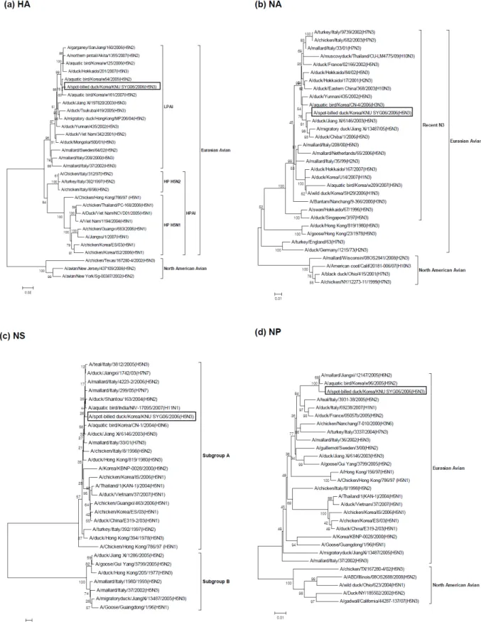
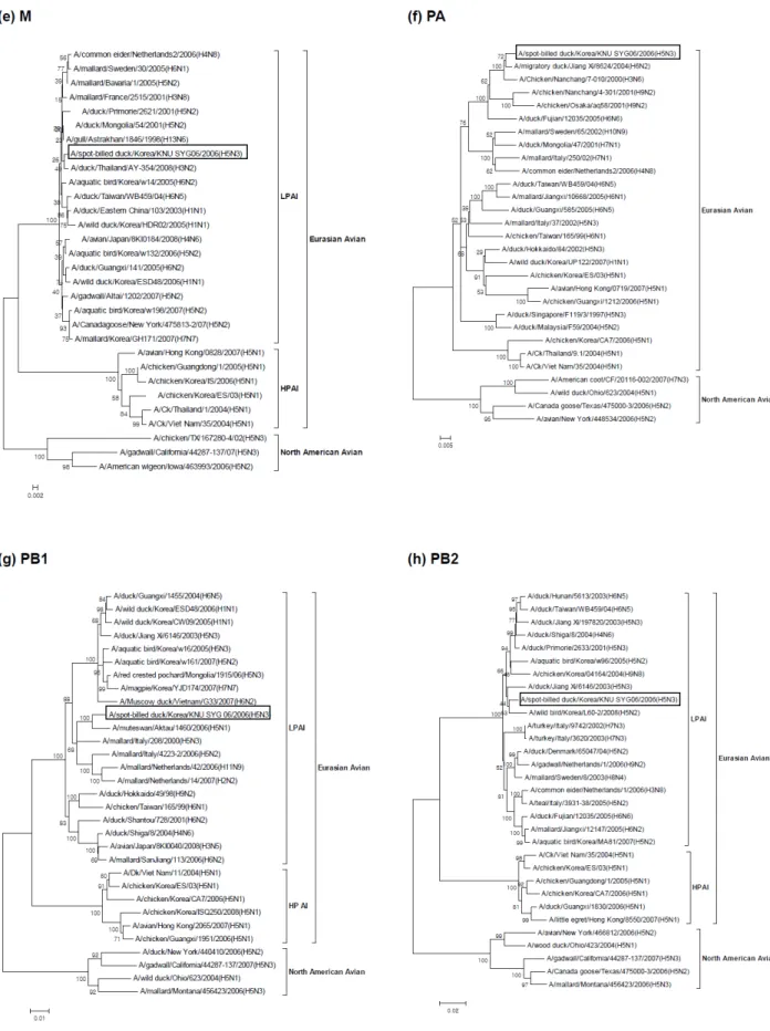
The sequences of the whole genome as well as the amino acids of all eight segments were determined to characterize the SBD/KR/KNU SYG06/06 virus. A phylogenetic analysis of the eight viral genes (Fig. 1a∼h) revealed that all seg- ments belonged to the Eurasian avian lineage. The percent similarities among the eight gene segments of the SBD/KR/
KNU SYG06/06 isolate with Eurasian avian viruses ranged from 98.2 ∼99.8% for nucleotide sequences and 98.3∼100%
for amino acid sequences (Table 1).
2. HA Gene
A phylogenetic analysis of the H5 gene revealed that the SBD/KR/KNU SYG06/06 viruses separated into Eurasian line- ages. Within the Eurasian lineage, at least two distinct clus- ters were observed (Fig. 1a). One cluster (HPAI) contained the
“HP H5N1” group of viruses, including human isolates as well as the “HP H5N2” viruses obtained from Italy from 1997 to 1998, while the other cluster contained the LPAI viruses. The SBD/KR/KNU SYG06/06 isolate belonged to the LPAI sub- group. Phylogenetically, the South Korean HPAI H5N1 stains isolated in 2003 and 2006 were clearly different from the SBD/KR/KNU SYG06/06 isolate, and they belonged to the
“HP H5N1” subgroup.
The SBD/KR/KNU SYG06/06 isolate shared the highest
sequence identity with the A/aquatic bird/Korea/w54/2005 (H5N2) isolate, with 99.2% nucleotide and 99.3% amino acid sequence identities (Table 1).
3. NA Gene
A phylogenetic analysis of the N3 gene showed that the NA gene from the SBD/KR/KNU SYG06/06 isolate also belong ed to the Eurasian avian lineage (Fig. 1b). In addition, the N3 gene of the SBD/KR/KNU SYG06/06 isolate belonged to the
"Recent N3" subgroup and was closely related to H5N3 viruses recently isolated in China and Japan. The N3 gene of the SBD/KR/KNU SYG06/06 isolate shared 98.2% nucleotide identity and 98.3% amino acid identity with A/ duck/Chiba/
1/2006 (H5N3) (Table 1).
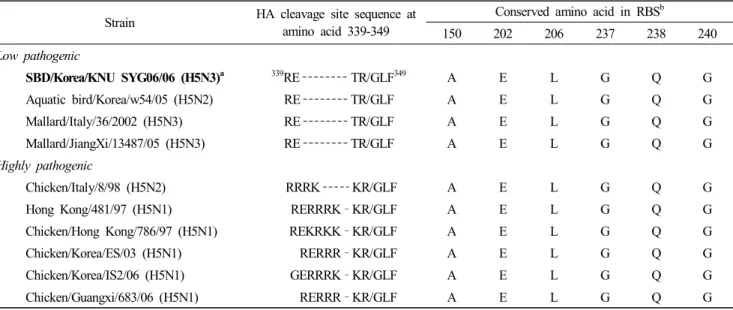
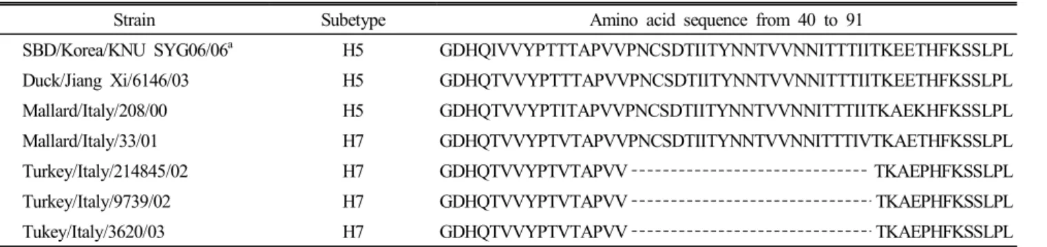
4. Internal Genes
The phylogenetic tree for the other six internal genes (NP, NS, M, PA, PB1, and PB2) showed similar clustering in the Eurasian avian lineage and exhibited no close relationships with HPAI viruses (Fig. 1c∼h). The phylogenetic tree based on the NS gene readily identified the two previously described subtypes (A and B) of the NS gene. The NS gene of the SBD/
KR/KNU SYG06/06 isolate, along with Hong Kong H5N1,
Italy H5N2, and South Korea H5N1 highly pathogenic
viruses, belonged to subgroup A, whereas the Italy H5N3
viruses isolated in 2002 and China H5N3 and H5N2 viruses
isolated in 2005 belonged to subgroup B (Fig. 1c). The
phylogenetic trees of the M, PB1, and PB2 genes showed two
distinct clusters within the Eurasian lineage, HPAI and
Fig. 1. Phylogenetic trees based on nucleotide sequences of HA (a), NA (b), NP (c), NS (d), M (e), PA (f), PB1 (g), PB2 (h) genes.
The tree was generated by neighbour-joining method with MEGA 4.0 program, using bootstrap replication (500 bootstraps).
An H5N3 isolate (SBD/KR/KNU SYG06/06) in this study was boxed.
Fig. 1. Continued.
Table 2. Comparison of amino acid sequence of different H5 viruses
Strain HA cleavage site sequence at amino acid 339-349
Conserved amino acid in RBS
b150 202 206 237 238 240
Low pathogenic
SBD/Korea/KNU SYG06/06 (H5N3)
a 339RE TR/GLF
349A E L G Q G
Aquatic bird/Korea/w54/05 (H5N2) RE TR/GLF A E L G Q G
Mallard/Italy/36/2002 (H5N3) RE TR/GLF A E L G Q G
Mallard/JiangXi/13487/05 (H5N3) RE TR/GLF A E L G Q G
Highly pathogenic
Chicken/Italy/8/98 (H5N2) RRRK KR/GLF A E L G Q G
Hong Kong/481/97 (H5N1) RERRRK –KR/GLF A E L G Q G
Chicken/Hong Kong/786/97 (H5N1) REKRKK –KR/GLF A E L G Q G
Chicken/Korea/ES/03 (H5N1) RERRR –KR/GLF A E L G Q G
Chicken/Korea/IS2/06 (H5N1) GERRRK –KR/GLF A E L G Q G
Chicken/Guangxi/683/06 (H5N1) RERRR –KR/GLF A E L G Q G
a
The isolate in boldface is the SBD/KR/KNU SYG06/06 (H5N3) isolate analyzed in this study.
b
RBS : receptor binding site.
LPAI. The SBD/KR/KNU SYG06/06 isolate belonged to the LPAI group.
The highest identities of nucleotide and amino acid se- quences are indicated in Table 1. The PB1 gene shared the highest sequence identity with the low pathogenic A/mute swan/Aktau/1460/2006(H5N1) isolate.
5. Molecular Characterization
Based on the amino acid sequence at the HA1-HA2 con- necting peptide, the SBD/KR/KNU SYG06/06 isolate exhi- bited a RETR/GLF motif. (Table 2). All isolates analyzed in this study exhibited the conserved amino acid residues at positions 150, 202, 206, 237, 238, and 240 (position 138, 190, 194, 225, 226, and 228 in H3 numbering), which define the receptor binding site and are related to the preferential binding of sialic acids joined to the sugar chain through an α-2,3 linkage. (Table 2) (Matrosovich et al., 1997).
The N3 gene was compared with other available N3 se- quences with differing HA subtypes. We observed that the Italy H7N3 isolates from 2002 to 2003 had a 23 amino acid deletion in the stalk region (position 56 to 78) that was not found in other Eurasian N3 genes, including the SBD/KR/
KNU SYG06/06 isolate (Table 3).
The four C-terminal amino acids of the NS1 gene bind to the protein interaction domain and perform localized signaling
at particular subcellular locations (Obenauer et al., 2006). The SBD/KR/KNU SYG06/06 isolate analyzed in this study exhi- bited the C-terminal motifs, ESEV. Within the NS1 gene, amino acid changes at position 149 (V149A) have been asso- ciated with virulence and resistance to antiviral cytokines in chickens (Li et al., 2006). The SBD/KR/KNU SYG06/06 iso- late had 149A.
Within the PB2 protein, the mutations E627K and D701N have previously been identified as determinants of virulence in mammalian hosts (Steel et al., 2009). These mutations were absent in the SBD/KR/KNU SYG06/06 isolate.
The NA protein of the SBD/KR/KNU SYG06/06 isolate possessed residue H276, which is a known marker for sensi- tivity to the neuraminidase inhibitor oseltamivir (Hayden, 2006).
The amino acid sequence analysis of the M2 protein revealed that the SBD/KR/KNU SYG06/06 isolate contained no muta- tions responsible for resistance to amantadine, the M2 inhibitor (Cheung et al., 2006).
6. Replication in MDCK Cells
We detected the virus titers with HA reaction after MDCK
cells were inoculated with the SBD/KR/KNU SYG06/06
isolate. The HA activity of the virus was observed in MDCK
cells with the addition of trypsin; however, there were no
detectable viruses in MDCK cultures without trypsin.
Table 3. Aligment of the NA stalk region of N3 viruses
Strain Subetype Amino acid sequence from 40 to 91
SBD/Korea/KNU SYG06/06
aH5 GDHQIVVYPTTTAPVVPNCSDTIITYNNTVVNNITTTIITKEETHFKSSLPL Duck/Jiang Xi/6146/03 H5 GDHQTVVYPTTTAPVVPNCSDTIITYNNTVVNNITTTIITKEETHFKSSLPL
Mallard/Italy/208/00 H5 GDHQTVVYPTITAPVVPNCSDTIITYNNTVVNNITTTIITKAEKHFKSSLPL
Mallard/Italy/33/01 H7 GDHQTVVYPTVTAPVVPNCSDTIITYNNTVVNNITTTIVTKAETHFKSSLPL
Turkey/Italy/214845/02 H7 GDHQTVVYPTVTAPVV TKAEPHFKSSLPL
Turkey/Italy/9739/02 H7 GDHQTVVYPTVTAPVV TKAEPHFKSSLPL
Tukey/Italy/3620/03 H7 GDHQTVVYPTVTAPVV TKAEPHFKSSLPL
a