Copyright © 2017 The Korean Society for Bone and Mineral Research
This is an Open Access article distributed under the terms of the Creative Commons Attribution Non-Commercial Li- cense (http://creativecommons.org/licenses/by-nc/4.0/) which permits unrestricted non-commercial use, distribu- tion, and reproduction in any medium, provided the original work is properly cited.
Coronary Artery Calcium Score and Bone
Metabolism: A Pilot Study in Postmenopausal Women
Pooneh Salari1, Abbasali Keshtkar2, Shapour Shirani3, Leila Mounesan4
1Medical Ethics and History of Medicine Research Center, Tehran University of Medical Sciences, Tehran;
2Osteoporosis Research Center, Endocrinology and Metabolism Clinical Sciences Institute, Tehran University of Medical Sciences, Tehran;
3Head of Imaging Center, Tehran Heart Center, Tehran University of Medical Sciences, Tehran;
4 Department of Epidemiology, Center for Academic and Health Policy, and Knowledge Utilization Research Center, Tehran University of Medical Sciences, Tehran, Iran
Background: Since 1991 many studies evaluated the link between cardiovascular dis- eases and osteoporosis, two age-related conditions, but the main common pathologic pathway has not been determined yet. The histological similarity between arterial calci- fied plaque and bone matrix and involvement of similar cells and mediators provide a special field of research. Therefore in the present study, we aimed to evaluate the rela- tionship between coronary artery calcium score (CACS) as a surrogate marker of athero- sclerosis and bone mediators and parameters in postmenopausal women. Methods:
Eleven postmenopausal women who had CACS higher than 80 were enrolled into the study and underwent bone densitometry. In addition, their serum and urine samples were taken for measuring osteoprotegerin, osteocalcin, and β cross laps. Patients’ 10- year probability of fracture was calculated by the World Health Organization fracture- risk assessment tool (FRAX). Results: The regression analysis of our results showed the association between CACS and OC (std β=0.66, 95% confidence interval [CI] 5.47-72.27, P=0.027), femoral bone density (std β=-0.6, 95% CI -6864.34-14.27, P=0.05) and T-score (std β=-0.6, 95% CI -773.08-1.28, P=0.05) which remained significant after adjustment for age, weight, years since menopause and body mass index. No association was found between CACS and osteoprotegerin, spinal bone density and FRAX score. Conclusions:
In conclusion, this pilot study with small sample size showed the potential association between CACS and osteocalcin, femoral bone density and T-score. However, the rela- tionship between CACS and osteoprotegerin, receptor activator of nuclear factor-kappa B ligand, FRAX score and other bone parameters remain to be clarified in larger sample size studies.
Key Words: Bone density, Calcium, Coronary artery disease, Osteocalcin, Osteoprotegerin
INTRODUCTION
Special attention has been paid to the link between atherosclerosis and osteo- porosis since 1991 and it is assumed to be regulated by biological mechanisms.
Both diseases are considered to be associated with aging. Although at the first glance osteoporosis and atherosclerosis seem to be independent, atherosclerotic plaques and bone tissue have similarities and the process of vascular calcification Corresponding author
Pooneh Salari
Medical Ethics and History of Medicine Research Center, Tehran University of Medical Sciences, 23# 16 Azar Ave, Keshavarz Blvd, Tehran, Iran
Tel: +98-21-6641-9661 Fax: +98-21-6641-9661 E-mail: [email protected] Received: December 3, 2016 Revised: December 25, 2016 Accepted: December 28, 2016
No potential conflict of interest relevant to this article was reported.
is mediated by cells and mediators involved in bone for- mation.[1] In this regard several common inflammatory and proinflammatory mediators including, bone markers and cytokines including interleukin (IL)-1, IL-6, IL-11, IL-12, IL-15, IL-17, tumor necrosis factor-alpha (TNF-α), osteopro- tegerin, bone morphogenetic proteins (BMPs), C-reactive protein (CRP) have been suggested but still the main com- mon causative agent or pathway is not fully understood.[1]
Histological findings demonstrated resorptive and remod- eling sites in atherosclerotic plaques similar to bone. Actu- ally these plaques have abundant numbers of different types of inflammatory cells inducing several types of me- diators. Vascular endothelium and vascular smooth muscle cells produce osteoprotegerin and receptor activator of nuclear factor-kappa B ligand (RANKL) that is expressed in atherosclerotic lesions.[2] Osteoprotegerin acts as a mem- ber of TNF-α superfamily and inhibits osteoclast differenti- ation and bone resorption by binding to RANKL. RANKL is involved in vascular calcification by inducing osteoclast formation in vascular smooth muscle cells and counteracts with osteoprotegerin.[3] The counteraction of osteoprote- gerin with RANKL regulates bone resorption. In addition osteoprotegerin controls bone pathological conditions in- cluding bone metabolism and inflammation. Human stud- ies indicated the possible direct association between os- teoprotegerin and severity of coronary artery disease (CAD).[1] In line with the evidence of involvement of osteo- protegerin, there are recent evidences which highlight the role of osteocalcin in osteoporosis and cardiovascular dis- ease (CVD). Osteocalcin is a bone matrix protein mainly produced by osteoblasts which is involved in bone miner- alization and formation.[4] Osteoblasts like cells in the vas- culature are responsible for secreting osteocalcin and cal- cifying vascular cells.[5]
There are recent evidences of the involvement of osteo- calcin in CAD.
One of the surrogate markers of atherosclerosis is coro- nary artery calcium (CAC) score (CACS) which can predict cardiovascular risk.[6] Data regarding the correlation be- tween CACS and bone metabolism are still paradox. Accor- dingly and in order to find out the relationship between CACS as a surrogate marker of atherosclerosis and bone biomarkers as well as bone density and other bone specific information, we conducted a pilot study to provide pre- sumptive data for more comprehensive assessment. Our
study is the first one which evaluated this association in post- menopausal women who had CACS higher than 80 which indicates the average risk of atherosclerosis and higher.
METHODS
Eleven postmenopausal women who underwent com- puted tomography scanning with (Dual Source Flash-128 slice) and had CACS higher than 80 were enrolled in the study. Based on the categories of CACS which was defined in previous studies,[7] we selected CACS ≥80 which is con- sidered as average risk of coronary artery events and higher.
The eligible patients were referred to the Diabetic Re- search Center for measuring bone density. Each subject underwent bone densitometry by dual energy X-ray ab- sorptiometry (DXA; Hologic Inc., Bedford, MA, USA) at fe- mur neck and lumbar vertebrae respectively. Blood and urine samples were obtained from study participants. All patients profile was evaluated and recorded including age, menopause state, smoking, alcohol consumption, history of fracture, parents fracture, and past medical history. Pa- tients with history of cancer, diabetes, acute infection, en- docrinologic disorders, use of special medications such as hormones, corticosteroids, gonadotropin releasing hor- mone (GnRH) analogs, anticonvulsant drugs, heparin, alu- minium containing antacids, thyroid hormones were ex- cluded. The study was conducted in Endocrinology and Metabolism Research Institute of Tehran University of Medi- cal Sciences after authorization by ethics committee of the institute. All patients signed the written informed consent after receiving thorough information about the study.
1. Laboratory measurement
Blood and urine samples were collected. Blood samples were centrifuged at 10,000 g for 10 min and kept in a fridge at -70°C until analysis. Serum concentration of osteoprote- gerin, RANKL, osteocalcin, and high-sensitivity CRP (hs-CRP) along with urine concentration of beta (c) cross labs were measured. Serum level of osteocalcin and osteoprotegerin were measured by N-MID®Osteocalcin enzyme-linked im- munosorbent assay (ELISA; Immuno Diagnostic Systems, Tyne & Wear, UK) and osteoprotegerin Human ELISA Kit (ab100617) respectively. Urine level of β-cross laps was mea- sured by Urine β-CrossLaps®ELISA (Immuno Diagnostic Systems). Serum concentration of hs-CRP and RANKL were
assessed by CRP Human ELISA Kit (ab99995) and Human RANKL ELISA Kit from DLdevelop respectively.
2. CACS
The CACS was calculated using the method of Agatston et al.[8] by an experienced radiologist.
3. Fracture-risk assessment tool (FRAX) score The FRAX score which was developed by World Health Organization (WHO) provides risk estimation for probabili- ty of fracture in the next 10 years. The score is calculating based on clinical risk factors and bone mineral density (BMD) at the femoral neck. This model of risk assessment has been developed by population studies for European, North Amer- ican, Asian countries and Australia.[9] Because there is no FRAX tool for Iranians, we used computer driven FRAX tool for Lebanese who seems to be more similar with Iranians.
4. Statistical analysis
One-sample Kolmogorov-Smirnov test was used to as- sess normal distribution of the data. All data were normally distributed and no transformation was made (P>0.05). To recognize the association between CACS and BMD, bone biomarkers and FRAX score, multiple linear regression mod- el was used. Several covariates for adjustment were chosen including age, weight, body mass index (BMI), and years since menopause.
All statistical analyses were performed with SPSS 18.0 (SPSS Inc., Chicago, IL, USA). The data were expressed as mean means±standard deviations (SD). All P-values of less than 0.05 were considered to indicate statistical signif- icance.
RESULTS
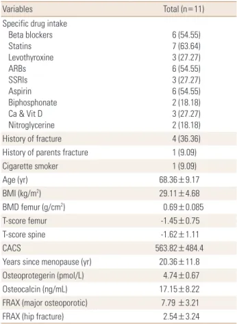
Eleven postmenopausal women who had CACS higher than 80 were enrolled in the study. The demographic data and past medical history of all study participants are sum- marized in Table 1.
The mean CACS was 563.82 (484.39). The mean serum concentration of osteocalcin was 17.15±8.22. The associa- tion between ca score and serum osteocalcin concentration was significant (std β=0.66, 95% confidence interval [CI]=
5.47-72.27, P=0.02). After adjustment for age, weight, BMI, and years since menopause the association remained sig-
nificant (std β=0.89, 95% CI=29.92-82.24, P=0.01).
The mean femoral BMD of all participants was 0.69±0.085.
Our analysis showed significant association between ca score and femoral BMD (std β=-0.6, 95% CI=-6,864.34-14.27, P=0.05) which remained significant after adjustment for age, weight, BMI, and years since menopause (std β=-1.3, 95% CI=-12,825.84-2,249.83, P=0.01).
Participants’ femoral T-score was -1.45±0.75. Significant association between CACS and femoral T-score was found (std β=-0.6, 95% CI=-773.08-1.28, P=0.05) which remained significant after adjustment for age, weight, BMI, and years since menopause (std β=-1.11, 95% CI=-1,379.91-49.78, P=0.04).
The mean serum concentration of osteoprotegerin in all participants was 4.74±0.67. No significant association was found between osteoprotegerin and calcium score (std β=
-0.05, 95% CI=-583.03-503.42, P=0.87).
Table 1. Patients’ characteristics
Variables Total (n=11)
Specific drug intake Beta blockers Statins Levothyroxine ARBs SSRIs Aspirin Biphosphonate Ca & Vit D Nitroglycerine
6 (54.55) 7 (63.64) 3 (27.27) 6 (54.55) 3 (27.27) 6 (54.55) 2 (18.18) 3 (27.27) 2 (18.18)
History of fracture 4 (36.36)
History of parents fracture 1 (9.09)
Cigarette smoker 1 (9.09)
Age (yr) 68.36±9.17
BMI (kg/m2) 29.11±4.68
BMD femur (g/cm2) 0.69±0.085
T-score femur -1.45±0.75
T-score spine -1.62±1.11
CACS 563.82±484.4
Years since menopause (yr) 20.36±11.8
Osteoprotegerin (pmol/L) 4.74±0.67
Osteocalcin (ng/mL) 17.15±8.22
FRAX (major osteoporotic) 7.79 ±3.21
FRAX (hip fracture) 2.54±3.24
The data is presented as mean±standard deviation or number (%).
ARBs, angiotensin receptor blockers; SSRIs, selective serotonin reup- take inhibitors; Ca, calcium; CACS, coronary artery calcium score; Vit D, vitamin D; BMI, body mass index; BMD, bone mineral density; FRAX, fracture-risk assessment tool.
The mean urine concentration of β cross laps was 0.51±
0.36. No significant association was detected between β cross laps and CACS (std β=0.29, 95% CI=-556.12-1,348.97, P=0.37). After adjustment for age, weight, BMI, and years since menopause the association found to be significant (std β=0.92, 95% CI=20.42-2,439.42, P=0.04).
The mean patients’ FRAX score (the ten year probability of fracture %) for major osteoporotic fracture was 7.79 (3.21) and for hip fracture was 2.54±3.24. No association between patients FRAX score and CACS was observed.
No more correlation was found between study variables.
Serum concentration of RANKL and hs-CRP was undetect- able except for one participant. Therefore we omitted those data from analysis.
DISCUSSION
This study conducted on postmenopausal women who had CACS higher than 80 and were considered to be at high risk of atherosclerosis. Our results showed that CACS is in- dependently associated with osteocalcin. Osteoclacin is a bone formation marker which originates from skeleton and indicates the osteoblasts potential for making bone matrix. It is a marker of bone mineralization and calcium homeostasis[1] and recently was recognized as a strong predictor of severity of coronary atherosclerosis.[10]
In agreement with our study Kim et al.[11] observed sig- nificant positive correlation between aortic CACS and os- teocalcin, vertebral fractures and total hip-trabecular BMD (tBMD). A recent cohort study in Chinese men indicated serum osteocalcin as a strong determinant of severity of CAD.[10] In contrast Awan et al.[12] reported a negative association between vascular calcification and serum os- teocalcin in familiar hypercholesterolemia and also Wilund et al.[13] found an inverse significant correlation between logarithmic value of CACS and plasma osteocalcin.
Chen et al.[14] observed significant lower levels of se- rum osteocalcin in patients with self-reported CVD in com- parison with middle-aged and elderly Chinese without CVD. In addition Sheng et al.[15] observed that reduced osteocalcin is associated with increased risk of carotid ath- erosclerosis plaques in type 2 diabetes patients. They indi- cated decreased serum concentration of osteocalcin as a high risk for carotid atherosclerotic plaques and presented osteocalcin as a promising candidate for risk assessment of
CVD. We included patients with CACS more than 80 which may explain the reason of the difference between our re- sults with those who reported a converse relationship be- tween CACS and osteocalcin. It was suggested that osteo- calcin may have preventive effect on arteriosclerosis[16]
while our patients CACS was high enough to show athero- sclerosis. So there is the possibility that the severity of ath- erosclerosis (based on CACS) determines serum osteocal- cin level.
Although the mean serum concentration of osteoprote- gerin in our study was higher than normal, no association was found between CACS and osteoprotegerin serum con- centration even after adjustment for age, weight, BMI and years since menopause. It was observed that the elevated level of osteoprotegerin is associated with severity of CAD.
[1] It has been reported that osteoprotegerin is higher in patients with 2 to 3 vessels atherosclerosis while its level in patients with 1 vessel atherosclerosis is similar to those with- out CAD.[17] The positive correlation of the level of osteo- protegerin and CAC was demonstrated in diabetic patients whose mean serum osteoprotegerin concentration was higher than our patients.[18] Asanuma et al.[19] observed an association between osteoprotegerin and CACS in pa- tients with rheumatoid arthritis. Also there are reports on the effect of angiotensin receptor blockers on reducing os- teoprotegerin secretion from carotid endarterectomy sam- ples taken from stroke patients[20] which may more explain our results. Furthermore chan ges in the local vascular ex- pression of osteoprotegerin may not be directly proportion- al to its circulating level.[21]
Bakhireva et al.[22] reported higher osteoprotegerin lev- els in women with CAC compared with no/minimal calcifi- cation which was disappeared after age adjustment. They found significant lower odds of having CAC with 1 SD in- crease in hip BMD. Other clinical studies also demonstrated the direct association between osteoprotegerin level and severity of CAD, vascular dysfunction and cardiovascular mortality.[23] According to the counteraction between os- teoprotegerin and RANKL, we measured serum RANKL in our patients but unfortunately we could not detect its lev- el. Therefore the interaction between osteoprotegerin and RANKL in association with CACS should be evaluated in fu- ture larger studies.
Our results showed an inverse association between CACS and BMD and T-score at femur (but not spine) which re-
mained significant after adjustment for age, weight, BMI and years since menopause. This is in line with other pre- vious studies which confirm this result with bone density at spine,[24-27] however to date no study evaluated T-score.
Choi et al.[28] observed an inverse correlation between CACS and BMD at femur which was stronger in women with a longer time since menopause and women with osteopo- rosis and osteopenia than normal BMD. Some researchers have indicated the site specific bone densitometry. In con- trast to our study the association of lower spine volumetric BMD (vBMD) with high aortic calcification but not to CAC was reported in a cross sectional study,[29] however the CACS of our study participants was higher than those. In middle-aged postmenopausal women with extensive cor- onary plaque burden Lin et al.[30] did not find an associa- tion between coronary calcification and low BMD. In addi- tion the association between vertebral bone density and CACS was determined by Filgueira et al.[31] in non-dialyzed chronic kidney disease (CKD) patients. Accordingly Carr et al.[32] demonstrated an association between vertebral tra- becular bone density and vascular calcification. They ex- plained their finding by introducing vertebral trabecular bone as the active metabolic bone which its density pro- vides a measurement of volumetric BMD. In agreement with our study bone densitometry at spinal sites failed to show association with vascular calcification.[33]
Bakhireva et al.[34] reported no age-independent asso- ciation between BMD at any site and CACS in men and wom- en not using hormone therapy (HT) while the association was significant in HT users and they suggested that estro- gen may mediated the association. Ramsey-Goldman and Manzi [35] observed an inverse significant correlation be- tween CACS and lumbar spine BMD and hip BMD in a pilot study in young women with Lupus.
Based on the importance of the serum concentration of hs-CRP in atherosclerosis and CVDs and former reports in- dicating higher hs-CRP levels in osteopenic and osteopo- rotic patients[36] we proposed to measure its level in our patients and its association with other markers and scores, but we could not detect it in our patients.
We evaluated the association between CACS and FRAX which was not significant. To date this study was the first one provided FRAX and assessed its association with CACS which have to be precisely re-assessed in larger study.
Our results showed that after adjustment for age, weight,
years since menopause and BMI the urine concentration of β cross laps have significant positive association with CACS.
It may assumed that in patients with high CACS and high risk of CAD the rate of bone metabolism is high, so we ob- served the positive association between CACS and bone formation marker (osteocalcin) and bone resorption mark- er (β cross laps).
The study is a pilot observational study with limitations that has to be taken into account. The small sample size, not assessing patients with low CACS (lower than 80), and enrolling only patients with high CACS are limitations of this study. However, future larger studies with new meth- ods of BMD and calcium scoring are necessitated. The study participants were chosen from patients referring for com- puted tomography-angiography therefore they are not representative of general population. The sample size is small enough to extrapolate the data to general popula- tion, but provides evidence for further extensive studies in larger sample size.
Taken together the association between CACS and os- teocalcin, femoral BMD and T-score were strong enough to be observed, however, the potential associations between RANKL/ osteoprotegerin system and CAC needs to be clari- fied in larger studies because finding the way of antago- nizing this pathway may benefit both cardiovascular sys- tem and bone. The underlying mechanisms of the above mentioned associations are still under question. Although we considered the most potential contributors, still the pos- sible effect of the other underlying diseases and medica- tions is not completely understood. Therefore further pop- ulation based studies are highly recommended.
ACKNOWLEDGEMENT
The study was approved and financially supported by Endocrinology and Metabolism Research Institute of Teh- ran University of Medical Sciences. The authors would like to thank Dr. Farideh Razi, Miss. Sara Shirazi, Dr. Patrishia Khashayar, Dr. Neda Mehrdad and Mrs. Espandbod for their great help and assistance. Also we thank all patients’ par- ticipation.
REFERENCES
1. Salari P, Abdollahi M. A comprehensive review of the shared
roles of inflammatory cytokines in osteoporosis and car- diovascular diseases as two common old people problem:
actions toward development of new drugs. Int J Pharma- col 2011;7:552-67.
2. Nadra I, Mason JC, Philippidis P, et al. Proinflammatory ac- tivation of macrophages by basic calcium phosphate crys- tals via protein kinase C and MAP kinase pathways: a vi- cious cycle of inflammation and arterial calcification? Circ Res 2005;96:1248-56.
3. Min H, Morony S, Sarosi I, et al. Osteoprotegerin reverses osteoporosis by inhibiting endosteal osteoclasts and pre- vents vascular calcification by blocking a process resem- bling osteoclastogenesis. J Exp Med 2000;192:463-74.
4. Khosla S, Amin S, Orwoll E. Osteoporosis in men. Endocr Rev 2008;29:441-64.
5. Dhore CR, Cleutjens JP, Lutgens E, et al. Differential expres- sion of bone matrix regulatory proteins in human athero- sclerotic plaques. Arterioscler Thromb Vasc Biol 2001;21:
1998-2003.
6. Greenland P, Bonow RO, Brundage BH, et al. ACCF/AHA 2007 clinical expert consensus document on coronary ar- tery calcium scoring by computed tomography in global cardiovascular risk assessment and in evaluation of patients with chest pain: a report of the American College of Cardi- ology Foundation Clinical Expert Consensus Task Force (ACCF/AHA Writing Committee to Update the 2000 Expert Consensus Document on Electron Beam Computed To- mography). Circulation 2007;115:402-26.
7. Krajnc M, Pečovnik-Balon B, Hojs R, et al. Comparison of coronary artery calcification and some coronary artery calcification risk factors in patients on haemodialysis and in patients with type 2 diabetes. J Int Med Res 2011;39:
1006-15.
8. Agatston AS, Janowitz WR, Hildner FJ, et al. Quantification of coronary artery calcium using ultrafast computed to- mography. J Am Coll Cardiol 1990;15:827-32.
9. Kanis JA, McCloskey EV, Johansson H, et al. Development and use of FRAX in osteoporosis. Osteoporos Int 2010;21 Suppl 2:S407-13.
10. Bao Y, Zhou M, Lu Z, et al. Serum levels of osteocalcin are inversely associated with the metabolic syndrome and the severity of coronary artery disease in Chinese men.
Clin Endocrinol (Oxf) 2011;75:196-201.
11. Kim KJ, Kim KM, Park KH, et al. Aortic calcification and bone metabolism: the relationship between aortic calcification,
BMD, vertebral fracture, 25-hydroxyvitamin D, and osteo- calcin. Calcif Tissue Int 2012;91:370-8.
12. Awan Z, Alwaili K, Alshahrani A, et al. Calcium homeosta- sis and skeletal integrity in individuals with familial hyper- cholesterolemia and aortic calcification. Clin Chem 2010;
56:1599-607.
13. Wilund KR, Tomayko EJ, Evans EM, et al. Physical activity, coronary artery calcium, and bone mineral density in el- derly men and women: a preliminary investigation. Me- tabolism 2008;57:584-91.
14. Chen L, Li Q, Yang Z, et al. Osteocalcin, glucose metabo- lism, lipid profile and chronic low-grade inflammation in middle-aged and elderly Chinese. Diabet Med 2013;30:
309-17.
15. Sheng L, Cao W, Cha B, et al. Serum osteocalcin level and its association with carotid atherosclerosis in patients with type 2 diabetes. Cardiovasc Diabetol 2013;12:22.
16. Ogawa-Furuya N, Yamaguchi T, Yamamoto M, et al. Serum osteocalcin levels are inversely associated with abdominal aortic calcification in men with type 2 diabetes mellitus.
Osteoporos Int 2013;24:2223-30.
17. Jono S, Ikari Y, Shioi A, et al. Serum osteoprotegerin levels are associated with the presence and severity of coronary artery disease. Circulation 2002;106:1192-4.
18. Ishiyama M, Suzuki E, Katsuda J, et al. Associations of cor- onary artery calcification and carotid intima-media thick- ness with plasma concentrations of vascular calcification inhibitors in type 2 diabetic patients. Diabetes Res Clin Pract 2009;85:189-96.
19. Asanuma Y, Chung CP, Oeser A, et al. Serum osteoprote- gerin is increased and independently associated with cor- onary-artery atherosclerosis in patients with rheumatoid arthritis. Atherosclerosis 2007;195:e135-41.
20. Golledge J, McCann M, Mangan S, et al. Osteoprotegerin and osteopontin are expressed at high concentrations with- in symptomatic carotid atherosclerosis. Stroke 2004;35:
1636-41.
21. Quercioli A, Luciano Viviani G, Dallegri F, et al. Receptor activator of nuclear factor kappa B ligand/osteoproteger- in pathway is a promising target to reduce atherosclerotic plaque calcification. Crit Pathw Cardiol 2010;9:227-30.
22. Bakhireva LN, Laughlin GA, Bettencourt R, et al. Does os- teoprotegerin or receptor activator of nuclear factor-kap- paB ligand mediate the association between bone and cor- onary artery calcification? J Clin Endocrinol Metab 2008;
93:2009-12.
23. Collin-Osdoby P. Regulation of vascular calcification by os- teoclast regulatory factors RANKL and osteoprotegerin.
Circ Res 2004;95:1046-57.
24. Barengolts EI, Berman M, Kukreja SC, et al. Osteoporosis and coronary atherosclerosis in asymptomatic postmeno- pausal women. Calcif Tissue Int 1998;62:209-13.
25. Hyder JA, Allison MA, Criqui MH, et al. Association between systemic calcified atherosclerosis and bone density. Calcif Tissue Int 2007;80:301-6.
26. Celik C, Altunkan S, Yildirim MO, et al. Relationship between decreased bone mineral density and subclinical athero- sclerosis in postmenopausal women. Climacteric 2010;13:
254-8.
27. Lee SH, Park SJ, Kim KN, et al. Coronary calcification Is re- versely related with bone and hair calcium: the relation- ship among different calcium pools in body. J Bone Metab 2016;23:191-7.
28. Choi SH, An JH, Lim S, et al. Lower bone mineral density is associated with higher coronary calcification and coronary plaque burdens by multidetector row coronary computed tomography in pre- and postmenopausal women. Clin Endocrinol (Oxf) 2009;71:644-51.
29. Farhat GN, Cauley JA, Matthews KA, et al. Volumetric BMD and vascular calcification in middle-aged women: the Study of Women's Health Across the Nation. J Bone Miner Res 2006;21:1839-46.
30. Lin T, Liu JC, Chang LY, et al. Association between coronary artery calcification using low-dose MDCT coronary angi- ography and bone mineral density in middle-aged men and women. Osteoporos Int 2011;22:627-34.
31. Filgueira A, Carvalho AB, Tomiyama C, et al. Is coronary ar- tery calcification associated with vertebral bone density in nondialyzed chronic kidney disease patients? Clin J Am Soc Nephrol 2011;6:1456-62.
32. Carr JJ, Register TC, Hsu FC, et al. Calcified atherosclerotic plaque and bone mineral density in type 2 diabetes: the diabetes heart study. Bone 2008;42:43-52.
33. van der Klift M, Pols HA, Hak AE, et al. Bone mineral densi- ty and the risk of peripheral arterial disease: the Rotterdam Study. Calcif Tissue Int 2002;70:443-9.
34. Bakhireva LN, Barrett-Connor EL, Laughlin GA, et al. Differ- ences in association of bone mineral density with coro- nary artery calcification in men and women: the Rancho Bernardo Study. Menopause 2005;12:691-8.
35. Ramsey-Goldman R, Manzi S. Association of osteoporosis and cardiovascular disease in women with systemic lupus erythematosus. Arthritis Rheum 2001;44:2338-41.
36. Koh JM, Khang YH, Jung CH, et al. Higher circulating hsCRP levels are associated with lower bone mineral density in healthy pre- and postmenopausal women: evidence for a link between systemic inflammation and osteoporosis.
Osteoporos Int 2005;16:1263-71.