Background and Purpose The magnetic resonance imaging in multiple sclerosis (MAG- NIMS) group recently proposed guidelines to replace the existing dissemination-in-space criteria in McDonald 2010 magnetic resonance imaging (MRI) criteria for diagnosing multi- ple sclerosis. There has been insufficient research regarding their applicability in Asians. Ob- jective of this study was to determine the sensitivity, specificity, accuracy, positive predictive value (PPV), and negative predictive value (NPV) of McDonald 2010 and MAGNIMS 2016 MRI criteria with the aim of verifying their applicability in Sri Lankan patients.
Methods Patients with clinically isolated syndrome diagnosed by consultant neurologists were recruited from five major neurology centers. Baseline and follow-up MRI scans were performed within 3 months from the initial presentation and at one year after baseline MRI, respectively.
McDonald 2010 and MAGNIMS 2016 MRI criteria were applied to all MRI scans. Patients were followed-up for 2 years to assess the conversion to clinically definite multiple sclerosis (CDMS). The sensitivity, specificity, accuracy, PPV, and NPV for predicting the conversion to CDMS were calculated.
Results Forty-two of 66 patients converted to CDMS. Thirty-seven fulfilled the McDonald 2010 MRI criteria, and 33 converted to CDMS. MAGNIMS 2016 MRI criteria were fulfilled by 29, with 28 converting to CDMS. The sensitivity, specificity, accuracy, PPV, and NPV were 78%, 83%, 64%, 89%, and 69%, respectively, for the McDonald 2010 criteria, and 67%, 96%, 77%, 96%, and 62% for the MAGNIMS 2016 MRI criteria.
Conclusions MAGNIMS 2016 MRI criteria were superior to McDonald 2010 MRI criteria in specificity, accuracy, and PPV, but inferior in sensitivity and NPV.
Key Words multiple sclerosis, McDonald 2010 magnetic resonance imaging criteria, magnetic resonance imaging in multiple sclerosis 2016 magnetic resonance imaging criteria, McDonald 2017 magnetic resonance imaging criteria, performance, Asia.
Applicability of McDonald 2010 and
Magnetic Resonance Imaging in Multiple Sclerosis
(MAGNIMS) 2016 Magnetic Resonance Imaging Criteria for the Diagnosis of Multiple Sclerosis in Sri Lanka
INTRODUCTION
Multiple sclerosis (MS) is a challenging disease to diagnose despite medical advances due to the complexities of its clinical presentation.1 MS is heterogeneous in terms of its clinical and radiological manifestations, disabilities, prognoses, and intrathecal antibody responses in different populations of the world,2-6 making confirmatory diagnoses even more difficult.
Early diagnosis is crucial for effective treatment, and magnetic resonance imaging (MRI) plays a major role in the early diagnosis of MS at the stage of clinically isolated syndrome (CIS).
Sujani Madhurika Kodagoda Gamagea
Indunil Wijeweerab Priyangi Wijesinghec Sanjaya Bandara Adikaria Katharina Finkd
Herath Mudiyanselage Ajith Sominandaa
a Department of Anatomy, Faculty of Medicine, University of Peradeniya, Peradeniya, Sri Lanka
b Neurology Unit, Teaching Hospital, Kandy, Sri Lanka
c Radiology Unit,
Sirimavo Bandaranayaka Specialized Children’s Hospital, Peradeniya, Sri Lanka
d Department of Neurology, Karolinska University Hospital, Stockholm, Sweden
pISSN 1738-6586 / eISSN 2005-5013 / J Clin Neurol 2018;14(3):339-344 / https://doi.org/10.3988/jcn.2018.14.3.339
Received December 18, 2017 Revised March 9, 2018 Accepted March 12, 2018 Correspondence
Herath Mudiyanselage Ajith Sominanda, MBBS, MPhil, PhD
Department of Anatomy, Faculty of Medicine, University of Peradeniya, Peradeniya 20400, Sri Lanka Tel +94712411139 Fax +94812389106
E-mail [email protected]
cc This is an Open Access article distributed under the terms of the Creative Commons Attribution Non-Com- mercial License (http://creativecommons.org/licenses/by-nc/4.0) which permits unrestricted non-commercial use, distribution, and reproduction in any medium, provided the original work is properly cited.
JCN
Open Access ORIGINAL ARTICLEMultiple Sclerosis in Sri Lanka
JCN
McDonald 2010 MRI criteria use magnetic resonance im- aging in multiple sclerosis (MAGNIMS) dissemination in space (DIS) criteria to identify DIS.7 However, in 2016 the MAGNIMS group proposed altering the MRI guidelines for diagnosing MS based on the intervening developments of MRI techniques and the availability of new data on using MRI to determine dissemination in time (DIT) and DIS cri- teria.8 The new guidelines proposed modifications to the DIS criteria but not to the DIT criteria, and to include optic-nerve lesions, to increase the number of periventricular lesions from one to three, and to consider cortical and subcortical lesions collectively under one category. That group has also pro- posed removing the distinction between asymptomatic and symptomatic lesions.8 The latest development is the intro- duction of McDonald 2017 MRI criteria, which considers the MAGNIMS 2016 MRI criteria and the availability of new data from diverse MS populations. The expert panel aimed to simplify the McDonald 2010 MRI criteria and promote earli- er diagnoses while preserving their specificity.9
McDonald 2010 MRI criteria have been developed using data gathered from adult Caucasians of European and North American populations and there has been less research and practical experience regarding their application in Asian populations. The need for new research in this area has been repeatedly emphasized by experts7,10 since the criteria were published in 2010. However, in the latest McDonald 2017 MRI criteria it is stated that the applicability of the McDon- ald 2010 MRI criteria has been studied in MS patients from Canada, Italy, the Netherlands, Spain, and Russia.9 A few studies conducted in East Asia, Russia, and Argentina have concluded that McDonald 2010 MRI criteria can also be ap- plied to these populations with significant confidence.11-14 In addition, studies of the applicability of the 2010 McDonald MRI criteria in Asian, Middle Eastern, and Latin American populations have been reported since 2010. Even though these studies were small, all of them have concluded that Mc- Donald 2010 MRI criteria are applicable to these diverse MS populations.9
Research on the applicability of MAGNIMS 2016 and Mc- Donald 2017 MRI criteria in diverse populations is high- lighted as a major requirement for future revisions of the Mc- Donald 2017 MRI criteria.9 One recent study in South Korea evaluated the performance of MAGNIMS DIS criteriain CIS patients.15 A study in the USA evaluated the performance of both criteria in a cohort of primary progressive MS patients.16 In addition, a multicenter European study reported in early 2018 compared the prediction of MS diagnosis in CIS patients using McDonald 2010 and MAGNIMS 2016 MRI criteria.17 However, there have been no studies of the applicability of the McDonald 2010 and MAGNIMS 2016 MRI criteria in South
Asia, and especially in Sri Lanka, where there is a paucity of research on MS.3
In this background, the present study assessed the sensitiv- ity, specificity, accuracy, positive predictive value (PPV), and negative predictive value (NPV) of McDonald 2010 MRI cri- teria and proposed MAGNIMS 2016 MRI criteria with the objective of determining their applicability in the Sri Lank- an MS population.
METHODS
Patients with CIS diagnosed by consultant neurologists were recruited for the study from 2012 to 2015 from five major neurology referral centers in Sri Lanka. Ethical approval for the study was granted by the Ethical Subcommittee, Faculty of Medicine, University of Peradeniya, Sri Lanka (IRB No. 2012/
EC114). All of the included patients provided informed writ- ten consent to participate in the study. The following inclu- sion criteria were applied: 1) CIS with clinical features sug- gestive of MS, 2) aged 18–60 years, since MS rarely presents in the childhood and elderly periods and may be challenging to diagnose,9,18 3) follow-up period of at least 2 years, 4) availabili- ty of spinal-cord MRI data if presenting with spinal-cord syn- drome, and 5) negativity for aquaporin-4 immunoglobulin.
The exclusion criteria were 1) receiving disease-modifying treatment (DMT), 2) having neuromyelitis optica spectrum disorder as defined by the 2015 neuromyelitis optica spec- trum disorders criteria,19 and 3) presence of atypical clinical features suggestive of other demyelinating disorders that mimic MS, including intractable hiccup, nausea, vomiting for more than 2 days, febrile illness preceding the onset of symptoms, myelopathy associated with spinal-cord lesions in- volving more than three spinal segments and the central part of the spinal cord on axial sections, severe bilateral optic neuritis associated with swollen optic-nerve chiasma lesion, presence of space-occupying lesions, or contrast enhancement of all the lesions.
Baseline brain and spinal-cord MRI was performed with T1-weighted, T2-weighted, and FLAIR pulse sequences and contrast enhancement within 3 months from the initial pre- sentation of the disease. The follow-up MRI scans were per- formed within 1 year from the baseline MRI.
All patients were followed up for at least 2 years to assess the conversion to clinically definite multiple sclerosis (CDMS), defined as at least two typical MS attacks and clinical evidence of at least two lesions (CDMS A1) according to the Poser cri- teria.20 The initial CIS patient recruitment and assessment of conversion to CDMS were performed by two experienced consultant neurologists. Both McDonald 2010 MRI criteria7 and MAGNIMS 2016 MRI criteria8 were applied retrospec-
Gamage SMK et al.
JCN
tively to all MRI scans by a single experienced investigator who was not blinded of the clinical presentation of the pa- tient, since it is recommended that MRI scans be interpreted by an experienced person who is aware of the patients clini- cal and laboratory findings.10 The sensitivity, specificity, ac- curacy, PPV, and NPV of both McDonald 2010 and MAG- NIMS 2016 MRI criteria for predicting the conversion to CDMS were calculated at the end of the follow-up period as follows:
Sensitivity=[TP/(TP+FN)]×100 Specificity=[TN/(TN+FP)]×100
(TP+TN) Accuracy= (TP+FP+TN+FN) ×100 PPV=[TP/(TP+FP)]×100
NPN=[TN/(TN+FN)]×100
True positive (TP), true negative (TN), false positive (FP), false negative (FN). All descriptive statistics were analyzed using GraphPad Prism software (GraphPad Software, La Jolla, CA, USA).
RESULTS
This study included 66 patients, of which 42 (64%) converted to CDMS during the follow-up period of 2.2±0.4 years (mean±SD). The female-to-male ratio was 2.2:1, and the
mean age at onset was 33.2 years (Table 1).
Periventricular lesions were the most common in both baseline and follow-up MRI, followed by juxtacortical, in- fratentorial, and spinal-cord lesions (Table 2).
Of the 66 patients, 37 fulfilled McDonald 2010 MRI cri- teria, most (89%) of whom converted to CDMS during the follow-up period. TP and TN for the McDonald 2010 MRI criteria were 50% and 30%, respectively. Similarly, 44% of patients fulfilled the MAGNIMS 2016 MRI criteria, with all but one of them converting to CDMS. TP and TN for the MAGNIMS 2016 MRI criteria were 42% and 35%, respec- tively (Table 3).
The sensitivity and NPV were higher for McDonald 2010 MRI criteria than for MAGNIMS 2016 MRI criteria, while MAGNIMS 2016 MRI criteria had higher specificity, accura- cy, and PPV (Table 4).
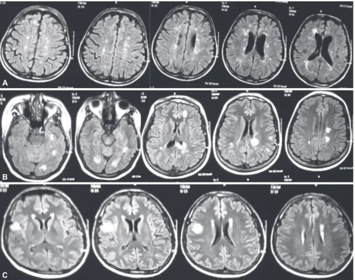
Example MRI scans of TP, FP, and FN patients when ap- plying McDonald 2010 MRI criteria are shown in Fig. 1. Both McDonald 2010 TP and TN patients predominantly pre- sented with cerebral motor manifestations, followed by op- tic neuritis and cerebral sensory manifestations, while both FP and FN patients predominantly had cerebral sensory manifestations in their first presentation, followed by cere- bral motor and cerebellar manifestations. The MAGNIMS 2016 TP group mainly presented with optic neuritis mani- festations, whereas most of the MAGNIMS 2016 TN patients had cerebral motor manifestations at their presentation. The single MAGNIMS 2016 FP patient had optic neuritis, whereas Table 1. Demographic characteristics of the patients
Follow-up period (years) 2.2±0.4
Conversion to CDMS 42 (64)
Time for conversion to CDMS (days) 356±30
Female-to-male ratio 2.2:1
EDSS (range) 2.0 (1–3.5)
Time of baseline MRI from disease onset (days) 69±20 Time of follow-up MRI from baseline MRI (days) 283±46 Number of attacks during follow-up (range) 3 [2–7]
CIS
After first 3 months (%) 57 (86)
After follow-up period (%) 24 (36)
Data are n (%), mean±SD, mean (range), or median [range] values.
CDMS: clinically definite multiple sclerosis, CIS: clinically isolated syn- drome, EDSS: Expanded Disability Status Scale, MRI: magnetic reso- nance imaging.
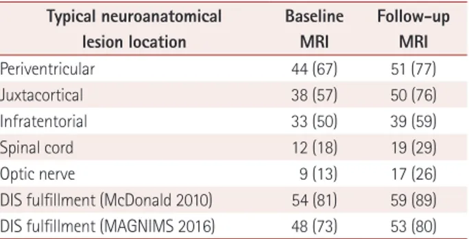
Table 2. Central nervous system lesion characteristics in baselineand follow-up MRI
Typical neuroanatomical lesion location
Baseline MRI
Follow-up MRI
Periventricular 44 (67) 51 (77)
Juxtacortical 38 (57) 50 (76)
Infratentorial 33 (50) 39 (59)
Spinal cord 12 (18) 19 (29)
Optic nerve 9 (13) 17 (26)
DIS fulfillment (McDonald 2010) 54 (81) 59 (89) DIS fulfillment (MAGNIMS 2016) 48 (73) 53 (80) Data are n (%) values. Dissemination in time fulfillment is the same for both McDonald 2010 and MAGNIMS 2016 MRI criteria.8
DIS: dissemination in space, MAGNIMS: magnetic resonance imaging in multiple sclerosis, MRI: magnetic resonance imaging.
Table 3. Results of application of McDonald 2010 and MAGNIMS 2016 MRI criteria after 2 years of follow-up
Positive Negative True positives False positives True negatives False negatives
McDonald 2010 MRI criteria 37 (56) 29 (44) 33 (50) 4 (6) 20 (30) 9 (14)
MAGNIMS2016 MRI criteria 29 (44) 37 (56) 28 (42) 1 (2) 23 (35) 14 (21)
Data are n (%) values.
MAGNIMS: magnetic resonance imaging in multiple sclerosis, MRI: magnetic resonance imaging.
Multiple Sclerosis in Sri Lanka
JCN
cerebral sensory manifestations predominated in the MAG- NIMS 2016 FN group.
The majority of patients across all the categories had typical periventricular lesions in MRI, followed by cortical/subcorti- cal, infratentorial, and optic-nerve lesions, respectively (Fig. 2).
DISCUSSION
This study found that all of the performance parameters (i.e., sensitivity, specificity, accuracy, PPV, and NPV) had high values when the McDonald 2010 and MAGNIMS 2016 MRI criteria were applied to Sri Lankan patients, which confirms the validity of both MRI criteria in our setting.
A similar study involving a Russian MS cohort found that a sensitivity of 74% for McDonald 2010 MRI criteria,14 while Swanton et al.21 found a sensitivity of 77%, and the sensitivi- ty was 68% in a study of Taiwanese MS patients.12 Thus, the sensitivity of McDonald 2010 MRI criteria in the present Sri Lankan MS cohort (78.5%) appears to be higher than that in Taiwanese patients and lower than that in Russian patients.
The accuracy of McDonald 2010 MRI criteria was consider- ably lower in the present study (64%) than those in the stud- ies of Belova et al.14 (82%), and Swanton et al.21 (86%), and Hsueh et al.12 (73.8%).
Table 4. Validation parameters of McDonald 2010 and MAGNIMS 2016 MRI criteria for the diagnosis of multiple sclerosis
McDonald 2010 MRI criteria (%)
MAGNIMS 2016 MRI criteria (%)
Sensitivity 78.5 66.7
Specificity 83.4 95.8
Accuracy 63.7 77.2
Positive predictive value 89.0 96.5
Negative predictive value 68.9 62.1
MAGNIMS: magnetic resonance imaging in multiple sclerosis, MRI: mag- netic resonance imaging.
Fig. 1. Magnetic resonance imaging scans of McDonald 2010 true-positive (A), false-positive (B), and false-negative (C) patients.
A
B
C
Gamage SMK et al.
JCN
A diagnostic tool with a high specificity will tend to ex- clude FP cases.12 The specificity of McDonald 2010 MRI cri- teria was 83% in our study population, which is higher than those of MS patient populations in Argentina13 (80%) and Taiwan12 (80%), but lower than that reported in central Rus- sia14 (93%).
The PPV of McDonald 2010 MRI criteria in the present cohort of Sri Lankan MS patients is comparable but slightly lower than the values reported in the literature for other MS populations, such as 94% for central-Russia MS patients14 and 96% for Argentinian MS patients.13 The NPVs of these two MS patient samples were 70%14 and 46%13 respectively, indicating that the NPV of McDonald 2010 MRI criteria for the present Sri Lankan MS patients was higher than that for central-Russia patients but lower than that for Argentinian patients.
This is the first study to have determined the applicability of the new MAGNIMS 2016 MRI criteria in a South Asian MS patient population. A multicenter European study that evaluated the prediction of an MS diagnosis in CIS patients concluded that McDonald 2010 and MAGNIMS 2016 MRI criteria provide similar accuracy.17 In the present study, the sensitivity of MAGNIMS 2016 MRI criteria (66.7%) was lower than that of McDonald 2010 MRI criteria (78.5%), whereas the specificity was higher for MAGNIMS 2016 MRI criteria (95.8% vs. 83.4%). These results are consistent with Lamas Pérez22 finding that MAGNIMS 2016 MRI criteria
had better specificity than McDonald 2010 MRI criteria in a group of 161 CIS patients. The lower sensitivity and higher specificity of MAGNIMS 2016 MRI criteria in both of these studies may be attributable to the required number of periven- tricular lesions increasing from one to three. However, in an- other recent study of 170 Korean patients with CIS, the sensi- tivity of new MAGNIMS DIS criteria was found to be superior to that of McDonald 2010 MRI criteria, whereas the specific- ity was lower than that of McDonald 2010 MRI criteria. The diversity of the findings for CIS cases might be attributable to differences in study methodologies. The total rate of con- version to CDMS was higher in the present study (64%) than in these two previous studies. This difference might have been due to our CIS patients not receiving DMT until they experi- enced their second clinical episode, whereas proportion of the patients in both of the other studies were receiving DMT.
This would have affected the conversion rate to CDMS since DMT slows the development of CDMS.15 This aspect may have also affected the differences in performance parameters.
Furthermore, it is recommended for interpreters of MRI scans to be aware of the clinical findings of patients to ensure accu- rate MRI interpretations in MS,10 and so the investigator who applied MRI criteria was not blinded of the clinical features and investigation findings of the patients in the present study, which contrasts with the approach taken by Hyun et al.15
The present study has the limitation of its sample being smaller than those in both previous studies. This is mainly
20 18 16 14 12 10 8 6 4 2 0
Number of patients
True positives (n=33)
McDonald 2010 MAGNIMS 2016
True positives (n=28) True negatives
(n=20) True negatives
(n=23) False negatives
(n=9) False negatives
(n=14) False positives
(n=4) False positives
(n=1)
Cerebral motor Cerebral sensory Cerebellar Autonomic Spinal cord Optic neuritis Brainstem/cranial nerves Fig. 2. Presenting clinical manifestations in the study population. MAGNIMS: magnetic resonance imaging in multiple sclerosis.
Multiple Sclerosis in Sri Lanka
JCN
due the expected low prevalence of MS in the South Asian region. The sample size was further restricted by the high cost and limited availability of MRI facilities. In addition, in revisions of McDonald 2010 MRI criteria it was suggested to perform spinal-cord MRI both in patients with spinal-cord syndrome, as well as those who present with non-spinal-cord CIS when their brain MRI did not fulfill the DIS criteria.7 Al- though spinal-cord MRI scans were available in patients with spinal-cord syndrome, we were unable to perform spinal-cord MRI in all other patients due to facility and financial limita- tions.
This study found that TP MS cases could be diagnosed more accurately using McDonald 2010 MRI criteria than new MAGNIMS 2016 MRI criteria in a cohort of Sri Lankan MS patients. However, the specificity, accuracy, and PPV were higher for the MAGNIMS 2016 MRI criteria. The very high specificity and PPV indicate that the new MAGNIMS con- sensus guidelines are more effective in excluding MS that mimics MRI lesions than are the McDonald 2010 MRI cri- teria. Thus, the new MAGNIMS consensus guidelines de- creased the probability of false diagnoses of MS in our study sample, and their high PPV further improves the ability to select a well-refined sample of true MS patients.
Thus, new MAGNIMS consensus guidelines had better specificity, accuracy, and PPV than McDonald 2010 MRI cri- teria in our sample of the Sri Lankan MS population. How- ever, McDonald 2010 MRI criteria had superior sensitivity and NPV compared to MAGNIMS 2016 MRI criteria in this population.
Conflicts of Interest
The authors have no financial conflicts of interest.
Acknowledgements
We thank the National Research Council of Sri Lanka (Grant12-106) and the University Grants Commission of Sri Lanka (Grant UGC/DRIC/
PG/2014MAY/PDN/01) for their financial support. We are also very grateful to Associate Professor Anna Fogdell-Hahn of the Karolinska In- stitute, Stockholm, Sweden and Professor R.P.V.J. Rajapakse of the Facul- ty of Veterinary Medicine and Animal Sciences, University of Peradeniya for their constant support and guidance. Ms Merja Kanerva and Ms Faezeh Vejdani are acknowledged for their valuable assistance.
REFERENCES
1. Brownlee WJ, Hardy TA, Fazekas F, Miller DH. Diagnosis of multiple sclerosis: progress and challenges. Lancet 2017;389:1336-1346.
2. Piccolo L, Kumar G, Nakashima I, Misu T, Kong Y, Wakerley B, et al.
Multiple sclerosis in Japan appears to be a milder disease compared to the UK. J Neurol 2015;262:831-836.
3. Gamage SMK, Wijeweera I, Adikari SB, Fink K, Hillert J, Fogdell- Hahn A, et al. Multiple sclerosis patients with markedly low intrathe- cal antibody response in Sri Lanka. Mult Scler Int 2018;2018:5342936.
4. Singhal B. Multiple sclerosis-Indian perspective. Neurol India 2015;63:
824-825.
5. Li T, Xiao H, Li S, Du X, Zhou J. Multiple sclerosis: clinical features and MRI findings in Northern China. Eur J Med Res 2014;19:20.
6. Viswanathan S, Rose N, Masita A, Dhaliwal JS, Puvanarajah SD, Rafia MH, et al. Multiple sclerosis in Malaysia: demographics, clinical fea- tures, and neuroimaging characteristics. Mult Scler Int 2013;2013:614716.
7. Polman CH, Reingold SC, Banwell B, Clanet M, Cohen JA, Filippi M, et al. Diagnostic criteria for multiple sclerosis: 2010 revisions to the McDonald criteria. Ann Neurol 2011;69:292-302.
8. Filippi M, Rocca MA, Ciccarelli O, De Stefano N, Evangelou N, Kap- pos L, et al. MRI criteria for the diagnosis of multiple sclerosis: MAG- NIMS consensus guidelines. Lancet Neurol 2016;15:292-303.
9. Thompson AJ, Banwell BL, Barkhof F, Carroll WM, Coetzee T, Comi G, et al. Diagnosis of multiple sclerosis: 2017 revisions of the McDon- ald criteria. Lancet Neurol 2018;17:162-173.
10. Rovira À, Wattjes MP, Tintoré M, Tur C, Yousry TA, Sormani MP, et al. Evidence-based guidelines: MAGNIMS consensus guidelines on the use of MRI in multiple sclerosis-clinical implementation in the diagnostic process. Nat Rev Neurol 2015;11:471-482.
11. Huh SY, Kim SH, Kim W, Lee SH, Park MS, Ahn SW, et al. Evaluation of McDonald MRI criteria for dissemination in space in Korean pa- tientswith clinically isolated syndromes. Mult Scler 2014;20:492-495.
12. Hsueh CJ, Kao HW, Chen SY, Lo CP, Hsu CC, Liu DW, et al. Com- parison of the 2010 and 2005 versions of the McDonald MRI criteria for dissemination-in-time in Taiwanese patients with classic multiple sclerosis. J Neurol Sci 2013;329:51-54.
13. Patrucco L, Rojas JI, Miguez JS, Cristiano E. Application of the Mc- Donald 2010 criteria for the diagnosis of multiple sclerosis in an Ar- gentinean cohort of patients with clinically isolated syndromes. Mult Scler 2013;19:1297-1301.
14. Belova AN, Shalenkov IV, Shakurova DN, Boyko AN. Revised Mc- Donald criteria for multiple sclerosis diagnostics in central Russia:
sensitivity and specificity. Mult Scler 2014;20:1896-1899.
15. Hyun JW, Huh SY, Kim W, Park MS, Ahn SW, Cho JY, et al. Evalua- tion of 2016 MAGNIMS MRI criteria for dissemination in space in patients with a clinically isolated syndrome. Mult Scler 2017 May 1 [Epub] available from: https://doi.org/10.1177/1352458517706744.
16. Gajofatto A, Nourbakhsh B, Benedetti MD, Waubant E. Performance of 2010 McDonald criteria and 2016 MAGNIMS guidelines in the di- agnosis of primary progressive multiple sclerosis. J Neurol Neurosurg Psychiatry 2017 Sep 22 [Epub]. available from: https://doi.org/10.1136/
jnnp-2017-316911.
17. Filippi M, Preziosa P, Meani A, Ciccarelli O, Mesaros S, Rovira A, et al.
Prediction of a multiple sclerosis diagnosis in patients with clinically isolated syndrome using the 2016 MAGNIMS and 2010 McDonald criteria: a retrospective study. Lancet Neurol 2018;17:133-142.
18. SadakaY, Verhey LH, Shroff MM, Branson HM, Arnold DL, Naray- anan S, et al. 2010 McDonald criteria for diagnosing pediatric multi- ple sclerosis. Ann Neurol 2012;72:211-223.
19. Wingerchuk DM, Banwell B, Bennett JL, Cabre P, Carroll W, Chitnis T, et al. International consensus diagnostic criteria for neuromyelitis optica spectrum disorders. Neurology 2015;85:177-189.
20. Poser CM, Paty DW, Scheinberg L, McDonald WI, Davis FA, Ebers GC, et al. New diagnostic criteria for multiple sclerosis: guidelines for research protocols. Ann Neurol 1983;13:227-231.
21. Swanton JK, Fernando K, Dalton CM, Miszkiel KA, Thompson AJ, Plant GT, et al. Modification of MRI criteria for multiple sclerosis in patients with clinically isolated syndromes. J Neurol Neurosurg Psychi- atry 2006;77:830-833.
22. Lamas Pérez R. Comparison between the 2010 McDonald and 2016 MAGNIMS MRI criteria for dissemination in space in patients with a clinically isolated syndrome. Does the recent recommendation regard- ing the current criteria improve diagnostic accuracy? ECTRIMS On- line Library 2017;200244.