338
females approximately 2 to 12. It is histologically similar to
mucoepidermoid carcinoma and squamous cell carcinoma but is a benign disease that spontaneously heals within 4 to 10 weeks. Recurrence is very rare3,4. In the present case,
nec-rotizing sialometaplasia of the hard palate re-occurred on the contralateral side after 5 months.
II. Case Report
A 36-year-old woman was referred to the Gangnam Sever-ance Hospital (Seoul, Korea) by a private practice endodon-tist. The patient stated that her palate felt uncomfortable and that her throat had been swollen for a week. She had a history of smoking more than 40 cigarettes a day for 20 years and de-nied any other medical history. Endodontic treatment of the upper left first premolar under local anesthesia with 2% lido-caine and 1:100,000 epinephrine was completed by the endo-dontist before the patient’s arrival to our hospital. A crater-like ulceration of 3 cm diameter, with necrotic tissue inside, was found on the left hard palate. No spontaneous hemor-rhage or bony exposure of the hard palate was observed.(Fig.
I. Introduction
Necrotizing sialometaplasia was first described by Abrams et al.1 in 1973, and since then, other similar cases have been
reported. Necrotizing sialometaplasia is an inflammatory, ul-cerative lesion of the small salivary glands that usually occurs on one side of the hard palate, although it can also occur on the lower lip, posterior molar area, buccal mucosa, the parot-id gland, tongue, and in the upper airway2,3. The average age
of occurrence is 46 years. Necrotizing sialometaplasia occurs more frequently in males, with the ratio of affected males to
CASE REPORT
Jong-Ki Huh
Department of Oral and Maxillofacial Surgery, Gangnam Severance Hospital, Yonsei University College of Dentistry, 211 Eonju-ro, Gangnam-gu, Seoul
06273, Korea
TEL: +82-2-2019-4560 FAX: +82-2-3463-4052 E-mail: [email protected]
ORCID: http://orcid.org/0000-0002-7381-3972
This is an open-access article distributed under the terms of the Creative Commons Attribution Non-Commercial License (http://creativecommons.org/licenses/by-nc/4.0/), which permits unrestricted non-commercial use, distribution, and reproduction in any medium, provided the original work is properly cited.
CC
Contralateral recurrence of necrotizing sialometaplasia of
the hard palate after five months: a case report
Chan-Woo Jeong1, Taegyun Youn1, Hyun Sil Kim2, Kwang-Ho Park1, Jong-Ki Huh1 1Department of Oral and Maxillofacial Surgery, Gangnam Severance Hospital, Yonsei University College of Dentistry, 2Department of Oral Pathology and Oral Cancer Research Institute, Yonsei University College of Dentistry, Seoul, Korea
Abstract(J Korean Assoc Oral Maxillofac Surg 2015;41:338-341)
Necrotizing sialometaplasia usually heals within 4 to 10 weeks with conservative treatment, and rarely recurs. When necrotizing sialometaplasia is present on the hard palate it may occur unilaterally or bilaterally. In this case, necrotizing ulceration occurred on the left hard palate of a 36-year-old woman after root canal treatment of the upper left first premolar under local anesthesia. After only saline irrigation the defect of the lesion completely healed and filled with soft tissue. After 5 months, however, a similar focal necrosis was found on the contralateral hard palate without any dental treat-ment having been performed on that side and progressed in similar fashion as the former lesion. We conducted an incisional biopsy and obtained a final pathological diagnosis for the palatal mass of necrotizing sialometaplasia. At the 3-year follow-up, the patient’s oral mucosa of the hard palate was nor-mal, without any signs and symptoms of the condition. We report a case of a second occurrence of necrotizing sialometaplasia on the contralateral side from the first, with a time lapse between the first and second occurrence.
Key words: Necrotizing sialometaplasia, Palate
[paper submitted 2015. 6. 26 / revised 2015. 9. 23 / accepted 2015. 10. 3]
Copyright Ⓒ 2015 The Korean Association of Oral and Maxillofacial Surgeons. All rights reserved.
http://dx.doi.org/10.5125/jkaoms.2015.41.6.338 pISSN 2234-7550·eISSN 2234-5930
Contralateral recurrence of necrotizing sialometaplasia of the hard palate after five months
339 of Yonsei University Gangnam Severance Hospital (IRB # 3-2015-0114), and informed consent was obtained.
III. Discussion
The cause of necrotizing sialometaplasia is unknown, but it is thought to result from infarctions of the salivary gland tissue1,2,5. The causes of such infarctions may be direct injury,
effects of vasoconstrictors, or injuries from needles used in local anesthesia, ill-fitting dentures, drinking, smoking, co-caine use, radiation, infection of the upper airway or allergy, intubation, surgical treatment, and systemic diseases such as sickle cell disease, Buerger’s disease, Raynaud’s disease, and AIDS2-5. Shigematsu et al.6 reported that
histopatho-logical changes similar to those associated with necrotizing sialometaplasia may be observed when local anesthesia is repeatedly injected into the hard palate of mice. Necrotiz-ing sialometaplasia with ulceration may occur when tissues in the salivary glands are infarcted and the acinar cells are subsequently necrotized. Brannon et al.2 reported that the
risk of necrotizing sialometaplasia in smokers is about 10% 1. A) The lesion was first diagnosed as either tissue necrosis
caused by local injection of lidocaine containing epinephrine or necrotizing sialometaplasia, and was treated palliatively. It was not possible to perform a biopsy at that time because the tissue was already necrotized.(Fig. 1. B) After 3 weeks, a stable healing state was observed clinically.(Fig. 1. C)
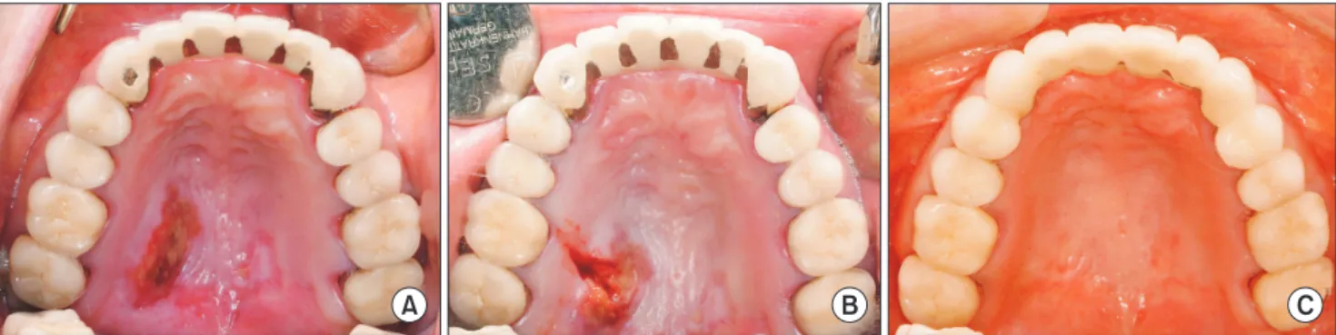
After 5 months, the patient returned to the hospital for re-currence of the same symptoms on the contralateral side of the hard palate without any particular dental event.(Fig. 2. A) We conducted an incisional biopsy to confirm the diagnosis. (Fig. 2. B) Microscopically, the palatal lesion showed dif-fuse necrosis of acini and ducts of palatal salivary glands; however, the overall lobular structure of the involved gland was preserved. There was mucin present with an associated inflammatory response.(Fig. 3) Based on histological analy-ses, the final pathological diagnosis for the palatal mass was necrotizing sialometaplasia.
At the 3-year follow-up, the patient’s oral mucosa of the hard palate was normal, without any signs or symptoms.(Fig. 2. C)
This study was approved by Institutional Review Board
A B C
Fig. 1. A. Ulceration on left hard palate of the patient at the first visit. B. Necrotic tissue detached from the lesion spontaneously. C. Normal
healing of the lesion three weeks after the first visit.
Chan-Woo Jeong et al: Contralateral recurrence of necrotizing sialometaplasia of the hard palate after five months: a case report. J Korean Assoc Oral Maxillofac Surg 2015
A B C
Fig. 2. A. Recurrent lesion on the right hard palate five months after the initial visit. The lesion on the left side had healed well. B. Incisional
biopsy was performed. C. Complete healing of bilateral hard palate necrotizing sialometaplasia at 3-year periodic check-up.
J Korean Assoc Oral Maxillofac Surg 2015;41:338-341
340
fully cured, necrotizing sialometaplasia rarely recurs2-4.
Necrotizing sialometaplasia is histopathologically divided into infarction, sequestration, ulceration, and reparative stages4,5. The stages are not clearly separated and can
prog-ress from occurrence to full recovery without any treatment. When salivary gland tissues become infarcted and are necro-tized and separated, the necronecro-tized tissues collapse, creating a crater-shaped ulcer. The surrounding mucous membranes show secondary healing and require approximately 4 to 10 weeks for complete recovery2-4,7. Pain may or may not be
present; however, if there is pain in the first period of ulcer formation, the use of an analgesic is recommended. Because necrotizing sialometaplasia appears as an inflammatory ulcer, some doctors treat it with antibiotics or steroids, but with no reported effects on clinical symptoms7.
Our patient case had a negative medical history with the ex-ception of smoking and local anesthesia for dental treatment. Thus, in the preliminary diagnosis, we diagnosed our patient with necrotized tissue caused by the blockage of the terminal blood vessels from complications of smoking and the use of lidocaine with epinephrine. After 3 weeks of follow-up, the necrotized tissues were no longer present and secondary healing of the soft tissues was apparent. Five months after the first occurrence of the lesion, we observed recurrence on the contralateral side of the hard palate without any particular factors other than smoking. The second lesion was still within the initial period of necrotizing when observed, and we there-fore conducted an incisional biopsy including both normal (7/69). Smoking could cause infarctionsin the salivary gland
by continuously stimulating the palate and inducing vascular stenosis. In a previous study, the researchers were unable to determine the cause of necrotizing sialometaplasia in 44% of the patient sample (30/69), and the rate of recurrence was 0% (0/14)2. A number of reports have concluded that the risk of
recurrence is normally 0%, and that instances of recurrence on the opposite side are rare.
Necrotizing sialometaplasia is a benign disease, which is usually only treated palliatively. However, histologically, tissues in the salivary glands show infarction, inflammation, granulation tissue, and squamous metaplasia of the salivary duct. Because such squamous metaplasia is often observed, it may be misdiagnosed as squamous cell carcinoma, and because it is similar to mucoepidermoid carcinoma in that the squamous metaplasia is surrounded by a faint outline of necrotized acinar cells, it is easily misdiagnosed as malig-nant1,2,7,8. However, despite similarities of infection and
ne-crosis, necrotizing sialometaplasia is distinguished from ma-lignancies because its ductal structure is preserved3,7. When
necrotizing sialometaplasia is misdiagnosed as a malignancy, irreversible treatment such as radical resection may follow. It is therefore essential to differentiate necrotizing sialometa-plasia from malignancies. In contrast, some malignancies can be misdiagnosed as necrotizing sialometaplasia and subse-quently go untreated. Therefore, Bascones-Martínez et al.5
reported that incisional biopsy is recommended for accurate diagnosis, with periodic treatment until full recovery9. When
A B
Fig. 3. Histopathologic findings. A. The lobular structure of the salivary gland is preserved, but the salivary gland showed diffuse necrosis
of acini and duct (arrowhead). A mucin pool was also identified (arrow) (H&E staining, ×40). B. Chronic inflammatory cell infiltrates around the necrotic acini and ducts (H&E staining, ×200).
Contralateral recurrence of necrotizing sialometaplasia of the hard palate after five months
341
ORCID
Chan-Woo Jeong, http://orcid.org/0000-0003-4118-075X Taegyun Youn, http://orcid.org/0000-0003-1409-3300 Hyun Sil Kim, http://orcid.org/0000-0003-3614-1764 Kwang-Ho Park, http://orcid.org/0000-0003-1942-2986 Jong-Ki Huh, http://orcid.org/0000-0002-7381-3972
References
1. Abrams AM, Melrose RJ, Howell FV. Necrotizing sialometaplasia. A disease simulating malignancy. Cancer 1973;32:130-5.
2. Brannon RB, Fowler CB, Hartman KS. Necrotizing sialometapla-sia: a clinicopathologic study of sixty-nine cases and review of the literature. Oral Surg Oral Med Oral Pathol 1991;72:317-25. 3. Newland J. Bilateral presentation of necrotizing sialometaplasia: a
case report. Dent Update 2007;34:586-8.
4. Imbery TA, Edwards PA. Necrotizing sialometaplasia: literature review and case reports. J Am Dent Assoc 1996;127:1087-92. 5. Bascones-Martínez A, Muñoz-Corcuera M, Cerero-Lapiedra R,
Bascones-Ilundáin J, Esparza-Gómez G. Case report of necrotizing sialometaplasia. Med Oral Patol Oral Cir Bucal 2011;16:e700-3. 6. Shigematsu H, Shigematsu Y, Noguchi Y, Fujita K. Experimental
study on necrotizing sialometaplasia of the palate in rats. Role of local anesthetic injections. Int J Oral Maxillofac Surg 1996;25:239-41.
7. Ylikontiola L, Siponen M, Salo T, Sándor GK. Sialometaplasia of the soft palate in a 2-year-old girl. J Mich Dent Assoc 2010;92:38-40.
8. Kim JW, Kim MJ, Woo SS. Salivary duct carcinoma of the minor salivary gland in hard palate. J Korean Assoc Oral Maxillofac Surg 1993;19:567-72.
9. Keogh PV, O'Regan E, Toner M, Flint S. Necrotizing sialometapla-sia: an unusual bilateral presentation associated with antecedent an-aesthesia and lack of response to intralesional steroids: case report and review of the literature. Br Dent J 2004;196:79-81.
and necrotized tissues to confirm the diagnosis of necrotizing sialometaplasia.
Necrotizing sialometaplasia occurs unilaterally in 71% of previously-reported cases, and on both sides of the median suture line of the hard palate in 12% of cases2. However, in
our case, there was a second occurrence on the contralateral side with a time lapse between the first and second occurrenc-es, a presentation that is thought to be quite rare. Our patient was not definitively diagnosed upon the second occurrence, but the second lesion may have been caused by the patient’s heavy smoking habit, because she had experienced no dental events such as the use of local anesthesia.
Conflict of Interest
No potential conflicts of interest relevant to this article are reported.
Acknowledgements
We would like to thank Ji-hye Moon and Chan-heok Gong for improving the use of English in the manuscript.