Received October 24, 2013, Revised November 20, 2013, Accepted November 25, 2013 Corresponding author: Jinyoung Moon
Deptartment of Acupoint, College of Korean Medicine, Dongguk University, 814-9, Siksa-dong, Goyang 410-773, Korea Tel: +82-31-961-5830, Fax: +82-31-961-5823, E-mail: [email protected]
This work was supported by the Dongguk University research Fund of 2013.
CCThis is an open access article distributed under the terms of the Creative Commons Attribution Non-Commercial License (http://creativecommons.org/licenses/ by-nc/3.0) which permits unrestricted non-commercial use, distribution, and reproduction in any medium, provided the original work is properly cited.
대계 약침액의 C6 신경교종 세포에 대한 이주 억제 효과
박주연ㆍ이강파1ㆍ장해룡1ㆍ문진영
동국대학교 한의과대학 경혈학교실, 1동국대학교 과학기술대학 세포유전학 실험실
The Anti-Migratory Effect of Cirsium japonicum Pharmacopuncture in C6 Glioma Cell
Juyeon Park, Kangpa Lee1, Haeryong Chang1, Jinyoung Moon
Deptartment of Acupoint, College of Korean Medicine, Dongguk University,
1Division of Bio Science, College of Science and Technology, Dongguk University
Objectives : Cirsium japonicum is a traditional Korean medicine that has been used in the treatment of inflammatory diseases such as appendicitis, hepatitis, pulmonary abscess and tumor. The aim of study was to elucidate anti-migratory activity of CJP(Cirsium japonicum pharmacopuncture) through regulation of inflammatory mediators in C6 glioma cell. Methods : Nitric oxide(NO) production was determined by using nitrite assay. The cell migration was analyzed by wound-healing assay and Boyden chamber assay. The expression levels of iNOS, and protein kinase C(PKC)-α were measured by western blotting assay. Results : CJP showed a significant decrease on NO production. Moreover, glioma cell migration was effectively suppressed by CJP. Furthermore, CJP inhibited the expressions of iNOS and PKC-α in C6 glioma cells. Conclusions : These results suggest that CJP inhibits glioma cell migration and iNOS expression through regulation of PKC-α. Therefore, it is expected that CJP could be an effective agents for blocking malignant progression of glioma.
Key words : cirsium japonicum, migration, iNOS, PKC-α, C6 glioma cells
서 론
신경교종은 이환율과 사망률이 높으며, 침습성이 강한 특징 때문 에 치료가 어렵고 예후가 불량하다1,2). 특히 악성 뇌신경교종은 인근 의 정상세포로 이주(migration)하는 특성이 있으며, 신속한 증식과 침윤(invasion)을 일으키며 방사능 및 화학요법에 저항성을 나타내 므로 기존의 암치료 요법으로는 치료가 어려우며3,4), 신생혈관 억제 (anti-angiogenesis) 및 세포자멸사(apoptosis)의 유도와 같은 치료 법도 환자의 생명연장에 크게 도움이 되지 않는 실정이다5,6).
한편 만성적인 염증반응은 DNA 손상과 염색체의 불안정을 초래
하여 암의 발생을 유도하는데7), 이 과정에는 cytokines, chemo- kines, cyclooxygenase-2(COX-2), prostaglandins(PGs), induc- ible nitric oxide synthase(iNOS) 및 nitric oxide(NO)와 같은 다양 한 염증 매개인자들이 관여한다8). 또한 염증 매개인자들의 과발현, 과잉생산 및 비정상적인 활성화는 암세포의 증식을 야기하고 세포자 멸사에 대한 저항성을 증가시켜 암 발생을 촉진하며, 신생혈관 형성 (angiogenesis)과 조직 변화(tissue remodeling)와 같은 자극을 통해 암세포가 정상 조직으로 이주하여 침윤과 전이(metastasis)가 일어나 도록 유도한다9,10).
대계(Cirsium japonicum De Candole)는 국화과의 여러해살이
풀 엉겅퀴의 全草로서, 性味는 甘苦, 凉하며 養精保血, 凉血止血, 散瘀消腫하는 효능이 있어 충수염, 폐농양, 화상, 종기, 간염 및 癰 腫 등과 같은 염증성 질환의 치료에 사용되어 왔다11,12).
한편, 대계의 효능에 관한 실험적 연구로 항산화 효능13), cy- tochrome P450 억제를 통한 항동맥경화 효능14), GABA receptor 작용을 통한 불안 완화효과15) 등이 규명된 바 있으며, 대계의 아종 인 var. maackii와 그 구성성분 luteolin 5-O-glucoside가 Raw 264.7 세포에서 iNOS 발현 억제를 통하여 NO 생성을 줄인다는 보고16)가 있다.
또한 본 약물의 항암효능과 관련한 연구로 쥐의 간암세포와 육 종에 대한 항암효과17,18), 사람 유방암 세포에 대한 증식 억제효과 및 세포자멸사 유도효과19) 등이 보고된 바 있으나, 아직까지 뇌종 양과 관련된 연구는 접할 수 없는 실정이다. 이에 본 연구에서는 신경교종에 대한 새로운 치료 전략으로 염증제어 및 암세포 이주 억제능을 지닌 약물이 신경교종의 악성 진행을 차단함으로써 환자 의 생명연장에 기여할 수 있을 것이라는 점에 착안하여, 대계 약침 액의 항염증 효능 및 신경교종 세포의 이주에 대한 영향을 중심으 로 연구를 진행하였다. 이에 흰쥐 C6 신경교종 세포에서 항염증효 능 및 세포 이주에 대한 효능을 관찰하여 유의한 결과를 얻었기에 보고하는 바이다.
재료 및 방법
1. 시약
세포배양에 사용된 Dulbecco’s Modified Eagle Medium(DMEM) 과 phosphate buffered saline(PBS)은 HyClone사(Logan, UT, USA), fetal bovine serum(FBS)은 WelGENE사(Seoul, Korea), penicillin-streptomycin과 trypsin-ethylene diamine tetraacetic acid(EDTA)는 Gibco사(Carlsbad, California, USA)에서 구입하였다.
Cell viability 측정에 사용된 Ez-cytox assay kit는 Daeil Lab Service(Seoul, Korea)에서, 단백질 정량에 사용된 Pierce BCA protein assay kit는 Thermo Scientific(Rockford, IL, USA)에서, LPS와 Diff Quick stain은 Sigma사(St. Louis, MO, USA)에서 구입하 여 사용하였다. 한편 primary antibody와 secondary antibody는 Cell signaling Technology(Beverly, MA, USA) 및 Santa Cruz Biotechnology(Santa Cruz, MA, USA)에서 구입하여 사용하였다.
2. 약재 및 약침액 조제
대계는 동국대학교 한의과대학 부속한방병원(경북, 경주시)에서 구입한 것을 정선하여 사용하였다. 본 실험에서 대계 약침액은 水 劑-알콜 沈法에 따라 제조하였다. 먼저 60 g의 약재를 작은 크기로 잘라 원저 flask에 넣고 500 ml의 증류수를 가하여 3시간 동안 80oC에서 추출한 후, Whatman paper로 여과하여 상층액을 수거 하였다. 여과액을 rotary evaporator(EYELA N-1000, Japan)로 감 압농축하여 전량을 50 ml로 농축한 다음 99.9% ethanol을 가하여 교반하고 저온에서 방치하여 상층액을 수거하였다. 위와 같은 방법 으로 차례로 75%, 85%, 95% ethanol 용액이 되도록 하면서 각각 의 단계에서 혼합액을 교반하고 저온에서 방치하여 침전물을 제거 하였다. 이후, 상층액을 수거하고 감압농축하여 에탄올 성분을 제 거하였다. 이를 동결건조기(LABCONCO FreeZone 6, USA)에서 동 결건조 한 다음, 최종 3.04 g의 분말을 회수(회수율 w/w:5.10%)하 여 대계 약침액(Cirsium japonicum pharmacopuncture:CJP)으 로 사용하였다. 한편, 회수된 분말은 실험에 사용하기 전까지 냉동 건조한 환경에서 보관하였으며, voucher specimen은 동국대학교 한의과대학 경혈학교실(경기도, 고양시)에 보관되어 있다.
3. 세포배양
흰쥐 신경교종 세포주인 C6 glioma cell은 한국세포주은행 (KCLB, Seoul, Korea)에서 분양받았으며, 10% FBS, 1% anti- biotics(penicillin-streptomycin)이 포함된 DMEM 배지에서 37oC, 5% CO2의 조건으로 배양하였고, 세포의 성장주기를 고려 하여 2∼3일마다 100 mm dish에서 subculture하여 실험에 사 용하였다.
4. 세포생존율 측정
CJP가 세포생존율에 미치는 영향을 관찰하기 위하여, C6 gli- oma 세포를 96 well-plate에 5×10³ cells/well의 농도로 조절하 여 분주한 다음, CO2 incubator에서 24시간 배양하였다. CJP를 농도별로 각각 0, 1, 2, 3, 4 및 5 mg/ml의 농도가 되도록 처리한 후, 18 시간 동안 배양하였다. 세포 독성 여부를 확인하기 위하여 Ez-cytox Assay reagent를 10 μl씩 처리하고 30분간 배양한 후 microplate reader(Molecular Devices, Synnyvale, CA, USA)를 이용하여 450 nm 파장에서 측정하였다.
5. Nitric oxide 소거능 측정
CJP의 nitric oxide(NO) 소거능을 알아보기 위하여 본 실험에서 는 Griess reagent를 이용하여 정량하였다. 24-well plate에 세포
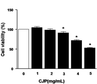
Fig. 1. Effect of CJP on rat C6 glioma cell viability.
To estimate cell viability on C6 glioma cells of CJP, the viability was assessed by using Ez-cytox assay kit. C6 glioma cells were incubated with 0, 1, 2, 3, 4 and 5 mg/ml of CJP for 18 h. Each value of results represents the mean±SD of independent experiments(n=3, *p<0.05 compared with the control group).
를 5×10⁵ cells/well의 농도로 분주하여 세포가 80% 이상 성장하 였을 때 무혈청 배지로 교환한 다음, 24시간 동안 배양하였다. 그 후, CJP를 농도별로 30분 전처리한 다음, LPS를 1 μg/ml의 농도 로 처리하였다. 이후 상층액을 수거하여 96-well plate에 농도별로 100 μl씩 처리하고 1% sulfanilamide(in 5% phosphoric acid)를 50 μl씩 처리하고 실온에서 10분간 배양하였다. 그 후, 0.1%
N-1-naphthylethylenediamine dihydrochloride(NED)를 50 μl 씩 처리한 다음, 실온에서 10분간 반응을 유도하였으며, micro- plate reader를 이용하여 540 nm 파장에서 흡광도를 측정하였다.
6. Wound healing assay
C6 신경교종 세포를 6-well plate에 1×10⁵ cells/well의 농도 로 분주한 다음, 24시간 동안 배양하였다. 세포가 단일층으로 균일 하게 배양된 것을 확인한 후 무혈청배지로 교환하여, 다시 24시간 을 추가 배양하고 PBS로 세척하였다. 그 후, FBS가 첨가된 배지로 교환한 다음, 농도별 CJP를 처리하였다. 멸균 피펫팁으로 단일 세 포층 위에 scratch를 가한 다음, 24시간 동안 세포가 이주되는 현 상을 현미경으로 관찰하였다.
7. Boyden chamber assay
CJP가 C6 신경교종 세포의 이주에 미치는 효능을 Boyden chamber assay를 이용하여 검토하였다. Boyden chamber는 상층과 하층으로 구분되는 48-well chemotaxis chamber(Neuro-Probe, Gaithersburg, Maryland, USA)로 이들 2개의 chamber는 type I collagen으로 코팅된 8 μm pore 크기의 polycarbonate filter membrane(Neuro-Probe)을 사이에 두고 나뉘어져 있다. C6 세포는 무혈청 배지를 이용해 5×10⁵ cells/ml의 농도로 조절하여 상단 chamber의 각 well에 50 μl씩 분주하였고, 하단 chamber에는 FBS 와 농도별 CJP를 가한 뒤, 37oC, 5% CO2 incubator에서 3시간 동안 배양함으로써 세포의 이주를 유도하였다. 그 후, filter membrane을 분리하여 상단에 남아있는 세포는 면봉으로 제거하였고, membrane 을 통과하여 이주된 하단의 세포를 메탄올로 고정하여 Diff Quick stain(Sigma, St. Luis, MO, USA)으로 염색한 후, 현미경을 이용해 이주된 세포의 정도를 관찰하였다.
8. Western blot analysis
CJP가 iNOS와 PKC-α의 단백질 발현에 미치는 영향을 확인하 기 위하여 Western blotting을 시행하였다. 세포를 ice-cold PBS 로 2회 세척한 후 3,000 rpm에서 5분간 원심분리하여 pellet을 회수하였다. PRO-PRET™ Protein Extraction Solution(iNtRON
Biotechnology)를 첨가하여 세포를 반응시킨 후 13,000 rpm에서 5분간 원심분리하여 상층액을 회수하였다. BCA protein assay kit 를 이용하여 단백질의 양을 정량한 다음 30 μg의 단백질을 100oC 에서 5분간 변성을 유도한 뒤, 변성이 완료된 단백질은 7.5%
polyacrylamide gel에서 전기영동하였다. 이후 Polyvinylidene fluoride(PVDF) membrane에 transfer하여 비특이적 단백질이 반 응하는 것을 방지하기 위하여 5% skim milk가 포함된 TBST(20 mM Tris, 500 mM NaCl, 0.05% Tween 20)로 1시간 동안 blocking하고 1차 antibody(iNOS, PKC-α)를 3% skim milk가 포함된 TBST에 희석하여 4oC에서 overnight하였다. 실온에서 15 분씩 총 4회 TBST로 washing한 다음 2차 antibody를 2% skim milk가 포함된 TBST에 희석하여 2시간 동안 실온에서 반응시켰다.
실온에서 10분씩 총 3회 TBST로 washing하고 ECL용액에 반응시 켜 단백질의 발현정도를 분석하였다.
9. 통계적 처리
실험결과는 평균과 표준편차(mean±SD)로 표기하였으며, stu- dent’s t-test 방법을 통해 유의성 여부를 분석하였고, p<0.05일 경우에 한하여 유의성을 인정하였다.
Fig. 2. Inhibition of LPS-induced NO production on C6 glioma cell by CJP.
C6 glioma cells were treated with the demonstrated concentra- tions of LPS(A). C6 glioma cells were incubated with LPS(1 μg/ml) for the demonstrated times(B). Each cells were treated with CJP prior to addition of LPS, followed by additional incubation for 24 h with LPS(1 μg/ml). Nitrite accumulations in the media were accounted by using the Griess reagent(C).
Fig. 3. Inhibition effect of CJP on serum-induced migration in C6 glio- ma cell by wound-healing assay.
C6 glioma cells were scratched using 200 μl sterile pipette tip and migration of C6 cells was induced by FBS containing media. After 24 h, migration levels of C6 glioma cell were observed using the optical microscope and photographs were obtained. The ratio of migration compared with control was evalu- ated measuring the distance. Each values represented means±SD and performed at least three indepen- dent experiments. *p<0.05 com- pared with FBS-treated alone.
결 과
1. 세포 생존율에 대한 CJP의 영향
CJP가 흰쥐 신경교종 세포인 C6 glioma 세포에 대한 세포사멸 혹은 세포괴사의 기전이 아닌, 이주억제를 통한 항암 활성을 지니 는지 규명하고자, 먼저 농도별 CJP를 처리한 다음, 신경교종세포의 생존율을 관찰하였다. 그 결과, 아무런 처리도 하지 않은 대조군의 생존율을 100%로 계산하였을 때, CJP를 1 mg/ml, 2 mg/ml의 농 도로 처리한 실험군에서는 대조군과 거의 유사한 수준의 생존율을
나타내었으나, 3 mg/ml, 4 mg/ml, 5 mg/ml의 고농도 처리군에서 는 세포생존율이 각각 현저하게 감소하였다. 따라서 CJP를 3 mg/ml 이상의 농도로 처리할 경우, 본 실험에서 규명하고자 하는 신경교종 세포에 대한 항염증 효능 및 이주 억제능 검토에 적합하 지 않음을 알 수 있었다(Fig. 1).
2. NO 생성 억제 효능
본 실험에서는 C6 신경교종 세포에서 LPS를 처리로 유도된 NO 의 생성에 대한 영향을 검토하고자, 먼저 LPS를 농도별 및 시간대 별로 처리하여 실험에 적절한 LPS의 농도와 처리 시간을 결정하였 다. 그 결과, C6 세포에 LPS를 1 μg/ml의 농도로 24시간 동안 처리하였을 때, 정상군에 비해 NO의 생성이 현저하게 유도됨을 확 인할 수 있었다(Fig. 2A, 2B). 따라서 이와 동일한 조건으로 LPS를 처리한 다음, C6 신경교종 세포의 NO 생성에 대한 CJP의 효능을 관찰하였다. 그 결과, 아무런 처리도 하지 않은 정상군에 비해 LPS 를 단독으로 처리한 실험군에서 NO의 생성이 현저하게 증가하였 고, CJP를 2.5 mg/ml의 농도로 처리하였을 때 NO의 생성이 유의 성 있게 억제되는 것을 확인할 수 있었다(Fig. 2C).
3. Wound-healing assay에서 CJP의 세포이주 억제 활 성
CJP가 C6 신경교종 세포의 이주에 미치는 영향을 규명하기 위 하여 본 실험에서는 wound-healing assay를 통해 관찰하였다. 단 층으로 균일하게 자란 신경교종 세포에 멸균 피펫 팁으로 scratch 를 가하여 점선안의 세포를 모두 제거하였다. 그 후, 24시간 동안 배양하여 세포가 제거된 점선안의 영역으로 세포가 이주되는 정도 를 실험군별로 비교 관찰하였다. 그 결과, 세포의 이주를 유도하기 위하여 혈청을 단독 처리한 대조군에서 이주된 세포의 정도를
Fig. 5. Effect of CJP on iNOS, PKC-α expression in LPS-stimulated C6 glioma cell.
After C6 cells in absence or presence CJP(2, 2.5 mg/ml) were stimulated with LPS(1 μg/ml), cell lysates were obtained. Proteins were analyzed by using western blot and expression levels of protein were measured, respectively. GAPDH antibody used as a house-keeping gene. *p<0.05 compaed with LPS-treated alone.
Fig. 4. Evaluation of inhibitory effect on C6 glioma cell migration of CJP using the Boyden chamber assay.
Migration was induced with serum containing media in C6 glioma cells additional 24 h. After incubation, C6 cells in different concentration of CJP(1, 1.5, 2, 2.5 mg/ml) that passed through the membrane were counted after staining and photographs were taken. The marked graph shows the percentage of migrated cells relative to control which is incubated with non-serum media. *p<0.05 com- paed with FBS-treated alone.
100%로 계산하였을 때, CJP를 1, 1.5, 2, 2.5 mg/ml을 처리한 결과, C6 신경교종 세포의 이주가 농도 의존적으로 감소하였다(Fig. 3).
4. Boyden chamber assay에서 CJP의 세포이주 억제 활성
Wound-healing assay를 통하여 혈청으로 유도된 C6 신경교종 세포의 이주를 효과적으로 억제함을 확인하였으므로, 본 실험에서 는 이러한 효능을 보다 심도 있게 규명하기 위하여 Boyden ch- amber assay를 통해 이주 억제능을 재확인하였다. 먼저 하단
chamber에 혈청을 처리함으로서 상단 chamber에 주입된 신경교 종 세포가 filter membrane을 통과하여 이주 하도록 유도한 다음, 이주된 세포를 염색하여 현미경으로 관찰하였다. 그 결과, 혈청을 처리하지 않은 정상군의 세포 이주율을 100%로 계산하였을 때, 혈 청을 단독 처리한 실험군에서 세포의 이주가 약 360% 수준으로 3.6배 증가하였다. 반면, CJP를 2, 2.5 mg/ml의 농도로 처리한 실 험군에서는 혈청으로 유도된 신경교종 세포의 이주가 혈청을 처리 하지 않은 정상군의 수준으로 감소하였다(Fig. 4).
5. iNOS 및 PKC-α의 단백질 발현에 미치는 영향
이전의 실험결과를 살펴 볼 때, CJP는 C6 신경교종 세포에서 NO 생성과 세포의 이주를 현저하게 억제하였다. 따라서 본 실험에 서는 먼저 NO 생성 감소효과가 iNOS의 발현억제를 통한 작용인지 를 확인하고자 하였다. C6 신경교종 세포에 LPS(1 μg/ml)를 처리 한 결과, 정상군에 비해 iNOS의 발현이 증가하였으나 CJP의 처리 로 인해 LPS로 유도된 iNOS의 발현이 억제되었다. 이 결과를 바탕 으로 CJP가 LPS로 유도된 iNOS의 발현을 억제한 기전을 검토하기 위하여 PKC-α 발현에 대한 영향을 관찰하였다. 그 결과, LPS를 단독 처리한 실험군에서 정상군에 비해 PKC-α의 발현이 증가하 였으나, CJP의 처리로 인해 PKC-α의 발현이 현저하게 억제되었 다(Fig. 5).
고 찰
신경교종은 원발성(primary) 뇌종양의 가장 흔한 유형으로 성장 이 매우 빠르게 일어나고 높은 이환율과 사망률을 지니는데, 복잡 한 세포 구성으로 인하여 광범위한 침습성을 보이므로 난치성 질환
이며 불량한 예후를 지닌다1,2). 신경교세포(migrolial cell)는 뇌에 서 특정한 병리적 상황에 처해진 경우, 가지를 낸 모양(휴지기)으로 부터 아메바모양으로 형태적 변화가 초래되는데 활성화된 신경교 세포는 호르몬을 분비시키고, 항면역인자로 작용하여 뇌의 퇴행과 발달을 조절하며20), 신경교종은 신경교세포에서 기원하는 종양으 로21), 주변 세포로 강한 이주와 침윤을 일으키는 특징이 있다3,4). 신경교종의 치료에 있어서 일반적으로 수술이 일차적인 치료로서 고려되며 그와 더불어 방사능요법, 화학요법, 종양 신생혈관 억제 및, 세포자멸사 등과 같은 일반적인 항암 치료는 환자들에게 단지 수개월간의 생존기간을 유예해주는 것에 지나지 않는다5,6). 따라서 신경교종 치료법과 관련한 연구로 최근에는 타겟 유전자를 이용하 기도 하며22,23), 기존의 항암 치료요법과 비교하였을 때 더욱 효과적 인 전략을 마련해야 할 필요성이 제기되고 있다. 한편, 만성적인 염증상태가 암으로 이어질 수 있다는 가설이 제시된 이후로 이를 뒷받침하는 연구 결과들이 점차적으로 보고24-26)되고 있으며, 실제 로 만성적인 감염이나 염증이 전 세계적으로 암 발생의 25%를 차
지27,28)한다는 사실은 향후 항암치료의 전(前) 단계에 있어서 염증
반응에 대한 적극적인 제어 및 치료가 필요하다는 바를 시사한다.
이에 본 연구에서는 전통적으로 염증성 질환의 치료에 사용되어 온 대계를 재료로 약침액을 조제하여 대식세포 및 신경교종 세포에 서 항염증 효능을 검토하였고 염증 매개물질의 조절을 통하여 본 약물이 신경교종 세포의 이주에 대한 억제 효능을 나타내는지를 검토하였다. 먼저, CJP의 항염증 효능을 검토하고자 흰쥐 C6 신경 교종 세포에서 LPS로 유도된 NO의 과잉 생성에 대한 영향을 관찰 한 결과, 본 약물은 유의성 있는 억제효능을 나타내었다(Fig. 2).
이 결과는 CJP가 신경교종 세포의 염증 반응을 제어할 수 있음을 시사하고 있다. 다음으로 신경교종 세포의 이주에 미치는 CJP의 영향을 검토하고자 본 연구에서는 wound-healing assay와 Boy- den chamber assay를 통해 각각 신경교종 세포의 이주에 대한 저해능을 관찰하였다. 그 결과, 혈청을 첨가하였을 때 C6 신경교종 세포는 현저한 이주 현상을 보였으며, CJP의 처리로 인해 주변으로 의 이주를 강하게 저해하는 결과를 보였다(Fig. 3, 4). 이상의 연구 결과에서 CJP는 신경교종 세포에서 NO의 생성을 억제하며, 세포 의 이주를 저해하였으므로 이러한 항염증 효능 및 항이주 효과의 기전을 검토하기 위하여 염증 매개인자인 iNOS 단백질 발현에 대 한 영향을 관찰하였다.
Nitric oxide synthase(NOS)는 L-arginine으로부터 NO를 형성 하는 반응을 촉진하는 효소로서, 구조와 활성 특징에 따라서 NOS 는 nNOS(또는 1형), iNOS(또는 2형), eNOS(또는 3형)의 3가지 isoform으로 나뉘어지며29), 그 중 nNOS와 eNOS는 Ca2+ 의존적으
로, iNOS는 Ca2+에 비의존적인 방식으로 기능을 수행30)한다. iNOS 는 LPS나 TNF-α, IL-1β, IFN-γ 등의 cytokine 같은 염증 전 (pro-inflammatory) 매개체들에 의해서 활성화되며31,32), 특히 iNOS 자극에 의한 NO의 생성은 염증반응에서 주요한 신호를 전달하는 역 할을 수행함과 더불어33), 정상세포를 변형시키고 종양세포의 신생혈 관형성 유도를 통하여 뇌종양, 유방암, 폐암, 전립선암, 흑색종 등의 악성 종양으로의 이행34,35)을 일으킨다고 알려져 있다. 본 연구에서 CJP는 LPS로 유도된 신경교종 세포의 iNOS 발현을 억제하였다 (Fig. 5). 이 결과로부터 CJP는 마우스 대식세포에서 뿐만 아니라, 신경교종 세포에서도 iNOS의 발현 억제를 통한 NO 생성 억제능이 있음을 확인할 수 있었다. 한편 protein kinase C(PKC) family는 conventional PKCs(α, β and γ), novel PKCs(δ, ε, η, and θ), atypical PKCs(μ, ξ and ι)로 나뉘는데36), 그 중에서도 PKC- α는 생리적인 스트레스에 의해서 활성화되며 염증반응을 촉진하는 중요한 인자로 알려져 있다37). 특히 C6 신경교종 세포에서 LPS에 의 해 유도된 iNOS 발현증가 및 NO 생성증가는 PKC-α의 발현을 통해 일어난다고 보고되었다38,39). 더불어 C6 신경교종 세포에 혈청을 처 리함으로써 증가된 세포이주 또한 PKC-α의 발현을 통하여 발생 한다고 보고되었다40,41). 따라서 PKC-α는 신경교종 세포의 염증 반응과 이주에 핵심적인 역할을 하므로 본 실험에서 CJP의 iNOS/NO 억제효과 및 이주억제 효과에 대한 기전을 규명하고자 C6 신경교종 세포에 LPS를 처리한 다음, PKC-α 단백질 발현 양 상을 관찰하였다. 그 결과, CJP는 LPS로 유도된 PKC-α의 발현을 강하게 억제하였다(Fig. 5). 이상의 연구결과를 종합해 볼 때, CJP 는 C6 신경교종 세포에서 iNOS/NO를 억제함으로써 항염증 효능 을 보였으며, 혈청으로 유도된 신경교종 세포의 이주를 강하게 저 해하였다. 이러한 효능은 본 약물이 PKC-α의 발현을 억제함과 관련성이 있을 것으로 추론된다. 이와 같은 결과는 대계 약침액이 신경교종 환자의 염증반응 제어와 암세포 이주 억제를 통한 항암 활성이 있음을 시사하며, 이에 본 약물이 향후 신경교종에 대해 효 과적인 치료제로서의 개발 가능성이 있음을 확인할 수 있었다.
결 론
이상의 결과에서 대계 약침액은 염증성 신호전달에 관여하는 iNOS 및 PKC-α의 발현을 저해함으로써 신경교종 세포의 염증성 반응을 제어할 수 있음을 확인하였고, 특히 신경교종의 악성 진행에 결정적인 역할을 하는 암세포의 이주를 효과적으로 차단하였다. 따 라서 본 약물은 신경교종 환자의 염증 및 종양 악화를 제어할 수
있을 가능성이 제시되므로, PKC-α 하위 시그널에 대한 본 약물의 효과에 대하여 보다 심도 있는 연구가 필요할 것으로 판단된다.
감사의 글
This work was supported by the Dongguk University research Fund of 2013.
References
1. An YL, Nie F, Wang ZY, Zhang DS. Preparation and character- ization of realgar nanoparticles and their inhibitory effect on rat glioma cells. International Journal of Nanomedicine. 2011 ; 6 : 3187-94.
2. Westphal M, Lamszus K. The neurobiology of gliomas: from cell biology to the development of therapeutic approaches. Nat Rev Neurosci. 2011 ; 12(9) : 495-508.
3. Furnari FB, Fenton T, Bachoo RM, Mukasa A, Stommel JM, Stegh A, et al. Malignant astrocytic glioma: genetics, biol- ogy, and paths to treatment. Genes Dev. 2007 ; 21(21) : 2683-710.
4. Guo G, Yao W, Zhang Q, Bo Y. Oleanolic acid suppresses migra- tion and invasion of malignant glioma cells by inactivating mapk/erk signaling pathway. PLoS ONE. 2013 ; 8(8) : e72079.
5. Auffinger B, Thaci B, Ahmed A, Ulasov I, Lesniak MS. MicroRNA targeting as a therapeutic strategy against glioma. Curr Mol Med.
2013 ; 13(4) : 535-42.
6. Ciechomska IA, Gabrusiewicz K, Szczepankiewicz AA, Kaminska B. Endoplasmic reticulum stress triggers autophagy in malignant glioma cells undergoing cyclosporine a-induced cell death. Oncogene. 2013 ; 32(12) : 1518-29.
7. Lowe DB, Storkus WJ. Chronic inflammation and immuno- logic-based constraints in malignant disease. Immunotherapy.
2011 ; 3(10) : 1265-74.
8. Schetter AJ, Heegaard NH, Harris CC. Inflammation and cancer:
interweaving microRNA, free radical, cytokine and p53 pathways. Carcinogenesis. 2010 ; 31(1) : 37-49.
9. Kundu JK, Surh YJ. Emerging avenues linking inflammation and
cancer. Free Radical Biology & Medicine. 2012 ; 52(9) : 2013-37.
10. Grivennikov SI, Greten FR, Karin M. Immunity, Inflammation, and Cancer. Cell. 2010 ; 140(6) : 883-99.
11. Hur J. Dong-eui-bo-gam. Seoul : Bupin Publishing Co., Ltd.
2007 : 1980.
12. Ahn DK. Illustrated Book of Korean Medicinal Herbs. 331.
Seoul : Kyo-Hak Publishing Co., Ltd. 2006 : 518.
13. Lee JJ, Moon JY. Antioxidant property of Aqua-Acupuncture solution from Circium japonicum. Korean Journal of Acupuncture. 2005 ; 22(4) : 57-65.
14. Lee JJ, Kim H, Yi HS, Park WH, Moon JY. Suppression of Lipid Peroxidation and CYP Isozymes activities by Circium japoni- cum Herbal-acupuncture solution; Basic study for screening of medicinal herb on reactive oxygen radical and CYP-mediated atherosclerosis. Korean Journal of Acupuncture. 2006 ; 23(4) : 177-86.
15. Dela Peña IJ, Lee HL, Yoon SY, Dela Peña JB, Kim HK, Hong EY, et al. The ethanol extract of Cirsium japonicum increased chloride ion influx through stimulating GABA(A) receptor in human neuroblastoma cells and exhibited anxiolytic-like ef- fects in mice. Drug Discoveries & Therapeutics. 2013 ; 7(1) : 18-23.
16. Jung HA, Jin SE, Min BS, Kim BW, Choi JS. Anti-in- flammatoryactivity of Korean thistle Cirsium maackii and its major flavonoid, luteolin 5-O-glucoside. Food Chem Toxicol.
2012 ; 50(6) : 2171-9.
17. Liu S, Luo X, Li D, Zhang J, Qiu D, Liu W, et al. Tumor inhibition and improved immunity in mice treated with flavone from Cirsium japonicum DC. International Immunopharmacology.
2006 ; 6(9) : 1387-93.
18. Liu S, Zhang J, Li D, Liu W, Luo X, Zhang R, et al. Anticancer ac- tivity and quantitative analysis of flavone of Cirsium japonicum DC. Nat Prod Res. 2007 ; 21(10) : 915-22.
19. Kim DY, Kang SH, Ghil SH. Cirsium japonicum extract induces apoptosis and anti-proliferation in the human breast cancer cell line MCF-7. Mol Med Rep. 2010 ; 3(3) : 427-32.
20. Hanisch UK, Kettenmann H. Microglia: active sensor and versa- tile effector cells in the normal and pathologic brain. Nat Neurosci. 2007 ; 10(11) : 1387-94.
21. Zong H, Verhaak RG, Canoll P. The cellular origin for malignant
glioma and prospects for clinical advancements. Expert Rev Mol Diagn. 2012 ; 12(4) : 383-94.
22. Kim Y. Regulation of cell proliferation and migration in glio- blastoma: new therapeutic approach. Front Oncol. 2013 ; 3 : 53.
23. Gao Z, Cheng P, Xue Y, Liu Y. Vascular endothelial growth fac- tor participates in modulating the C6 glioma-induced migration of rat bone marrow-derived mesenchymal stem cells and upre- gulates their vascular cell adhesion molecule-1 expression.
Experimental and Therapeutic Medicine. 2012 ; 4(6) : 993-8.
24. Muntané J, la Mata MD. Nitric oxide and cancer. World J Hepatol. 2010 ; 2(9) : 337-44.
25. Rakoff-Nahoum S. Why Cancer and Inflammation. Yale J Biol Med. 2006 ; 79(3-4) : 123-30.
26. Demaria S, Pikarsky E, Karin M, Coussens LM, Chen YC, El-Omar EM, et al. Cancer and Inflammation: Promise for Biological Therapy. J Immunother. 2010 ; 33(4) : 335-51.
27. Hussain SP, Harris CC. Inflammation and cancer: An ancient link with novel potentials. Int J Cancer. 2007 ; 121 : 2373-80.
28. Wu Y, Antony S, Meitzler JL, Doroshow JH. Molecular mecha- nisms underlying chronic inflammation-associated cancers.
Cancer Letters. 2013.
29. Jiang Q, Zhou Z, Wang L, Wang L, Yue F, Wang J, et al. A Scallop Nitric Oxide Synthase (NOS) with Structure Similar to Neuronal NOS and Its Involvement in the Immune Defense. PLoS One.
2013 ; 8(7) : e69158.
30. Pautz A, Art J, Hahn S, Nowag S, Voss C, Kleinert H. Regulation of the expression of inducible nitric oxide synthase. Nitric Oxide. 2010 ; 23(2) : 75-93.
31. Lieb K, Engels S, Fiebich BL. Inhibition of LPS-induced iNOS and NO synthesis in primary rat microglial cells. Neurochemistry International. 2003 ; 42(2) : 131-7.
32. Xu X, Malave A. P38 MAPK, but not p42/p44 MAPK mediated inducible nitric oxide synthase expression in C6 glioma cells.
Life Sci. 2000 ; 67(26) : 3221-30.
33. Ganster RW, Taylor BS, Shao L, Geller DA. Complex regulation of human inducible nitric oxide synthase gene transcription by Stat 1 and NF-kappa B. Proc Natl Acad Sci USA. 2001 ; 98(15) : 8638-43.
34. Block ML, Zecca L, Hong JS. Microglia-mediated neurotoxicity:
uncovering the molecular mechanisms. Nat Rev Neurosci. 2007 ; 8(1) : 57-69.
35. Brown GC, Bal-Price A. Inflammatory neurodegeneration mediated by nitric oxide, glutamate, and mitochondria. Mol Neurobiol. 2003 ; 27(3) : 325-55.
36. Clerk A, Sugden PH. Untangling the Web: specific signaling from PKC isoforms to MAPK cascades. Circ Res. 2001 ; 89(10) : 847-9.
37. Lee SJ, Lim KT. Inhibitory effect of 30-kDa phytoglycoprotein on expression of TNF-alpha and COX-2 via activation of PKC-alpha and ERK 1/2 in LPS-stimulated RAW 264.7 cells. Mol Cell Biochem. 2008 ; 317(1-2) : 151-9.
38. Chen CC, Wang JK, Lin SB. Antisense Oligonucleotides Targeting Protein Kinase C-α, -βⅠ, or -δ But Not –η Inhibit Lipopolysaccharide-Induced Nitric Oxide Synthase Expression in RAW 264.7 Macrophages: Involvement of a Nuclear Factor kB-Dependent Mechanism. The Journal of Immunology. 1998 ; 161 : 6206-14.
39. Lin TH, Kuo HC, Chou FP, Lu FJ. Berberine enhances inhibition of glioma tumor cell migration and invasiveness mediated by arsenic trioxide. BMC Cancer. 2008 ; 8 : 58.
40. Hu JG, Wang XF, Zhou JS, Wang FC, Li XW, Lü HZ. Activation of PKC-alpha is required for migration of C6 glioma cells. Acta Neurobiol Exp (Wars). 2010 ; 70 : 239-45.
41. Lee KG, Cho HJ, Bae YS, Park KK, Choe JY, Chung IK, et al. Bee venom suppresses LPS-mediated NO/iNOS induction through inhibition of PKC-alpha expression. J Ethnopharmacol. 2009 ; 123(1) : 15-21.