INTRODUCTION
Rab25, a member of the Rab11 small GTPase family, plays a central role in the accomplishment and maintenance of ep-ithelial polarity (Goldenring et al., 1993; Wang et al., 2000; Welz et al., 2014). Along with other Rab small GTPases, it regulates polarized membrane trafficking pathways for recy-cling of integrins or claudins, a feature critical for the initiation of cellular polarity and basolateral-to-apical differentiation. It is especially involved in the regulation of apical recycling en-dosomes (Casanova et al., 1999). The important roles played by Rab25 in the epithelial tissues have been demonstrated in various reports previously, suggesting its deficiency may pos-sibly augment malignancy of epithelial cancers, such as colon cancer (Nam et al., 2010; Goldenring and Nam, 2011), triple
negative breast cancer, mammary cancer, head and neck squamous cell carcinoma (Seven et al., 2015), and skin squa-mous cell carcinoma (Jeong et al., 2019).
Despite its well-established role in cancer development, little is known about its role in epithelial physiology and func-tion. The epidermis, a major epithelial tissue separating the external milieu from the inner body, is composed of 4 layers: stratum corneum, stratum granulosa, stratum spinosum, and stratum basale (Wickett and Visscher, 2006). The epidermal layer is well organized, with connective tissues and support-ing connective tissues, which promote hair growth as well as higher collagen density (Kim et al., 2017). The epidermis is composed of keratinocytes, proliferating and differentiating outwards from the basal layer. Corneocytes, at the terminal phase, form a physical and chemical barrier against exoge-Rab25, a member of the Rab11 small GTPase family, is central to achieving cellular polarity in epithelial tissues. Rab25 is highly expressed in epithelial cells of various tissues including breast, vagina, cervix, the gastrointestinal tract, and skin. Rab25 plays key roles in tumorigenesis, mainly by regulating epithelial differentiation and proliferation. However, its role in skin physiology is relatively unknown. In this study, we demonstrated that Rab25 knock-out (KO) mice show a skin barrier dysfunction with high trans-epidermal water loss and low cutaneous hydration. To examine this observation, we investigated the histology and epider-mal differentiation markers of the skin in Rab25 KO mice. Rab25 KO increased cell proliferation at the basal layer of epidermis, whereas the supra-basal layer remained unaffected. Ceramide, which is a critical lipid component for skin barrier function, was not altered by Rab25 KO in its distribution or amount, as determined by immunohistochemistry. Notably, levels of epidermal dif-ferentiation markers, including loricrin, involucrin, and keratins (5, 14, 1, and 10) increased prominently in Rab25 KO mice. In line with this, depletion of Rab25 with single hairpin RNA increased the expression of differentiation markers in a human keratinocyte cell line, HaCaT. Transcriptomic analysis of the skin revealed increased expression of genes associated with skin development, epidermal development, and keratinocyte differentiation in Rab25 KO mice. Collectively, these results suggested that Rab25 is involved in the regulation of epidermal differentiation and proliferation.
Key Words: Rab25, Skin, Epidermis, Epidermal differentiation, Skin proliferation
Abstract
*
Corresponding Author E-mail: [email protected]Tel: +82-2-2228-0754, Fax: +82-2-2227-8129
†The first two authors contributed equally to this work. Received Jul 22, 2019 Revised Aug 6, 2019 Accepted Aug 12, 2019
Published Online Sep 30, 2019
Copyright © 2019 The Korean Society of Applied Pharmacology
https://doi.org/10.4062/biomolther.2019.125
Open Access
This is an Open Access article distributed under the terms of the Creative Com-mons Attribution Non-Commercial License (http://creativecomCom-mons.org/licens- (http://creativecommons.org/licens-es/by-nc/4.0/) which permits unrestricted non-commercial use, distribution, and reproduction in any medium, provided the original work is properly cited.
www.biomolther.org
Rab25 Deficiency Perturbs Epidermal Differentiation and Skin
Barrier Function in Mice
Haengdueng Jeong
1,†, Kyung-Min Lim
2,†, James R. Goldenring
3and Ki Taek Nam
1,*
1Severance Biomedical Science Institute and Brain Korea 21 PLUS Project for Medical Science, Yonsei University College of Medicine, Seoul 03722,
2College of Pharmacy, Ewha Womans University, Seoul 03760, Republic of Korea
3Epithelial Biology Center and Department of Surgery, Vanderbilt University School of Medicine and the Nashville VA Medical Center, Nashville, TN 37232, USA
nous substances (Muroyama and Lechler, 2012). Each layer of the epidermis expresses distinctive markers, such as loric-rin, involucloric-rin, keratin family members, filaggloric-rin, and integrins, which are critical for epidermal differentiation and functional maturation of the skin barrier. Meanwhile, integrins α6, β4, and β1 are abundantly expressed in the basolateral side of basal cell layer bordering the dermis, where keratinocyte pro-liferation is highly active (Rodius et al., 2007), thus reflecting the essential roles of integrins in the differentiation and prolif-eration of keratinocytes, and their interaction with extracellular matrices on basal membrane.
Epidermal marker proteins including keratins, loricrin, invo-lucrin, and filaggrin are richly expressed and tightly regulated in keratinocyte differentiation (Bickenbach et al., 1995). They are synthesized as pro-protein and sequestered in keratohya-lin granules. They are eventually processed or cross-keratohya-linked to form structural support of the cornified envelope. Keratins 5 and 15 are expressed at the early differentiation stages in basal cell layer, where cell proliferation is active. On the other hand, keratin 1, keratin 10, involucrin, and loricrin appear at the late differentiation stage from the upper spinous cell layer to the cornified layer. During keratinocyte differentiation, integ-rin expression is down-regulated and structural matrix proteins are up-regulated. Deletion of β1 integrin results in severe epi-dermal inflammation (Brakebusch et al., 2000) and deficiency of α6 integrin leads to abnormal expression of differentiation genes (Rodius et al., 2007), reflecting the crosstalk between integrins and epidermal differentiation marker proteins.
Recently, we have demonstrated that Rab25 behaves as a tumor suppressor for skin squamous cell carcinoma devel-opment (Jeong et al., 2019). Deletion of Rab25 is known to impair the trafficking of integrins, suggesting an important role of Rab25 in the regulation of keratinocyte differentiation and proliferation. Here, we investigated the effects of Rab25 KO on epidermal differentiation and skin barrier function.
MATERIALS AND METHODS
Mice
All animal experiments were conducted in accordance with the Public Health Service Policy on Humane Care and Use of Laboratory Animals, and were approved by the IACUC of the Department of Laboratory Animal Resources of Yonsei University College of Medicine, an AAALAC-accredited unit (#001071). Five to nine-week-old 129/J background mice, maintained in specific pathogen-free conditions (SPF), were used for all experiments. We used same age of WT and Rab25 KO mice for all experiment. Rab25 KO mice were genotyped as previously described (Nam et al., 2010).
Establishment of Rab25 knockdown cell lines
Recombinant lentiviruses were commercially designed and synthesized using GIPZ lentiviral shRNA vector (Open Bio-systems, Huntsville, AL, USA). Lentiviruses were produced by transfection of 293T cells with packaging plasmids PMD2G and psPAX2, using a CalPhosTM Mammalian Transfection Kit (631312, Clontech, Mountain View, CA, USA) according to the manufacturer’s protocol. Knockdown of Rab25 in HaCaT cells was established by infection with recombinant lentivirus using a polybrene mixture. Stable clones expressing shRNA were selected by further incubation with puromycin (1 μg/ml) and
fluorescence of GFP.
Immunohistochemistry
For immunostaining, samples were de-paraffinized and sequentially rehydrated using a descending graded series (100%, 95%, and 70%) of ethanol. Antigen retrieval (S1699, DAKO, Carpinteria, CA, USA) was performed using a pres-sure cooker. After cooling on ice for at least 1 h, sections were incubated in 3% H2O2 for 30 min for blocking endogenous peroxidase. Sections were washed twice with PBS and incu-bated with protein block serum-free (X0909, DAKO) for 1-2 h at room temperature to reduce non-specific signals. Treat-ment with M.O.M (BMK-2202, Vector Laboratories, Burlin-game, CA, USA) reagent for 1 h was performed with mouse primary antibodies. Primary antibodies were incubated over-night at 4°C. After three washes in PBS, sections were incu-bated in HRP-conjugated secondary antibody (K4003, DAKO) (K4001, DAKO) for 15 min at room temperature. For immu-nohistochemistry, DAB (K3468, DAKO) was used for the de-velopment of antibodies, and Mayer’s hematoxylin (S3309, DAKO) was used for counterstaining. Each experiment was performed using an identical time for DAB development. For immunofluorescence, primary antibodies were detected with Cy3-conjugated anti-mouse IgG and Alexa488-conjugated anti-rabbit IgG. Immunofluorescence images were taken with an EVOS-FL.
For BrdU assay, BrdU (B5002, Sigma, St. Louis, MO, USA) was dissolved in PBS (20 mg/ml) at room temperature and immediately administered to wild-type (WT) and Rab25 knock-out (KO) mice by intraperitoneal injection (4 mg/20 g). After 48 h, the mice were sacrificed, and their skin samples were fixed in 4% paraformaldehyde. BrdU staining was performed follow-ing the immunohistochemistry protocol detailed above.
Antibodies
The following primary antibodies were commercially pur-chased: Rab25 (3F12, Novus, Rockford, IL, USA, 1:10K for WB and IHC), Kertin1 (ab185628, Abcam, Cambridge, Cambs, UK, 1:1K for WB and IHC), Keratin 5 (ab52635, Abcam, 1:500 for WB and IHC), Keratin 10 (ab76318, Abcam, 1:3K for WB and IHC), Keratin 14 (ab1851595, Abcam, 1:4K for WB and IHC), BrdU (033990, Novex, Frederick, MD, USA, 1:2K for IF), Involucrin (MA5-11803, Invitrogen, Waltham, MA, USA, 1:100 for WB and IHC), Loricrin (ab24722, Abcam, 1:500 for WB and IHC), Ceramide (ALX-804-196, Enzo, Farmingdale, NY, USA, 1:100 for IHC), and GAPDH (ab181602, Abcam, 1:10K for WB).
Western blotting
For immunoblot using the cell line, cells were harvested and incubated in protein lysis buffer (20 mM Tris (pH 7.4), 0.15 M NaCl, 2.5 mM EDTA, and 1% Triton X-100) for 40 min. For immunoblot using mouse skin, mice were shaved and euthanized in a CO2 chamber. The skin was carefully cut and collected in 1.5-ml microtubes. Proteins were extracted from the sections using protein lysis buffer (20 mM HEPES (pH 7.0), 0.15 M NaCl, 10% Glycerol, 1% Triton X-100, 1 mM EDTA, 1 mM EGTA, and 10 mM β-phosphoglycerate) along with protease and phosphatase inhibitor cocktails (Thermo, MA, USA). All lysates were collected by centrifugation (13,000 rpm, 15 min) and boiled in 1× SDS-PAGE sample buffer (62.5 mM Tris–HCl (pH 6.8), 2% SDS, 5% β-mercaptoethanol, 10%
glycerol, and 0.01% bromophenol blue) after measurement of protein concentration. Approximately 20 μg protein samples were separated by SDS-PAGE and transferred to a PVDF membrane (Millipore, Billerica, USA). The membranes were incubated with primary antibodies overnight at 4°C. Signal in-tensities were measured using the image analysis software Image J (National Institutes of Health, MD, USA).
Quantitative real-time PCR
RNA was extracted from tissues using TRIzol (Life Tech-nologies, Carlsbad, CA, USA). cDNAs were synthesized from 1-μg samples after treatment with DNase (Takara, Kusatsu, Shiga, Japan). It used the ImProm-IITM reverse transcription system (Promega, Madison, WI, USA). POWER SYBR Green Master Mix from Applied Biosystems (4367659, Life Technolo-gies) was used to perform real-time PCR. The specific se-quences of primers were as follows:
Rab25; Fw: CTTAAAAGCTGAGAGTTG, Rv: CTCGCCGATCAGC ACCAC Dlx3; Fw: GTGCCTTAGGGGTAAGGCTGTCAG, Rv: GGGACCT GCTTCTCTTGGTTGCTC Elf5; Fw: GTGGCATCAAGAGTCAAGACTGTC, Rv: CTCAGCT TCTCGTACGTCATCCTG Foxn1; Fw: GGCCCTCAATCCTTCCAAAATCGAC, Rv: GCTGGA TGCATTGGGTGCAGAGG
Measurement of skin physiology
The dorsal skin of mice was carefully shaven, without any injury, just 1 day before the experiment. TEWL and moisture content were measured by a vapometer (Delfin technologies, Kuopio, Finland) and moisture meter (Delfin technologies, Kuopio, Finland), respectively. Measurement was performed in SPF condition with identical humidity and temperature to reduce variability, and the calculator was perfectly attached to the dorsal skin of mice.
Affymetrix microarray
Mouse skin was shaved, and specimens were collected, with a biopsy punch (Kai medical, BP-40F), in a 1.5-ml coni-cal tube. RNAlater (AM7024, Invitrogen, Waltham, MA, USA) was supplemented in the conical tube immediately to stabi-lize the RNA, followed by 24-h incubation in the cold room. Total RNA was extracted with TRIzol (Life Technologies) as described above. GeneChip® Mouse Gene 2.0 ST Array was used for platform, and microarray was conducted by Macro-gen, Inc (Seoul, Korea). The acquired data were processed by Affymetrix® GeneChip Command Console® Software (AGCC, Thermo).
Statistical analysis
Data are presented as mean ± SEM. Statistical significance was determined using unpaired Student’s t-test or one-way ANOVA with Dunnett’s multiple comparison. p<0.05 was con-sidered significant. *p<0.05, **p<0.01, ***p<0.001.
RESULTS
Rab25 KO mice have normal skin morphology, but exhibit
skin barrier dysfunction
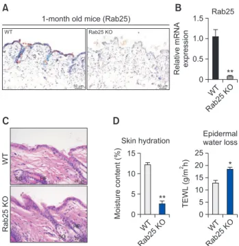
To investigate the expression of Rab25 on mouse skin, we conducted immunohistochemistry staining of the skin of 5-week old mice. As shown in Fig. 1A, Rab25 expression in WT mice was exclusively detected in the epidermis of skin; however, its expression was absent in the skin dermis or subcutis. As expected, Rab25 expression was absent in the epidermis of Rab25 knock-out (Rab25 KO) mice. In addition, the mRNA level of Rab25 in skin specimen was significantly reduced in Rab25 KO mice compared to that in WT mice (Fig. 1B). Interestingly, deficiency of Rab25 did not induce notable histopathological differences in the skin, and Rab25 KO mice exhibited relatively normal skin structure, with intact hair fol-licles (HF) and sebaceous glands (SG), compared to that in the skin of WT mice (Fig. 1C). Although abnormal features of epidermis were not apparent in Rab25 KO mice, moisture content and trans-epidermal water loss (TEWL) in the dorsal skin of Rab25 KO mice were altered significantly, revealing that Rab25 KO mice might have a dysfunctional skin barrier (Fig. 1D). Increased TEWL, with accompanying low skin hy-dration, can be observed in dry skin diseases, like diabetes mellitus (Park et al., 2011; Kim et al., 2019) or atopic dermatitis (Joo et al., 2015), reflecting that Rab25 KO mice might have xerotic skin.
A
B
0 WT Rab25 KO Relative mRNA expression 1.5 1.0 0.5 ** 0 WT Rab25 KO TEWL (g/m h) 2 25 20 15 10 5 *D
0 WT Rab25 KO Moisture content (%) 15 10 5 **C
WT Rab25 KO 50 m 50 m SG HF 20 m 20 m SG HF1-month old mice (Rab25) Rab25
Skin hydration Epidermalwater loss
WT
Rab25
KO
Fig. 1.
Histopathological and physiological features of skin inRab25 KO mice. (A) Representative immunohistochemistry im-ages for Rab25 in 5-week old WT and Rab25 KO mice. (B) mRNA expression of Rab25 was normalized to that of GAPDH. Graphs represent mean ± SEM (Unpaired student’s t-test, n=4, **p<0.01). (C) Representative hematoxylin and eosin (H&E) staining images of WT and Rab25 KO mouse skin (HF: hair follicle, SG: sebaceous gland). (D) Skin hydration and epidermal water loss were mea-sured by close attachment of Delfin’s vapometer and moistureme-ter on the dorsal skin of 9-week old mice. Graphs represent mean ± SEM (Unpaired student’s t-test, *p<0.05, **p<0.01).
Rab25 KO mice show increased keratinocyte proliferation
To identify the reason behind skin barrier dysfunction in Rab25 KO mice, we first investigated the proliferation of epi-dermal cells in Rab25 KO mice. Bromodeoxyuridine (BrdU), an S-phase proliferation marker, was pre-injected into Rab25 KO mice 2 h before sacrifice, and BrdU-positive cells were detected in the epidermis by immunofluorescence staining. BrdU-positive cells in the skin epidermis were significantly in-creased in Rab25 KO mice compared to that in control (Fig. 2A, 2B). The majority of BrdU-positive proliferating cells were progenitor cells of the basal cell layer, where keratins 5 and 14 were highly positive. As shown in Fig. 2C and 2D, the number of BrdU- and keratin 5 (K5)-double positive cells was remark-ably increased in Rab25 KO mice. Taken together, loss of Rab25 promoted proliferation of keratinocyte but did not in-duced alteration of its distribution on skin epidermis.Rab25 KO globally increases the expression of
keratinocyte differentiation markers
Intercellular lipids in stratum corneum play a critical role in skin barrier function, for the prevention of epidermal wa-ter loss and invasion of pathogen. Patients with defective skin barrier functions, like atopic dermatitis, show altered ceramide composition in the skin (Choi and Maibach, 2005; Park et al., 2012; Joo et al., 2015). To explore the relation between ce-ramide composition and barrier defects in Rab25 KO mice, we performed immunohistochemistry staining for ceramide in WT and Rab25 KO mice. Surprisingly, the staining intensity of ceramide in the epidermis barely changed in Rab25 KO mice,
and its expression and distribution were indistinguishable from that in WT mice (Fig. 3A).
We investigated other components of stratum corneum, such as, epidermal differentiation markers in KO mice. In-deed, aberrant or increased expression of early differentiation markers like keratins 5 and 14 (Fig. 3B), as well as of late dif-ferentiation markers like keratin 1, keratin 10, involucrin, and loricrin (Fig. 3C), was found in the epidermis of Rab25 KO mice. Interestingly, although these markers increased signifi-cantly in content, their distribution was not different from that in WT mice.
Rab25 KO is associated with remarkable alteration of
multiple biological processes in the skin of mice and in
human keratinocyte cell line HaCaT
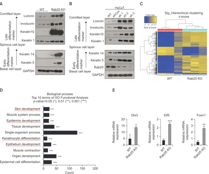
To clarify further the alteration of keratinocyte differentia-tion in Rab25 KO mice, we conducted western blot analysis for the differentiation markers in skin. As shown in Fig. 4A, early and late differentiation markers were all up-regulated in the skin of Rab25 KO mice at the protein level, which was in line with the immunohistochemistry findings (Fig. 3B, 3C). To identify whether this event can occur in human skin as well, Rab25 was knocked down using single hairpin RNA (shRNA)
Ceramide WT Rab25 KO Keratin 5 20 m Keratin 14
Early differentiation marker
20 m 20 m 20 m WT Rab25 KO Keratin 1 20 m Keratin 10
Late differentiation marker
20 m 20 m 20 m Involucrin 20 m 20 m Loricrin 20 m 20 m WT Rab25 KO 20 m 20 m 50 m 50 m
A
B
C
Fig. 3.
Characteristics of skin differentiation markers in 9-weekold Rab25 KO mice. (A) Representative immunohistochemistry images of ceramide. Lower panels indicate higher magnification of epidermis. (B, C) Immunohistochemistry images of representative keratinocyte differentiation markers. Both (B) early differentiation markers (kerain 5 and keratin 14) and (C) late differentiation mark-ers (kerain 1, keratin 10, involucrin, and loricrin) were exclusively stained in the epidermis of WT and Rab25 KO mice.
A
B
0 BrdU BrdU positive cells/ 40X field 8 6 4 2 ** WT Rab25 KO 20 m WT Rab25 KO BrdU DAPI 20 mC
D
0 K5-BrdU Double positive cells/ 40X field 10 6 4 2 ** WT Rab25 KO 20 m WT Rab25 KO BrdU Keratin 5 20 m 8Fig. 2.
Increment of proliferating cells in 9-week old Rab25 KOmouse skin. (A) Representative immunofluorescence images of BrdU-positive cells in the skin epidermis of WT and Rab25 KO. White arrow indicated positive cells. (B) Quantitation of BrdU-positive proliferating cells in 40×-magnification field of view. Graphs represent mean ± SEM (Unpaired student’s t-test, **p<0.01). (C) Dual immunofluorescence images of BrdU and keratin 5 (K5) in the epidermis of WT and Rab25 KO mice. (D) Quantitation of K5 and BrdU double-positive cells in 40×-magnification field of view. Graphs represent mean ± SEM (Unpaired student’s t-test, **p<0.01).
in HaCaT cells, a human keratinocyte cell line. Consistent with the findings in Rab25 KO mice, all skin markers, including ear-ly differentiation and late differentiation markers, were found to be up-regulated overall (Fig. 4B).
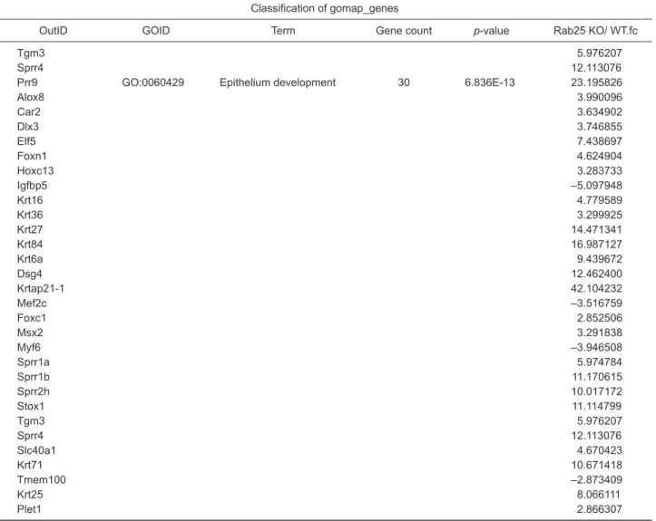
Rab25 appeared to be a key mediator of skin differentiation, as shown above. To clarify this finding, we performed tran-scriptomic analysis using gene-chip microarray for full dorsal skin specimens of Rab25 KO and WT mice. As presented in Fig. 4C, considerable differences in gene expression patterns and hierarchical clusters were seen. Moreover, significant up-regulation of the genes involved in processes such as skin development, epidermis development, keratinocyte differen-tiation, and epithelium development was observed (Fig. 4D,
Table 1), confirming the key role played by Rab25 in epider-mal differentiation. For the validation of expression data from gene-chip microarray, we investigated the mRNA level of rep-resentative transcription factors, which were involved in kera-tinocyte differentiation and epithelium development (Fig. 4D, Table 1). Importantly, we observed a significant up-regulation of Dlx3, Elf5, and Foxn1 expression in Rab25 KO mice com-pared to skin from WT mice (Fig. 4E).
DISCUSSION
This study demonstrated that Rab25 deficiency may lead
A
Early differentiation marker Late dif
ferentiation
marker
WT Rab25 KO
Cornified layer
Spinous cell layer
Basal cell layer Loricrin Involucrin Keratin10 Keratin 1 Keratin 14 Keratin 5 GAPDH
B
Sig_Hierarchical clustering z-scoreC
WT Rab25 KO Row Z-score 1.5 0.5 0.51.01.5 Biological processTop 10 terms of GO Functional Analysis -value<0.05 (*), 0.01 (**), 0.001 (***) p
Skin development Muscle system process Epidermis development Tissue development Single-organism process Keratinocyte differentiation Epithelium development Muscle contraction Organ development Epidermal cell differentiation
D
0 200 Count 50 100 150 *** *** *** *** *** *** *** *** *** ***E
0 WT Rab2 5K O Relative mRNA expression 3 2 1 *** Elf5 Early dif ferentiation marker Late dif ferentiation marker WT shConsh1sh2 sh3 Cornified layerSpinous cell layer
Basal cell layer Loricrin Involucrin Keratin10 Keratin 1 Keratin 14 Keratin 5 GAPDH HaCaT Rab25 0 WT Rab25 KO Relative mRNA expression 20 15 10 5 * Dlx3 0 WT Rab25 KO Relative mRNA expression 4 2 1 ** Foxn1 3 Color key
Fig. 4.
Alteration of skin differentiation in 9-week old Rab25 KO mice. (A) Images of western blots for representative keratinocytedifferenti-ation markers using skin specimens from WT and Rab25 KO mice. GAPDH was used for housekeeping control. (B) Images of western blots for representative keratinocyte differentiation marker. Knockdown-HaCaT cell (sh1, sh2, and sh3) line was constructed by infection with the recombinant pGIPZ vector, using polybrene mixture. Expression level of the knockdown cell was compared to that of non-infected cell (WT) and vector-conveying cell (shCon). GAPDH was used for housekeeping control. (C) Schematic image of cluster analysis-hierarchical clus-tering heat map acquired from Affymetrix GeneChip® Mouse Gene 2.0 ST Array (Thermo) using skin specimen of WT and Rab25 KO mice.
The heat map was represented and sorted by the normalized value of expression level using each probe. (D) Bar graph indicated top 10 biological processes with the highest p-value. GO functional analysis was based on gomap_stat. (E) mRNA expression was normalized with GAPDH. Graphs represent mean ± SEM (Unpaired student’s t-test, n=4, *p<0.05, **p<0.01, ***p<0.001).
Table 1.
Fold change of expression level on Affymetrix gene arrayClassification of gomap_genes
OutID GOID Term Gene count p-value Rab25 KO/ WT.fc
Prr9 GO:0043588 Skin development 23 1.354E-19 23.195826
Alox8 3.990096 Col1a1 –2.366208 Col1a2 –2.561034 Foxn1 4.624904 Hoxc13 3.283733 Igfbp5 –5.097948 Krt16 4.779589 Krt36 3.299925 Krt27 14.471341 Krt84 16.987127 Krt6a 9.439672 Dsg4 12.462400 Krtap21-1 42.104232 Msx2 3.291838 Ryr1 –4.751830 Sprr1a 5.974784 Sprr1b 11.170615 Sprr2h 10.017172 Tgm3 5.976207 Sprr4 12.113076 Krt71 10.671418 Krt25 8.066111
Prr9 GO:0008544 Epidermis development 20 4.825E-15 23.195826
Alox8 3.990096 Foxn1 4.624904 Hoxc13 3.283733 Igfbp5 –5.097948 Krt16 4.779589 Krt36 3.299925 Krt27 14.471341 Krt84 16.987127 Krt6a 9.439672 Dsg4 12.462400 Krtap21-1 42.104232 Msx2 3.291838 Sprr1a 5.974784 Sprr1b 11.170615 Sprr2h 10.017172 Tgm3 5.976207 Sprr4 12.113076 Krt71 10.671418 Krt25 8.066111
Prr9 GO:0030216 Keratinocyte differentiation 14 3.665E-13 23.195826
Alox8 3.990096 Foxn1 4.624904 Krt16 4.779589 Krt36 3.299925 Krt84 16.987127 Krt6a 9.439672 Dsg4 12.462400 Msx2 3.291838 Sprr1a 5.974784 Sprr1b 11.170615 Sprr2h 10.017172
to skin barrier dysfunction in KO mice, as determined by higher trans-epidermal water loss and lower cutaneous hy-dration than in wild type mice. While the skin morphology was observed to be relatively normal, aberrantly increased expression of epidermal differentiation markers such as loric-rin, involucloric-rin, and keratins 5, 14, 1, and 10 was observed in Rab25 KO mice, which might cause perturbation of epidermal physiology. In line with this, depletion of Rab25 with shRNA led to increased expression of differentiation markers in the human keratinocyte cell line, HaCaT, reflecting the critical role played by Rab25 in epidermal differentiation of human skin. Transcriptomic analysis of the skin revealed that Rab25 KO to have increased the expression of genes associated with skin development, epidermal development, and keratinocyte differentiation, thus suggesting that Rab25 is involved in the regulation of epidermal differentiation and proliferation.
Interestingly, Rab25 is linked with the activation of AKT/ PI3K pathway by binding and activating AKT (Cheng et al., 2012; Fan et al., 2015), which promotes the resistance of can-cer cells against metabolic stress through enhancing ATP gen-eration and glycogen synthesis. AKT/PI3K pathways are also known to be pivotal in keratinocyte differentiation (Calautti et
al., 2005). During keratinocyte differentiation, the activation of PI3K pathway, which depends on the activity of EGFR, Fyn/ Src kinases, and E-cadherin-mediated adhesion, actually initi-ates AKT activation, which in turn, promotes the growth arrest and differentiation of keratinocytes, as evidenced by increased expression of filaggrin, loricrin, keratin 1, and keratin 5. Inhibi-tion of PI3K activity with wortmannin or Ly294002 results in the suppression of these differentiation markers and results in cell death. In this regard, our results, showing that Rab25 KO leads to increased expression of differentiation markers, con-tradict the existing mechanisms underlying epidermal differ-entiation. It would therefore be worth examining the PI3K/AKT pathway in the skin of Rab25 KO mice to clarify this conflict.
Rab25 facilitates the transport of integrins to the plasma membrane and is critical in membrane recycling along with other Rab11 family proteins (Welz et al., 2014), including inte-grins or receptor tyrosine kinases like EGFR (Dozynkiewicz et al., 2012; De Franceschi et al., 2015). In line with this, we had previously demonstrated that Rab25 KO leads to the depletion of integrins, β1, β4, and α6 in the skin of mice (Jeong et al., 2019), thereby indicating that Rab25 deficiency leads to dys-regulation of integrins in keratinocytes. Rodius et al. (2007)
Table 1.
ContinuedClassification of gomap_genes
OutID GOID Term Gene count p-value Rab25 KO/ WT.fc
Tgm3 5.976207
Sprr4 12.113076
Prr9 GO:0060429 Epithelium development 30 6.836E-13 23.195826
Alox8 3.990096 Car2 3.634902 Dlx3 3.746855 Elf5 7.438697 Foxn1 4.624904 Hoxc13 3.283733 Igfbp5 –5.097948 Krt16 4.779589 Krt36 3.299925 Krt27 14.471341 Krt84 16.987127 Krt6a 9.439672 Dsg4 12.462400 Krtap21-1 42.104232 Mef2c –3.516759 Foxc1 2.852506 Msx2 3.291838 Myf6 –3.946508 Sprr1a 5.974784 Sprr1b 11.170615 Sprr2h 10.017172 Stox1 11.114799 Tgm3 5.976207 Sprr4 12.113076 Slc40a1 4.670423 Krt71 10.671418 Tmem100 –2.873409 Krt25 8.066111 Plet1 2.866307
reported that deletion of α6 integrin in keratinocytes increases the expression of keratins 1, 10, and 14, and loricrin, involu-crin, and filaggrin, which is in good agreement with our find-ings. Rodius et al. (2007) also reported that increased levels of c-Jun, c-Fos, and phospho-Jun, which ultimately activate AP-1 transcription factor, are attributable to the increased dif-ferentiation markers in α6 integrin-deficient keratinocytes.
Not only AP-1, but also other transcription factors have been reported to play a critical role in regulating skin homeo-stasis and keratinocyte differentiation (Park and Morasso, 1999; Tummala and Sinha, 2006; Hwang et al., 2011). Previ-ous reports had shown higher expression of ELF5 and DLX3 to occur during Ca2+-induced mouse keratinocyte differentia-tion in vitro (Park and Morasso, 1999; Tummala and Sinha, 2006). Moreover, Hwang et al. (2011) had discovered that de-letion of Dlx3 using K14-Cre (Dlx3Kin/f) mice leads to abnormal keratinocyte differentiation. Simialrly, activation of FOXN1, a transcription factor expressed in the suprabasal layer, pro-moted the expression of keratinocyte differentiation markers like involucrin and keratin 10, whereas inhibition of PI3K pre-vented FOXN1-induced differentiation (Janes et al., 2004). Moreover, Prowse et al. (1999) constructed a transgenic mouse, expressing FOXN1 under the involucrin promoter, and revealed that proliferation of basal keratinocyte was promoted in the transgenic mouse. These findings on FOXN1 were also consistent with our findings in Rab25 KO mice. In our gene-chip microarray, we found significant changes in the mRNA of transcription factors such as Dlx3, Elf5, Foxn1, Hoxc13, while there was no alteration for c-Jun or c-Fos. Indeed, we verified the increased mRNA expression of these transcription factors in the epidermis of KO mice by q-RT PCR. Interest-ingly, these transcription factors are critical in the hair follicle cycle, especially in the period of anagen (Lin et al., 2004), and gene ontology analysis revealed their strong association with epithelial development. Although further studies are required to clarify the underlying mechanism, it would be interesting to investigate the involvement of transcription factors in abnor-mal epiderabnor-mal differentiation in the skin of Rab25 KO mice.
Collectively, we demonstrated that Rab25 deficiency per-turbs epidermal differentiation and skin barrier function, lead-ing to high trans-epidermal water loss (TEWL) and low skin moisture. It is notable that although the expression of epider-mal differentiation markers increased, skin barrier function was significantly impaired in Rab25 KO mice, implying other factors are accountable for the barrier dysfunction. Further studies in this regard are recommended in the future.
CONFLICT OF INTEREST
The authors state no conflict of interest.
ACKNOWLEDGMENTS
This research was supported by The Myung-Sun Kim Me-morial Foundation (to KTN).
REFERENCES
Bickenbach, J. R., Greer, J. M., Bundman, D. S., Rothnagel, J. A. and
Roop, D. R. (1995) Loricrin expression is coordinated with other epidermal proteins and the appearance of lipid lamellar granules in development. J. Invest. Dermatol. 104, 405-410.
Brakebusch, C., Grose, R., Quondamatteo, F., Ramirez, A., Jorcano, J. L., Pirro, A., Svensson, M., Herken, R., Sasaki, T., Timpl, R., Werner, S. and Fassler, R. (2000) Skin and hair follicle integrity is crucially dependent on beta 1 integrin expression on keratinocytes.
EMBO J. 19, 3990-4003.
Calautti, E., Li, J., Saoncella, S., Brissette, J. L. and Goetinck, P. F. (2005) Phosphoinositide 3-kinase signaling to Akt promotes kera-tinocyte differentiation versus death. J. Biol. Chem. 280,
32856-32865.
Casanova, J. E., Wang, X., Kumar, R., Bhartur, S. G., Navarre, J., Woodrum, J. E., Altschuler, Y., Ray, G. S. and Goldenring, J. R. (1999) Association of Rab25 and Rab11a with the apical recycling system of polarized Madin-Darby canine kidney cells. Mol. Biol. Cell 10, 47-61.
Cheng, K. W., Agarwal, R., Mitra, S., Lee, J. S., Carey, M., Gray, J. W. and Mills, G. B. (2012) Rab25 increases cellular ATP and glycogen stores protecting cancer cells from bioenergetic stress. EMBO Mol. Med. 4, 125-141.
Choi, M. J. and Maibach, H. I. (2005) Role of ceramides in barrier func-tion of healthy and diseased skin. Am. J. Clin. Dermatol. 6,
215-223.
De Franceschi, N., Hamidi, H., Alanko, J., Sahgal, P. and Ivaska, J. (2015) Integrin traffic - the update. J. Cell Sci. 128, 839-852.
Dozynkiewicz, M. A., Jamieson, N. B., Macpherson, I., Grindlay, J., van den Berghe, P. V., von Thun, A., Morton, J. P., Gourley, C., Timpson, P., Nixon, C., McKay, C. J., Carter, R., Strachan, D., An-derson, K., Sansom, O. J., Caswell, P. T. and Norman, J. C. (2012) Rab25 and CLIC3 collaborate to promote integrin recycling from late endosomes/lysosomes and drive cancer progression. Dev.
Cell 22, 131-145.
Fan, Y., Wang, L., Han, X., Liu, X. and Ma, H. (2015) Rab25 is sponsible for phosphoinositide 3-kinase/AKTmediated cisplatin re-sistance in human epithelial ovarian cancer cells. Mol. Med. Rep.
11, 2173-2178.
Goldenring, J. R., Shen, K. R., Vaughan, H. D. and Modlin, I. M. (1993) Identification of a small GTP-binding protein, Rab25, expressed in the gastrointestinal mucosa, kidney, and lung. J. Biol. Chem. 268,
18419-18422.
Goldenring, J. R. and Nam, K. T. (2011) Rab25 as a tumour suppres-sor in colon carcinogenesis. Br. J. Cancer 104, 33-36.
Hwang, J., Kita, R., Kwon, H. S., Choi, E. H., Lee, S. H., Udey, M. C. and Morasso, M. I. (2011) Epidermal ablation of Dlx3 is linked to IL-17-associated skin inflammation. Proc. Natl. Acad. Sci. U.S.A.
108, 11566-11571.
Janes, S. M., Ofstad, T. A., Campbell, D. H., Watt, F. M. and Prowse, D. M. (2004) Transient activation of FOXN1 in keratinocytes induces a transcriptional programme that promotes terminal differentiation: contrasting roles of FOXN1 and Akt. J. Cell Sci. 117, 4157-4168.
Jeong, H., Lim, K. M., Kim, K. H., Cho, Y., Lee, B., Knowles, B. C., Ro-land, J. T., Zwerner, J. P., Goldenring, J. R. and Nam, K. T. (2019) Loss of Rab25 promotes the development of skin squamous cell carcinoma through the dysregulation of integrin trafficking. J Pathol. doi: 10.1002/path.5311 [Epub ahead of print].
Joo, K. M., Hwang, J. H., Bae, S., Nahm, D. H., Park, H. S., Ye, Y. M. and Lim, K. M. (2015) Relationship of ceramide-, and free fatty ac-id-cholesterol ratios in the stratum corneum with skin barrier func-tion of normal, atopic dermatitis lesional and non-lesional skins. J.
Dermatol. Sci. 77, 71-74.
Kim, J. H., Cho, E. Y., Kwon, E., Kim, W. H., Park, J. S., Lee, Y. S., Yun, J. W. and Kang, B. C. (2017) Gold thread implantation promotes hair growth in human and mice. Lab. Anim. Res. 33, 291-297.
Kim, M., Jeong, H., Lee, B., Cho, Y., Yoon, W. K., Cho, A., Kwon, G., Nam, K. T., Ha, H. and Lim, K. M. (2019) Enrichment of short-chain ceramides and free fatty acids in the skin epidermis, liver, and kid-neys of db/db mice, a type 2 diabetes mellitus model. Biomol. Ther. (Seoul) doi: 10.4062/biomolther.2018.214 [Epub ahead of print]. Lin, K. K., Chudova, D., Hatfield, G. W., Smyth, P. and Andersen, B.
(2004) Identification of hair cycle-associated genes from time-course gene expression profile data by using replicate variance.
Proc. Natl. Acad. Sci. U.S.A. 101, 15955-15960.
Muroyama, A. and Lechler, T. (2012) Polarity and stratification of the epidermis. Semin. Cell Dev. Biol. 23, 890-896.
Nam, K. T., Lee, H. J., Smith, J. J., Lapierre, L. A., Kamath, V. P., Chen, X., Aronow, B. J., Yeatman, T. J., Bhartur, S. G., Calhoun, B. C., Condie, B., Manley, N. R., Beauchamp, R. D., Coffey, R. J. and Goldenring, J. R. (2010) Loss of Rab25 promotes the develop-ment of intestinal neoplasia in mice and is associated with human colorectal adenocarcinomas. J. Clin. Invest. 120, 840-849.
Park, G. T. and Morasso, M. I. (1999) Regulation of the Dlx3 homeo-box gene upon differentiation of mouse keratinocytes. J. Biol.
Chem. 274, 26599-26608.
Park, H. Y., Kim, J. H., Jung, M., Chung, C. H., Hasham, R., Park, C. S. and Choi, E. H. (2011) A long-standing hyperglycaemic condi-tion impairs skin barrier by accelerating skin ageing process. Exp.
Dermatol. 20, 969-974.
Park, Y. H., Jang, W. H., Seo, J. A., Park, M., Lee, T. R., Park, Y. H., Kim, D. K. and Lim, K. M. (2012) Decrease of ceramides with very long-chain fatty acids and downregulation of elongases in a murine atopic dermatitis model. J. Invest. Dermatol. 132, 476-479.
Prowse, D. M., Lee, D., Weiner, L., Jiang, N., Magro, C. M., Baden, H.
P. and Brissette, J. L. (1999) Ectopic expression of the nude gene induces hyperproliferation and defects in differentiation: implications for the self-renewal of cutaneous epithelia. Dev. Biol. 212, 54-67.
Rodius, S., Indra, G., Thibault, C., Pfister, V. and Georges-Labouesse, E. (2007) Loss of alpha6 integrins in keratinocytes leads to an in-crease in TGFbeta and AP1 signaling and in expression of differen-tiation genes. J. Cell. Physiol. 212, 439-449.
Seven, D., Dogan, S., Kilic, E., Karaman, E., Koseoglu, H. and Buyru, N. (2015) Downregulation of Rab25 activates Akt1 in head and neck squamous cell carcinoma. Oncol. Lett. 10, 1927-1931.
Tummala, R. and Sinha, S. (2006) Differentiation-specific transcrip-tional regulation of the ESE-2 gene by a novel keratinocyte-restrict-ed factor. J. Cell. Biochem. 97, 766-781.
Wang, X., Kumar, R., Navarre, J., Casanova, J. E. and Goldenring, J. R. (2000) Regulation of vesicle trafficking in madin-darby canine kidney cells by Rab11a and Rab25. J. Biol. Chem. 275,
29138-29146.
Welz, T., Wellbourne-Wood, J. and Kerkhoff, E. (2014) Orchestration of cell surface proteins by Rab11. Trends Cell Biol. 24, 407-415.
Wickett, R. R. and Visscher, M. O. (2006) Structure and function of the epidermal barrier. Am. J. Infect. Control 34, S98-S110.