INTRODUCTION
The development of allergic rhinitis, conjunctivitis, and asthma with rabbit exposure is common among scientists, technicians, and laboratory animal handlers (1). There was a report of a case of occupational allergic rhinitis working for the woolen fabric factory with rabbit fur in Korea (2). In a large epidemiologic study in Japan, allergic reactions were reported in 30% of laboratory animal workers with exposure to rabbits (3). However, few studies have investigated the spe- cific allergens involved in sensitization to rabbits. Ory c 1, a 17-kDa glycoprotein found in saliva and fur, has previously been identified as a major rabbit allergen (4, 5). Rabbit serum albumin and Ag2 have also been identified as allergens (5).
Although rabbits are common domestic pets, the develop- ment of allergic asthma and/or rhinitis attributable to rab- bits at home is unusual, and few studies have evaluated the allergenic relationship between rabbits and other pets such as cats and dogs.
Here, we report the identification of serum-specific IgE and IgG4antibodies to rabbit epithelium and IgE binding components in three cases of allergic asthma and/or rhinitis resulting from rabbit exposure at home. Furthermore, to
elucidate the allergenic cross-reactivity between rabbits and other furry animals, we performed inhibition enzyme-linked immunosorbent assays (ELISA).
MATERIALS AND METHODS Case description
Case 1
A 20-yr-old woman presented with a history of severe bre- athing difficulty, coughing, and wheezing for several days. She had lived in a school dormitory during the past year without incident but experienced breathing difficulty and coughing whenever she visited her family’s house. Her family had kept two rabbits as pets during the past 3 yr. On admission, the patient’s forced expiratory volume in one second (FEV1) level was 50%, and the sputum eosinophil level was 80%. Her FEV1level improved to 100% after 3 days of treatment with systemic steroids and bronchodilators. These results suggest- ed bronchial asthma, so methacholine challenge test was not needed to confirm asthma in this patient. Allergy skin prick tests with 55 common inhalant and food allergens (rabbit epi-
Jeong Hee Choi, Hyun-Mi Kim, Hae Sim Park*
Department of Pulmonology and Allergy, Bundang Jesaeng General Hospital, Seongnam; Department of Allergy and Rheumatology*, Ajou University School of Medicine, Suwon, Korea
Current address of Choi JH: Department of Internal Medicine, Hallym University College of Medicine, Seoul.
Address for correspondence Hae-Sim Park, M.D.
Department of Allergy and Rheumatology, Ajou University School of Medicine, Wonchondong San-5, Yongtong-gu, Suwon 442-721, Korea
Tel : +82.31-219-5196, Fax : +82.31-219-5154 E-mail : [email protected]
*This study was supported by a grant of the Korean Health 21 R&D project, Ministry of Health & Welfare, R.O.K (A 050571).
820
Allergic Asthma and Rhinitis Caused by Household Rabbit Exposure:
Identification of Serum-Specific IgE and Its Allergens
Although rabbits are common domestic pets, severe respiratory allergic reactions to rabbits in households are unusual. Ory c 1, a 17-kDa glycoprotein found in sali- va and fur, has previously been identified as a major rabbit allergen. In this report, we describe the cases of three patients with rabbit allergy who presented with asth- ma and/or rhinitis while living in households with detectable levels of serum-specif- ic IgE and major IgE binding components. Three patients with rabbit allergy and 18 unexposed nonatopic healthy controls were enrolled. Enzyme-linked immunosor- bent assays (ELISA) for serum-specific IgE and IgG4to rabbit epithelium and inhi- bition ELISA were performed followed by sodium dodecye sulfate polyacrylamide gel electrophoresis (SDS-PAGE) and IgE immunoblotting. All three patients with rabbit allergy had high serum-specific IgE antibody levels compared with controls.
The results of the inhibition ELISA showed significant inhibition with the addition of rabbit epithelium, whereas no significant inhibition was noted with the addition of cat and dog epithelia. Two IgE-binding components with molecular weights of 16 kDa and 67.5 kDa were identified by IgE immunoblotting. In conclusion, rabbit exposure may induce IgE-mediated bronchial asthma and/or rhinitis in domestic settings.
Key Words : Rabbits, Animals; Domestic; Hypersensitivity; Allergens
Received : 14 September 2006 Accepted : 25 January 2007
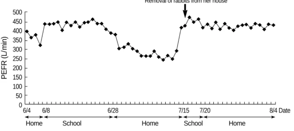
thelium not included) showed negative results. The patient had no history or family history of allergic diseases. To eval- uate the presence of rabbit-induced bronchial asthma, peak expiratory flow rate (PEFR) and serum-specific IgE to rabbit epithelium were measured. The results of PEFR monitoring showed significant decreases in PEFR values at home com- pared with those at school (Fig. 1), even she did not take asth- ma medication at school. Serum-specific IgE to rabbit epithe- lium was detected (31.9 kU/L; CAP system, Pharmacia, Upp- sala, Sweden). After removal of the rabbits from her family’s home, the patient’s respiratory symptoms improved without the need for medication.
Case 2
A 37-yr-old woman presented with allergic conjunctivitis and eczema, and a history of rabbit allergy 8 yr before. After breeding rabbits at her home during the past 6 months, she developed shortness of breath, wheezing, cough, rhinorrhea, and sneezing. After removal of the rabbits from her home, her asthma and rhinitis improved. A methacholine challenge test showed a positive reaction, with a PC20value of 0.094 mg/mL. Allergy skin prick tests with 55 common inhalant and food allergens showed positive reactions, with an A/H ratio of 4+ in Dermatophagoides pteronyssinus, 4+ in D. farine, 4+ in dog, and 2+ in cat. Serum-specific IgE was detected for rabbit, dog, and cat epithelia (41.0, 4.15, and 0.83 kU/
L, respectively). Her family history was significant for par- ents and a sister who had allergic rhinitis. She also bred dogs and birds but denied experiencing any symptoms on expo- sure to these animals.
Case 3
A 33-yr-old man with allergic rhinitis presented with a 2- month history of coughing, intermittent shortness of breath, and wheezing. He had kept a rabbit as a pet during the past 4 yr. After living with the rabbit for 2 yr, his rhinitis symp- toms became aggravated whenever he stayed at home or han- dled the animal. He denied experiencing any asthma symp- toms on exposure to the rabbit. His family history was sig- nificant for a grandfather with asthma. A methacholine chal- lenge test showed a positive reaction, with a PC20value of 1.25 mg/mL. Allergy skin prick tests with 55 common in- halant and food allergens showed positive reactions, with an A/H ratio of 3+ in D. pteronyssinus, 2+ in D. farine, 4+ in mugwort, and 5+ in ragweed. The serum-specific IgE to rabbit epithelium was 4.86 kU/L.
The three cases are summarized in Table 1.
ELISA and ELISA inhibition assays
We measured the levels of serum-specific IgE (or IgG4) to rabbit epithelium in three patients with rabbit allergy and in 18 healthy non-atopic controls. Microtiter plates (Costar, Corning, NY, U.S.A.) were Hamburg coated with 100 L of rabbit epithelium (Allergopharma, Hamburg, Germany) and stored at 4℃overnight. Plates were washed 3 times be- tween steps with 0.05% phosphate-buffered saline-Tween (PBS-T). Blocking was performed by using 10% fetal bovine serum albumin for 1 hr at room temperature. The patients’
sera (undiluted, 50 L/well) were added and incubated for 1 hr at room temperature. After washing, 100 L of 1:1,000
PEFR (L/min)
500 450 400 350 300 250 200 150 100 50
06/4 6/8 6/28 7/15 7/20 8/4 Date
Home School Home School Home
Removal of rabbits from her house
Fig. 1.PEFR values markedly dec- reased while at home with the rab- bit. PEFR, peak expiratory flow rate.
Sex/Age
Case Underlying allergic
disease
Specific IgE to rabbit, cat, and dog
(kU/L, CAP) Allergic symptoms
to rabbit Exposure
period FHx of aller-
gic disease Atopy
1 F/18 No No No 2 yr BA 31.9, 0, 0
2 F/37 BA, AR, AC, AD Yes Yes 6 mo BA, AR 41.0, 0.83, 4.15
3 M/33 BA, AR Yes Yes 2 yr AR 4.86, 0, 0
Table 1.Clinical characteristics and specific IgE levels to rabbit epithelium in study subjects
FHx, family history; BA, bronchial asthma; AR, allergic rhinitis; AC, allergic conjunctivitis; AD, atopic dermatitis.
biotin-labeled goat anti-human IgE (or IgG4) antibodies (Si- gma, St. Louis, MO) were added and incubated for 1 hr at room temperature. IgE reactivity was detected by colorimetric reaction using 3,3′, 5,5′-tetramethyl benzidine and H2O2as substrate. The absorbance at 450 nm was read using an auto- mated microplate reader. A competitive inhibition ELISA was performed to determine the specificity of the IgE bind- ing to rabbit epithelium and to identify allergenic cross-reac- tivity with cat and dog epithelia. A sera pool from two patients with high-specific IgE levels to rabbit epithelium was prein- cubated with 0, 1, 5, and 10 g/mL of rabbit, cat, dog epithe- lia (Allergopharma), and D. pteronyssinus. The inhibition per- centage of the specific IgE binding was expressed as 100-[(ab- sorbance of samples reincubated with allergens/ absorbance of samples pre-incubated with PBS)×100%].
SDS-PAGE and immunoblot analysis
Rabbit epithelium (0.75 g/well) was boiled in sample buffer (0.5 M Tris [pH 6.8], glycerol, 10% SDS, 0.5% bro- mophenol blue, 2.5% -mercaptoethanol) for 5 min. Stan- dard markers (4 to 250 kDa, Invitrogen, San Diego, CA, U.S.A.) and rabbit epithelia were loaded onto a 10% Tris- glycine gel for antigen separation. Electrophoresis was per- formed with the Mini Protean II cell (Bio-Rad Laboratories, Hercules, CA, U.S.A.) for 120 min at 125 V. The gel was fixed and stained with Coomasie Brilliant blue. Sodium dode- cyl sulfate polyacrylamide gel electrophoresis (SDS-PAGE)- separated proteins were transferred to a polyvinylidene difluo- ride (PVDF) membrane (Millipore Co., Bedford, MA, U.S.A.) at 200 mA for 2 hr. Nonspecific binding sites on the nitro- cellulose membrane were blocked by incubation with 5%
skim milk in Tris-buffered saline with Tween 20 (TBST) for 2 hr, and the membranes were incubated with sera from
patients and controls for 2 hr at room temperature. After washing 4 times with TBST, the membranes were incubat- ed with alkaline phosphatase-labeled conjugated goat anti- human IgE (or IgG4) antibody (Sigma) diluted 1:1,000 with 5% skim milk in TBST for 1 hr. After washing, the colori- metric reaction was developed with the BCIP/NBT alkaline phosphatase substrate (Sigma).
RESULTS Specific IgE/IgG4to rabbit epithelium
When the mean+3 SD was defined as the cut-off value for the controls, all three patients with rabbit allergy showed positive results for specific IgE antibody, and two of the pa- tients showed positive results for specific IgG4antibody (Fig.
2). In addition, the specific IgE levels were higher in the two patients with asthma than in the patient with rhinitis alone.
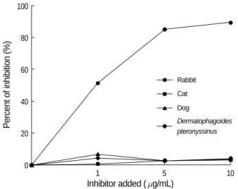
ELISA inhibition assays
Inhibition ELISA using the sera pool from the patients with rabbit allergy showed marked dose-dependent inhibi- tion with the addition of rabbit antigen, whereas minimal inhibition was noted with the addition of cat, dog, and D.
pteronyssinus antigens (Fig. 3).
SDS-PAGE and immunoblot analysis
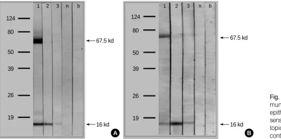
Rabbit epithelium was resolved into two bands, 16 kDa and 67.5 kDa, by SDS-PAGE and IgE immunoblot analy- sis; a 16-kDa protein band was noted in the sera of all three patients, and a 67.5-kDa band was noted in the sera of one patient (Fig. 4A). The IgG4immunoblot analysis showed a
Specific lgE and lgE4of rabbit (OD*1,000)
2,500 A
2,200
0 500 1,000
Patient Control Patient Control
(n=3) (n=18) (n=3) (n=18)
Fig. 2.Specific IgE (A) and IgG4 (B) binding to rabbit epithelium according to ELISA using sera from three patients with rabbit allergy ( ) and unexposed healthy controls ( ). Solid lines rep- resent mean + 3 SD, and dotted lines represent mean values of the healthy controls.
B
Percent of inhibition (%)
100
80
60
40
20
0
1 5 10
Inhibitor added ( g/mL)
Rabbit Cat Dog
Dermatophagoides pteronyssinus
Fig. 3.Inhibition ELISA using a sera pool from three patients with rabbit allergy with the addition of rabbit ( ), cat ( ), and dog ( ) epithelia and D. pteronyssinus ( ) extracts.
similar pattern (Fig. 4B).
DISCUSSION
IgE-mediated allergic sensitization is commonly experienced by individuals in regular contact with rabbits in laboratory and domestic settings (1, 2). Although rabbits are common domestic pets, there have been few reports of severe respiratory allergy resulting from household exposure to rabbits. One report described an atopic child who had developed anaphy- laxis following inhalant exposure to a rabbit at home (6), and another report described an 18-yr-old woman who had devel- oped severe respiratory allergy by indirect exposure to rabbit epithelia carried on the clothes of a colleague who kept a rab- bit as a pet (7). We described the cases of three patients with rabbit allergy who presented with asthma and/or rhinitis. Al- though allergy skin test with rabbit epithelium was not avail- able in our hospital, specific IgE antibodies to rabbit epithe- lium were detected in the sera of the patients by both ELISA and CAP. Furthermore, high serum-specific IgG4antibody levels were detected in two patients, which might have result- ed from a parallel immune response to rabbit allergen. Risk factors for the development of laboratory animal allergies in- clude atopy, familial history of allergic diseases, and environ- mental susceptibility (1). However, one case in the present study showed pure rabbit-induced asthma in a patient with- out a family history of allergic diseases, atopy, or underlying allergic diseases, indicating that even a non-atopic person with- out a family history of allergic diseases may develop severe animal-induced asthma from household exposure. Also, as rabbits are frequently kept as domestic pets theses days, rab- bit epithelium should be considered one of common inhalant animal allergens.
To date, several rabbit (Oryctolagus cuniculus) allergens have been identified in saliva, fur, urine, dander, and dust (4, 5, 8, 9). Ohman et al. reported allergens with molecular weights
ranging from 18 to 38 kDa from rabbit pelt extracts (8).
Longbottom et al. identified several allergens, including a 17-kDa glycoprotein, Ag R1 (referred to as Ory c 1), Ag2, serum albumin, and several other bands, using crossed immu- noelectrophoresis (4, 5). Recently, Baker et al. showed that 18-kDa and 21-kDa allergens (previously identified as Ory c 1 and Ag2, respectively) belong to the lipocalin superfam- ily and have significant sequence similarity to odorant bind- ing proteins (9). Lipocalins play a role in the binding and transport of small hydrophobic molecules such as pheromones (10). In this report, IgE immunoblot analysis showed two distinct proteins, 16 kDa and 67.5 kDa, which might have been Ag R1 and rabbit serum albumin, respectively. The 16-kDa band reacted with serum IgE and IgG4in all three patients, whereas the 67.5-kDa band reacted only in one patient. Consistent with previous reports, Ag R1 was iden- tified as a major rabbit allergen, and rabbit serum albumin did not appear to be a major allergen (4, 5, 9). This latter finding is in contrast to reports showing that serum albumin is an important allergen in cat, dog, and horse sera (1, 10).
It is well recognized that albumins from different species may cause allergenic cross-reactions (11, 12). In this report, the possibility of cross-reactivity between rabbit allergen and cat and dog allergens was shown to be very low. Although we used commercially available animal epithelia instead of crude extracts, we were able to find the 68-kDa glycoprotein band, which might have been serum albumin, in cat and dog epithelia by SDS-PAGE (data not shown). One explana- tion for this observation is the presence of specific IgE epi- topes for certain albumins. Furthermore, a significant vari- ability has been shown in IgE reactivities to variant albu- mins, despite a high degree of sequence similarity among the different albumins and overall cross-reactivity of IgE reactivities in patients with allergies (11). Additional stud- ies are required to elucidate the allergenic cross-reactivity among albumins and its clinical significance.
In conclusion, we described the cases of three patients who
Fig. 4.IgE (A) and IgG4(B) im- munoblot analysis for rabbit epithelium in sera from three sensitized patients (1-3), nona- topic controls (n), and buffer controls (b).
67.5 kd 124
1 2 3 n b
80
50
39
26
19
16 kd A
67.5 kd 124
1 2 3 n b
80
50
39
26
19
16 kd B
experienced rabbit-induced bronchial asthma and/or rhini- tis in the home environment. Serum-specific IgE and IgG4 antibodies to rabbit allergens were demonstrated, and a 17- kDa glycoprotein was confirmed as a major rabbit allergen.
As rabbits have become increasingly popular domestic pets, rabbit allergies experienced at home or at the workplace have become more common. Physicians should be aware that rabbit exposure may cause severe respiratory allergic reactions even in non-atopic individuals.
REFERENCES
1. Bush RK, Wood RA, Eggleston PA. Laboratory animal allergy. J Allergy Clin Immunol 1998; 102: 99-112.
2. Lee JC, Lee KS, Park EC, Kang MH, Kang SY. J Asthma Allergy Clin Immunol 1990; 10: 71-6.
3. Aoyama K, Ueda A, Manda F, Matsushita T, Ueda T, Yamauchi C.
Allergy to laboratory animals: an epidemiologic study. Br J Ind Med 1992; 49: 41-7.
4. Price JA, Longbottom JL. Allergy to rabbits. II. Identification and characterization of a major rabbit allergen. Allergy 1988; 43: 39-48.
5. Warner JA, Longbottom JL. Allergy to rabbits. III. Further identifi-
cation and characterization of rabbit allergens. Allergy 1991; 46:
481-91.
6. Prince E, Zacharisen MC, Kurup VP. Anaphylaxis to rabbit: a case report. Ann Allergy Asthma Immunol 1998; 81: 272-3.
7. Liccardi G, D’Amato G, Canonica GW, Dente B, Passalacqua G.
Severe respiratory allergy induced by indirect exposure to rabbit dander: a case report. Allergy 2004; 59: 1237-8.
8. Ohman JL Jr, Lowell FC, Bloch KJ. Allergens of mammalian origin.
II. Characterization of allergens extracted from rat, mouse, guinea pig, and rabbit pelts. J Allergy Clin Immunol 1975; 55: 16-24.
9. Baker J, Berry A, Boscato LM, Gordon S, Walsh BJ, Stuart MC.
Identification of some rabbit allergens as lipocalins. Clin Exp Aller- gy 2001; 31: 303-12.
10. Stewart GA, Robinson C. Allergen structure and function. In: Adkin- son NF, Jr., Yunginger JW, Busse WW, Bochner BS, Holgate ST, Simons FER, editors, Middleton’s allergy. Principles and practices.
6th ed. Philadelphia: Mosby Inc., 2003: 585-609.
11. Spitzauer S, Pandjaitan B, Soregi G, Muhl S, Ebner C, Kraft D, Valen- ta R, Rumpold H. IgE cross-reactivities against albumins in patients allergic to animals. J Allergy Clin Immunol 1995; 96: 951-9.
12. Hilger C, Kohnen M, Grigioni F, Lehners C, Hentges F. Allergic cross-reactions between cat and pig serum albumin. Study at the protein and DNA levels. Allergy 1997; 52: 179-87.
. . . .