INTRODUCTION
Ota’s nevus was first described in 1939 by Ota (1). It is usually characterized by unilateral, mottled, blue or dark brown macules occurring in the sclera and the surrounding skin innervated by the first and second branches of the trigemi- nal nerve. Ota’s nevus is usually congenital but may appear in early childhood or in puberty. Acquired, bilateral nevus of Ota-like macules (ABNOM) was first described in 1984 by Hori et al. (2). It is located bilaterally on the face and is blue- brown or slate-gray in color. These macules usually appear in the third or fourth decade of life in women and only rarely in men. Pigmented macules are absent in the mucous mem- branes of oral cavity, nose, or eyes.
There has been debate as to whether ABNOM is a part of the Ota’s nevus spectrum. We report here a case of ABNOM associated with Ota’s nevus to discuss the relations between two entities.
CASE REPORT
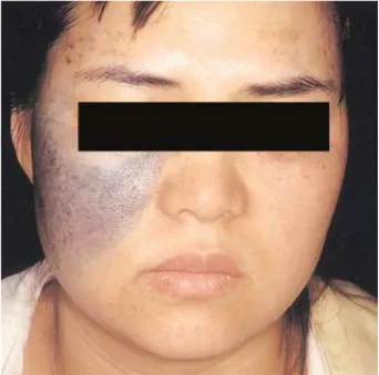
A 36-yr-old Korean women visited our clinic with dark bluish patch on the right cheek and right conjunctiva since
birth. She also had mottled brownish macules on both fore- head and both lower eyelids that have developed 3 yr ago (Fig. 1, 2). She was taking anti-hypertensive drugs for 1 yr.
There was no family history of abnormal cutaneous pigmen- tation. Skin biopsy specimens taken from the right cheek and left forehead all showed scattered, bipolar or irregular melanocytes in the dermis (Fig. 3, 4). Immunohistochemical study revealed melanocytes in the dermis were positive in Fontana-Masson stain and S-100 protein. By clinical and hi- stopathological findings, we diagnosed lesion on the right cheek area as Ota’s nevus and those on both forehead and both lower eyelids as ABNOM.
DISCUSSION
Ota’s nevus, originally described as nevus fuscocaeruleus ophthalmomaxillaris by Ota and Tanino in 1939, is a nevus of dermal melanocytes (1). It is more prevalent in orientals and is said to occur in up to 0.8% of the dermatologic out- patients in Japan but also occurs in Korean, Chinese, blacks, and whites (3, 4). The sex ratio is 1:4.8, women being more frequently involved than men (5). Ota’s nevus most often appears in the perinatal period or around puberty. A study
Jung-Hun Park, Mu-Hyoung Lee
Department of Dermatology, College of Medicine, Kyung Hee University, Seoul, Korea
Address for correspondence Mu-Hyoung Lee, M.D.
Department of Dermatology, College of Medicine, Kyung Hee University, 1 Hoegi-dong, Dongdaemoon-gu, Seoul 130-702, Korea Tel : +82.2-958-8501, Fax : +82.2-969-6538 E-mail : mhlee@khmc.or.kr
616 J Korean Med Sci 2004; 19: 616-8
ISSN 1011-8934
Copyright � The Korean Academy of Medical Sciences
Acquired, Bilateral Nevus of Ota-like Macules (ABNOM) Associated with Ota's Nevus
: Case Report
Ota’s nevus is mongolian spot-like macular blue-black or gray-brown patchy pigmen- tation that most commonly ocurrs in areas innervated by the first and second divi- sion of the trigeminal nerve. Acquired, bilateral nevus of Ota-like macules (ABNOM) is located bilaterally on the face, appears later in life, is blue-brown or slate-gray in color. It is not accompanied by macules on the ocular and mucosal membranes.
There is also debate as to whether ABNOM is part of the Ota’s nevus spectrum.
We report an interesting case of ABNOM associated with Ota’s nevus. A 36-yr-old Korean women visited our clinic with dark bluish patch on the right cheek and right conjunctiva since birth. She also had mottled brownish macules on both forehead and both lower eyelids that have developed 3 yr ago. Skin biopsy specimens taken from the right cheek and left forehead all showed scattered, bipolar or irregular me- lanocytes in the dermis. We diagnosed lesion on the right cheek area as Ota’s nevus and those on both forehead and both lower eyelids as ABNOM by clinical and his- tologic findings. This case may support the view that ABNOM is a separate entity from bilateral Ota’s nevus.
Key Words : Nevus of Ota; Acquired, Bilateral Nevus of Ota-like Macules (ABNOM)
Received : 8 August 2003 Accepted : 1 October 2003
ABNOM Associated with Ota’s Nevus 617
in Japan by Hidano et al. (5) of 240 patients revealed that onset of the nevus was at birth or in the first few months of life in 48%, between the ages of one and ten years in 11%, between eleven and twenty years in 36%, and between twen- ty-one and twenty-six years in only 5% of cases. It is usually unilateral and is located in the areas innervated by the first and second branches of trigeminal nerve. The pigmentation of Ota’s nevus is composed of flat, blue-black or slate-gray macules intermingled with brown, small, flat spots. The inten- sity of the pigmentation may be influenced by fatigue, men- struation, insomnia, and weather (5). Based on the distribu- tion and the extent of pigmentation, Ota’s nevus has been classified into four types (6): mild, Type I; moderate, Type II;
intensive, Type III; and bilateral, Type IV. Bilateral involve-
ment is about 10% (1, 7). Noncutaneous pigmentation, which is not addressed in this classification, may occur and often involves the conjunctiva, sclera, and tympanic membrane.
Less frequently, the nasal mucosa, palate, pharynx, cornea, iris, uveal tract, and fundus are involved. Histologic studies show bipolar to stellate melanocytes widely scattered in the reticular dermis.
Acquired, bilateral nevus of Ota-like macules (ABNOM), first described by Hori et al. in 1984, is classified in a group of circumscribed dermal melanoses (2). It is prevalent in Asian people. The incidence as reported by Sun et al. in Taiwan was 0.8% (8). It is usually appears in the third or fourth decade of life and showed a marked preponderance of females (2).
ABNOM is usually characterized by blue-brown or slate-gray
Fig. 1.Dark bluish patch on the right malar area, temple and cheek (Ota’s nevus).
Fig. 2.Mottled brownish macules on both forehead and both lower eyelids (ABNOM lesion).
Fig. 3. Skin biopsy from the right malar area (hematoxylin-eosin st- ain; ×200): Melano- cytes, are scattered in the dermis.
Fig. 4. Skin biopsy from the left forehead (he- matoxylin-eosin stain;
×200): Melanocytes, are scattered in the dermis.
618 J.-H. Park, M.-H. Lee
patches occurring bilaterally on the forehead, temples, eye- lids, cheeks and nose. Histopathologic studies showed dermal melanocytes, bipolar or oval in shape, scattered in the upper and middle portions of the dermis. ABNOM should be clin- ically and histologically differentiated from bilateral Ota’s nevus, Riehl’s melanosis and melasma. ABNOM differs clin- ically from bilateral Ota’s nevus in the following aspects (2, 9); (1) ABNOM is an acquired disease that age of onset ranges from 16 to 69 and averaged 36.3 yr while nevus of Ota usu- ally present at birth or develops within 1 yr of life or in ado- lescence: (2) There is no mucosal pigmentation in ABNOM, while bilateral Ota’s nevus may involve conjunctiva, oral, or nasal mucosa as well as tympanic membrane: (3) Pigmenta- tion of ABNOM is not as intensive as the bilateral Ota’s nevus.
Histologically, only dermal melanin and melanophages but no dermal melanocytes are found in the lesions of Riehl’s mela- nosis and melasma. This is contrast to ABNOM and Ota’s nevus as mentioned earlier.
There are also opinions that ABNOM is not a separate enti- tiy, but ‘‘symmetrical variety of bilateral Ota’s nevus’’ (10). In our case, we diagnosed lesion on the right cheek area as Ota’s nevus and those on both forehead and both lower eyelids as ABNOM by clinical and histologic findings. This case may support the view that ABNOM is a separate entity from bilat- eral Ota’s nevus. However, we could not exclude the possibili- ty that lesions on both forehead and both lower eyelids may be attributed to the activation of latent dermal melanocytes
in Ota’s nevus by unknown cause.
REFERENCES
1. Ota M. Nevus fusco-caeruleus ophthalmo-maxillaris. Jap J Dermatol 1939; 46: 369.
2. Hori Y, Kawashima M, Oohara K, Kukita A. Acquired, bilateral nevus of Ota-like macules. J Am Acad Dermatol 1984; 10: 961-4.
3. Fitzpatrick TB, Eisen AZ, Wolff K. Dermatology in general medicine 3rd ed. New York : McGraw-Hill; 1987.
4. Mishima Y, Mevorah B. Nevus Ota and nevus Ito in American Neg- roes. J Invest Dermatol 1961; 36: 133-54.
5. Hidano A, Kajima H, Ikeda S, Mizutani, Miyasato H, Niimura M.
Natural history of nevus of Ota. Arch Dermatol 1967; 95: 187-95.
6. Hori Y, Takayama O. Circumscribed dermal malanoses; Classifica- tion and histologic features. Dermatologic Clinics 1988; 6: 315-26.
7. Lowe NJ, Wieder JM, Sawcer D, Burrows P, Chalet M. Nevus of Ota:
Treatment with high energy fluences of the Q-switched ruby laser. J Am Acad Dermatol 1993; 29: 997-1001.
8. Sun CC, Lu YC, Lee EF, Nakagawa H. Naevus fusco-caeruleus zygo- maticus. Br J Dermatol 1987; 117: 545-53.
9. Hori Y. Acquired, bilateral nevus of Ota-like macules[reply]. J Am Acad Dermatol 1985; 12: 369.
10. Hidano A. Acquired, bilateral nevus of Ota-like macules[letter]. J Am Acad Dermatol 1985; 12: 368-9.