INTRODUCTION
Smooth muscle tumors of the ciliary body are extremely rare.
Jakobiec et al. insisted that the smooth muscle tumor of the ciliary body constitutes a new nosologic entity of myogenic neoplasia in 1977 (1). Embryologically, the ciliary muscle orig- inates from the neural crest (mesectoderm). So they proposed the new term, ‘mesectodermal leiomyoma’. Until now, 27 cases have been reported in the literature. We report a case of recurrent mesectodermal leiomyoma of the ciliary body in a 19-yr-old Korean woman.
CASE REPORT
A 12-yr-old Korean girl was referred to the ophthalmology department due to gradual decrease of visual acuity of right eye for several months. She was born premature at 34 weeks of gestational age with 1.75 kg of birth weight. Her elder brother died of congenital heart disease. On ophthalmologic examination, a bullous lesion was noted in the right ciliary body. Funduscopic examination showed retinal detachment.
Magnetic resonance imaging (MRI) revealed a 1.5×1×1 cm- sized, ovoid intraocular mass on upper outer quadrant of the orbit. Radiologic impression included melanoma, choroidal hemangioma, medulloepithelioma, and retinoblastoma. Exci-
sional biopsy of the mass was performed and we examined sev- eral fragmented tissues from mass. Histologically, the tumor was composed of relatively monotonous spindle-shaped cells and the fibrillary stroma with fine blood vessels. The tumor cells had round to oval nuclei with indistinct cellular border and vacuolar changes. There was no obvious nuclear atypia.
Immunohistochemical study with antibodies for S-100 pro- tein, synaptophysin, neurofilament, epithelial membrane anti- gen (EMA), glial fibrillary acidic protein (GFAP), and smooth muscle actin (SMA) was not contributory to the diagnosis due to nonspecific staining. The pathologist suggested the possi- bilities of glial tumor or smooth muscle tumor.
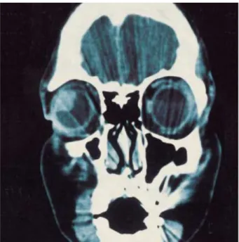
Seven years after the initial operation, the patient visited the ophthalmology department again due to progressive decrease of visual acuity of right eye. Ophthalmologic examination and computed tomography (CT) revealed regrowth of the tumor at the same region (Fig. 1). Enucleation of the right eyeball was performed under the clinical diagnosis of glioma or leio- myosarcoma. Grossly, a well demarcated, round to ovoid tu- mor was noted in the anterolateral portion of the ciliary body and measured 2.0×1.5×1.2 cm in size. The retina was shifted upwardly by the tumor. The cut surface of the tumor showed homogeneous yellowish white, firm and solid appearance (Fig.
2). Microscopically, the tumor was well demarcated and cov- ered by the ciliary epithelium. The transitional area from normal ciliary smooth muscle was noted (Fig. 3). The tumor
Seong Hwan Park, Ji-Hye Lee, Yang Seok Chae, Chul Hwan Kim
Department of Pathology, College of Medicine, Korea University, Seoul, Korea
Address for correspondence Chul Hwan Kim, M.D.
Department of Pathology, Korea University Guro Hospital, 97 Guro-dong, Guro-gu, Seoul 152-705, Korea
Tel : +82.2-818-6233, Fax : +82.2-818-6239 E-mail : [email protected]
614 J Korean Med Sci 2003; 18: 614-7
ISSN 1011-8934
Copyright � The Korean Academy of Medical Sciences
Recurrent Mesectodermal Leiomyoma of the Ciliary Body : A Case Report
A 19-yr-old woman with a previous history of a mass of the right ciliary body pre- sented with a decreased visual acuity of right eye. Clinicoradiologic examinations suggested a recurrent mass of the ciliary body. Enucleation of the right eye was performed under the impression of malignant tumor. On microscopic examination, the tumor was a mesectodermal leiomyoma of the ciliary body. On immunohisto- chemistry, the tumor cells were reactive to smooth muscle actin and vimentin, but not reactive to cytokeratin, S-100 protein, neurofilament, desmin, epithelial mem- brane antigen, HMB-45, glial fibrillary acidic protein, and synaptophysin. Electron microscopy revealed numerous thin longitudinally placed myofilaments and focal densities in the cytoplasms. In the review of the literature, only 27 cases of mesec- todermal leiomyoma of the ciliary body were reported, however, there was no report of recurrent cases. Mesectodermal leiomyoma should be differentiated from other orbital spindle-cell tumors such as amelanotic melanomas and glial tumors. Im- munohistochemical and electron microscopic studies may be useful for the correct diagnosis by showing smooth muscle differentiation in the tumor cells.
Key Words : Leiomyoma; Ciliary Body
Received : 12 July 2002 Accepted : 17 September 2002
Recurrent Mesectodermal Leiomyoma 615
Fig. 4. Polygonal tumor cells in the fibrillary stroma are noted (H&E stain, ×200).
Fig. 3. The tumor is covered by the ciliary epithelium and mixed with normal ciliary smooth muscle (H&E stain, ×40).
cells were polygonal to spindle in shape and had abundant eosinophilic, fibrillary cytoplasms (Fig. 4). The cellularity was moderate. There was no nuclear atypia or mitosis. Immunohis- tochemically, the tumor cells were reactive to vimentin and SMA (Fig. 5). They were negative for cytokeratin, S-100 pro- tein, neurofilament, desmin, EMA, HMB-45, GFAP, and syn- aptophysin. Electron microscopy (EM) revealed abundant mito- chondria, numerous longitudinally placed thin myofilaments, and focal densities in the cytoplasms (Fig. 6). The nuclear
membrane showed mild degree of irregularity. The nucleus showed inconspicuous nucleoli and chromatin clumping.
DISCUSSION
The mesectodermal leiomyoma is a rare variant of the be- nign smooth muscle tumor, which microscopically resembles a neurogenic rather than a myogenic tumor. The term mesec-
Fig. 1.Facial computed tomography, coronal view: a well demar- cated ovoid mass is noted in the right eye.
Fig. 2. A pale gray solid firm mass is located in the anterolateral por- tion of the ciliary body.
616 S.H. Park, J.-H. Lee, Y.S. Chae, et al.
Fig. 6. Electron microscopy reveals longitudinally placed thin fila- ments and focal densities in the cytoplasm (×10,000).
Fig. 5. Immunohistochemical stain for smooth muscle actin reveals positive reaction (×100).
Author (Reference) YearAge (yr) Sex Race Side Size (mm) Operation Clinical Impression Follow up
Blodi (2) 1950 40 F NA* Rt NA Enucleation NA NA
Dunbar (3) 1956 49 F NA Lt 7×5 Enucleation NA NA
Bonameur et al. (4) 1957 39 F NA NC NA NA NA NA
Meyer et al. (5) 1968 50 F NA Rt 9×7 Enucleation NA NA
Lowe & Greer (6) 1970 24 F NA Rt 8×6×5 Resection NA NA
Calmettes et al. (7) 1971 25 F NA Lt NA Enucleation NA NA
Jakobiec et al. (1) 1977 37 F NA Lt 6×5×5 Resection metastasis, melanoma Disease free 6 yrs
Jakobiec et al. (1) 1977 20 F NA Rt 9×7×2 Enucleation melanoma NA
Jakobiec & Iwamoto (8) 1978 28 F NA Rt NA Enucleation melanoma NA
Vogel et al. (9) 1978 55 F NA Rt 8×8×3 Resection NA NA
Gloor et al. (10) 1979 12 F NA Rt NA Resection NA NA
Sautter et al. (11) 1979 23 F NA Lt 6×6 Resection NA NA
Croxatto & Malbran (12) 1982 23 F White Lt 7×5×5 Resection melanoma, cyst NA
Orsoni et al. (13) 1985 18 F NA Rt 8×5 Enucleation uncertain NA
Takagi et al. (14) 1985 38 F Japanese Lt 13×7×5 Enucleation malignant tumor NA
Burk et al. (15) 1989 63 F NA NA NA NA NA NA
Ishigooka et al. (16) 1989 28 F Japanese Rt 9×8×3 Resection neurogenic or glial tumor NA
White et al. (17) 1989 38 M White Rt 8×7×5 Resection melanoma NA
Yu et al. (18) 1990 8 M White Lt NC Resection NA NA
Shields et al. (19) 1994 80 F White Lt 4×4×3 Resection melanoma, leiomyoma Death by another cause (2 yrs) Shields et al. (19) 1994 11 F White Rt 14×12×9 Resection melanoma Disease free (5 yrs) Shields et al. (19) 1994 29 F NC Rt NA Resection atypical staphyloma Disease free (4 yrs) Shields et al. (19) 1994 20 F NC Rt 16×14 Resection leiomyoma Disease free (4 yrs) Shields et al. (19) 1994 68 M White Lt 15×10 Enucleation melanoma Disease free (3 yrs) Shields et al. (19) 1994 54 F White Lt 9×9×4 Resection melanoma, leiomyoma, NA
neurilemmoma
Shields et al. (19) 1994 24 M White Lt 13×12×8 Resection melanoma, leiomyoma, NA neurilemmoma
Campbell et al. (20) 1997 47 F NA NA NA NA NA NA
Present case 2003 19 F Korean Rt 20×15×12 Enucleation melanoma, glioma Recurrent 7 yrs
after resection after resection
Table 1.Summary of leiomyomas of the ciliary body reported in the literature
*NA: not available.
Recurrent Mesectodermal Leiomyoma 617
todermal leiomyoma was suggested by Jakobiec et al. in 1977 (1). Embryologically, the cells of the neural crest that contri- bute to the formation of bone, cartilage, connective tissue, and smooth muscle in the regions of the head and neck have been called mesectoderm (1). They proposed that the unusual neu- ral appearance of ciliary leiomyoma was a reflection of their probable origin from the mesectodermal smooth muscle of the ciliary body.
Mesectodermal leiomyomas resemble ganglionic, astrocytic, and peripheral nerve tumors because of their fibrillary neuro- genic appearance. Therefore, histologically, the differential di- agnosis of this unusual tumor includes melanoma, glioma, peripheral nerve tumor, or paraganglioma. We initially diag- nosed this tumor as glial tumor or smooth muscle tumor.
The final diagnosis of a mesectodermal leiomyoma was sup- ported by the electron microscopic demonstration of thin filaments with focal densities and smooth muscle actin reac- tivity by immunohistochemistry.
To our knowledge, 27 cases of mesectodermal leiomyoma in the ciliary body have been reported in the literature (Table 1). Twenty-three patients were female and four were male.
Thirteen cases were right, eleven were left, and three were un- known. The tumor size ranged from 0.4 to 1.6 cm. Enucle- ation was performed in 9 cases and resection was performed in 15 cases. The type of operation was not documented in 3 cases.
Among seven cases with follow-up data, there was no recur- rence or metastasis. The maximum follow-up period was 6 yr.
In the present case, the tumor was 2 cm in its greatest dimen- sion, which is the largest one in the reported cases and is the only case that recurred after simple resection, which was not iridocyclochoroidectomy but just local resection by curettage.
Although the tumor in the present case recurred because the initial tumor removal was incomplete, it was thought to be benign on light microscopic appearance because of its well cir- cumscription, moderate cellularity, and lack of mitosis. Based on our literature review, the mesectodermal leiomyoma can exhibit slowly progressive enlargement and can produce a large mass with complications that may require enucleation. How- ever, in most reported cases, the local resection of the tumor such as modified partial lamellar sclerouvectomy was perfo- rmed. Enucleation seems inappropriate for this tumor, and radiotherapy generally has little effect on benign tumor.
REFERENCES
1. Jakobiec FA, Font RL, Tso MO, Zimmerman LE. Mesectodermal leiomyoma of the ciliary body: a tumor of presumed neural crest ori- gin. Cancer 1977; 39: 2102-13.
2. Blodi FC. Leiomyoma of the ciliary body. Am J Ophthalmol 1950; 33:
939-42.
3. Dunbar JC. Leiomyoma of the ciliary body: report of a case exhibiting a significant uptake of radioactive phosphorus. Am J Ophthalmol 1956; 42: 204-7.
4. Bonamour MM, Bonnet JL, Jambon M. Leiomyome du corps ciliare;
quelques connsiderations a propos du diagnostic et du traitement des tumeurs benignes de l'iris et du corps ciliare. Bull Soc Ophthalmol Fr 1961; 74: 158-67.
5. Meyer SL, Fine BS, Font RL, Zimmerman LE. Leiomyoma of the cil- iary body. Electron microscopic verification. Am J Ophthalmol 1968;
66: 1061-8.
6. Lowe RF, Greer CH. Leiomyoma of the ciliary body. A clinico-patho- logical case report. Br J Ophthalmol 1970; 54: 383-7.
7. Calmettes MML, Deodatti F, Bec P. Leiomyome du corps ciliare. Bull Soc Ophthalmol Fr 1961; 74: 158-67.
8. Jakobiec FA, Iwamoto T. Mesectodermal leiomyoma of the ciliary body associated with a nevus. Arch Ophthalmol 1978; 96: 692-5.
9. Vogel M, Spitznas M, Waubke TN. Leiomyoma of the ciliary body.
Albrecht von Graefes Arch Klin Exp Ophthalmol 1978; 209: 89-98.
10. Gloor BP, Daicker B, Gafner F. Leiomyom des Zilliakorpers-Durch Fehldiagnose auf ungewohnliche Weise erfolgreich operiert. Klin Monatsbl Augenheilkd 1979; 175: 760-6.
11. Sautter H, Boke W, Domarus D, Demeler U. Leiomyom des Zilliako- rpers: klinischer, fluoreszenzangiographischer und histologischer Befundbericht. Klin Monatsbl Augenheilkd 1979; 175: 704-10.
12. Croxatto JO, Malbran ES. Unusual ciliary body tumor: mesectodermal leiomyoma. Ophthalmology 1982; 89: 1208-12.
13. Orsoni JG, Daicker B, Cardillo Piccolino F. Mesectodermal leiomy- oma of the ciliary body extending into the anterior chamber. Oph- thalmologica 1985; 191: 127-9.
14. Takagi T, Ueno Y, Matsuya N. Mesectodermal leiomyoma of the ciliary body: an ultrastructural study. Arch Ophthalmol 1985; 103:
1711-4.
15. Burk RO, Volcker HE, Daus W, Born IA. Mesectodermal leiomyoma of the ciliary body: clinical aspects, surgery and immunohistochem- istry. Fortschr Ophthalmol 1989; 86: 631-5.
16. Ishigooka H, Yamabe H, Kobashi Y, Magata M. Clinical and patho- logical status of mesectodermal leiomyoma of the ciliary body. A case report and review of the literature. Graefes Arch Clin Exp Ophthalmol 1989; 227: 101-5.
17. White V, Stevenson K, Garner A, Hungerford J. Mesectodermal leiomy- oma of the ciliary body: case report. Br J Ophthalmol 1989; 73: 12-8.
18. Yu DY, Cohen SB, Peyman G, Tso MO. Mesectodermal leiomyoma of the ciliary body: new evidence for neural crest origin. J Pediatr Oph- thalmol Strabismus 1990; 27: 317-21.
19. Shields JA, Shields CL, Eagle RC Jr, De Potter P. Observations on seven cases of intraocular leiomyoma: the 1993 Byron Demorest lec- ture. Arch Ophthalmol 1994; 112: 521-8.
20. Campbell RJ, Min KW, Bolling JP. Skeinoid fibers in mesectodermal leiomyoma of the ciliary body. Ultrastruct Pathol 1997; 21: 559-67.