서 론
반코마이신 내성 장구균(vancomycin-resistant entero- cocci, VRE)은 1987년 유럽에서 처음 출현한 이래 세계적으로 원내감염의 중요한 병원균이 되었다[1, 2]. 국내에서 1992년 처 음 보고되었고 1990년대 중반부터 균주 분리가 급격히 증가하 였다[3, 4]. 현재 중환자실에서 분리되는Enterococcus faeci- um의 30-45%에서 반코마이신에 내성을 보인다[5]. VRE에 대 한 적절한 감염관리가 필요한 상황이다.
신속하고 정확한 검출은 VRE의 감염관리에 매우 중요하다.
VRE 선별 고형배지에 접종하거나 선별 액체배지에 증균 후 고 형배지에 접종하는 통상의 검출방법은 결과 보고까지 2-5일이 걸린다. VanB형과 VanC형 VRE는 검출이 어려운 경우도 있다 [6]. 이에 중합효소연쇄반응법(PCR)과 실시간-중합효소연쇄반 응법(real-time PCR) 등 glycopeptides 내성유전자 검사법이 VRE 감시배양에 사용되었다[7-13]. 실시간-중합효소연쇄반응 법은 중합효소연쇄반응법보다 VRE를 더 빠르게 검출하고 업무 량도 적다[12]. 반응시간이 짧고 실시간으로 반응산물이 모니터 링되어 전기영동이 필요하지 않기 때문이다. 하지만 염기서열 에 특이한 소식자를 사용하는 경우 시약 비용이 증가하는 단점 이 있다.
본 연구에서 저자들은 염기서열에 특이한 소식자를 사용하지 않고 융해곡선(melting curve) 분석을 이용하여 VRE를 검출 하는 다중 실시간-중합효소연쇄반응법(multiplex real-time PCR)을 개발하여 유용성을 평가하였다.
138 138 138 138 Received :October 17, 2009 Manuscript No :KJLM09-122 Revision received :February 21, 2010
Accepted :March 10, 2010
Corresponding author :Jeong Uk Kim, M.D.
Department of Laboratory Medicine, Gangneung Asan Hospital, University of Ulsan College of Medicine, 415 Bangdong-ri, Sacheon-myeon, Gangneung 210-711, Korea
Tel : +82-33-610-3446, Fax : +82-33-610-3449 E-mail : [email protected]
Background : We developed and evaluated the utility of a multiplex real-time PCR assay that uses melting curve analysis and allows simultaneous identification of vancomycin-resistant genotypes and clinically relevant enterococci.
Methods : The specificity of the assay was tested using 4 reference strains of vancomycin-resis- tant enterococci (VRE) and 2 reference strains of vancomycin-susceptible enterococci. Ninety-three clinical isolates of enterococci with different glycopeptide-resistant phenotypes were genotyped and identified using a multiplex real-time PCR assay and melting curve analysis.
Results : Representative melting curves were obtained for Enterococcus faecium, Enterococcus faecalis, vanA-containing E. faecium, vanB-containing E. faecalis, Enterococcus gallinarum, and Enterococcus casseliflavus. Phenotypic and genotypic analysis of the isolates revealed same results for 82 enterococcal isolates, while in 4 isolates, the glycopeptide-resistant phenotypes were incon- sistent with the glycopeptide-resistant genotypes and in the 4 other isolates, species could not be accurately identified. Three isolates with mixed strains, which were detected by the PCR assay, could not be correctly identified using phenotypic methods.
Conclusions : VRE genotyping and identification of clinically relevant enterococci were rapidly and correctly performed using multiplex real-time PCR assay and melting curve analysis. (Korean J Lab Med 2010;30:138-46)
Key Words : Multiplex real-time PCR, Melting curve analysis, Vancomycin-resistant enterococci
Detection of Vancomycin-resistant Enterococci using Multiplex Real-time PCR Assay and Melting Curve Analysis
Choong-Hwan Cha, M.D., Hae Kyong An, M.T., and Jeong Uk Kim, M.D.
Department of Laboratory Medicine, Gangneung Asan Hospital, University of Ulsan College of Medicine, Gangneung, Korea
재료 및 방법
1. 대상 균주본원 진단검사의학과에 의뢰된 임상검체에서 분리된 93주의 장구균(E. faecium72주, Enterococcus faecalis13주, Ente- rococcus avium3주, Enterococcus durans2주, Entero- coccus gallinarum2주, Enterococcus casseliflavus1주)을 대상으로 하였다. 표준 균주로는E. faeciumATCC 700221 (vanA), E. faecalisATCC 51299 (vanB), E. gallinarum ATCC 49573 (vanC1), E. casseliflavusATCC 25788 (vanC2/
C3), E. faecalisATCC 29212, E. faecium ATCC 19434를 사용하였다.
2. 장구균의 동정검사 및 항균제 감수성검사
대상 균주는 반코마이신 6 mg/mL를 첨가하여 자가제조한 Enterococcosel agar (EA) (Becton Dickinson, Sparks, MD, USA)와 반코마이신을 첨가하지 않은 EA에서 선별되었다. 배 양 1일과 2일째 검은색으로 변한 집락을 혈액한천배지에 계대 배양하여 균 동정과 항균제 감수성검사를 시행하였다. 균 동정 은 MicroScan (Dade Behring, West Sacramento, CA, USA) 과 API 20 STREP (bioMerieux, Hazelwood, MO, USA)을 사용하였으며, 반코마이신과 타이코프라닌의 최소억제농도 (minimum inhibitory concentration, MIC)는 E-test (AB Biodisk, Solna, Sweden)로 측정하였다. 항균제별 MIC의 해 석은 CLSI의 기준[14]을 따랐다.
3. DNA 추출
중탕가열(boiling)법으로 DNA를 추출하였다. EA에서 자란 검은색 집락 적당량을 면봉에 묻혀 증류수 0.5 mL가 들어있는 1.5 mL microtube에 풀고 10분 동안 끓은 물에 중탕시켰다.
13,000 rpm으로 3분간 원심 분리한 후 상청액을 사용하였다.
상청액의 DNA 농도는 20-90 ng/mL가 되도록 멸균증류수로 조절하였다.
4. 다중 실시간-중합효소연쇄반응법
네 종류의 glycopeptide 내성 유전자(vanA, vanB, vanC1, vanC2/C3)와E. faecalis와E. faecium 균종에 특이한 D-
alanine-D-alanine ligase 유전자(ddl)를 검출부위로 하는 시 발체를 Primer3 프로그램을 이용하여 설계하였다[15]. 각 시발 체의 염기서열, 반응산물의 크기, 온도(Tm)는 Table 1과 같다.
Type-it HRM PCR 키트(QIAGEN Inc., Germantown, MD, USA)를 사용하여 실시간-중합효소연쇄반응을 실시하였 다. 5 mL의 2× HRM PCR Master Mix, 0.8 mL의 시발체 혼 합액 (E. faecalis시발체, 0.6 mM; E. faecium시발체, 0.3 mM;
vanA시발체, 0.8 mM; vanB시발체, 0.2 mM; vanC1시발체, 0.2 mM; vanC2/C3시발체, 0.2 mM), 3.2 mL의 증류수에 1 mL 의 DNA를 넣어 반응액 10 mL를 제조하여 중합효소연쇄반응을 실시하였다.
중합효소연쇄반응은 Rotor-gene 6000 (QIAGEN Inc., Ger- mantown, MD, USA)를 이용하였다. 95℃에서 5분 반응 후 95℃에서 10초, 53℃에서 30초, 63℃에서 20초의 반응을 40 회 반복하였다. 마지막 PCR 반응이 끝난 후 65℃에서 89℃까 지 초당 0.2℃의 속도로 온도를 증가시키면서 융해곡선을 모니 터링하였다.
결 과
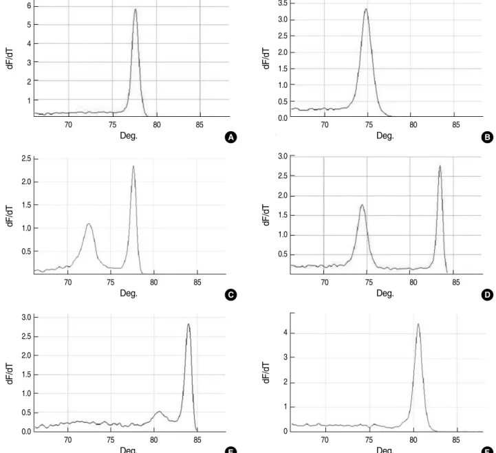
저자들은 융해곡선을 분석하여 네 종류의 glycopeptide 내성 유전자의 검출과E. faecalis와E. faecium의 동정이 가능한 다중 실시간-중합효소연쇄반응법을 개발하였다. ATCC 표준균 주들의 융해곡선은 Fig. 1과 같다. 융해곡선분석이 용이하도록 반응산물 간에 Tm 차이가 나도록 시발체를 설계하였다. 가능 한 2℃ 이상 차이가 나도록 제작하였지만vanB와vanC1반응 Abbreviations: Tm, melting temperature; ddl, gene encoding D-alanine- D-alanine ligase; E. faecalis, Enterococcus faecalis; E. faecium, Ente- rococcus faecium.
Amplified gene Primer sequences (5′-3′)
Size of the PCR product (bp)
Product Tm (℃)
vanA TATTGACTTCGTTCAGTACA 53 72.7±0.3
TGTGGATATGTTTTTACAAG
vanB CAGACCCTGTATCGCACCAT 195 83.7±0.3
AACGGCGTATGGAAGCTATG
vanC1 TGCTTGTGATGCGATTTCTC 204 84.1±0.4 ATCGCTCCTTGATTGGTGAC
vanC2/C3 GGGAAGATGGCAGTATCCAA 102 80.6±0.3 GCAGCAGCCATTTGTTCATA
ddl E. faecalis GTGGCTTAAGTCGCTGTGAT 74 74.9±0.3 AGGCATGGTGTTCAATTCAT
ddl E. faecium TTTACAAGCTGCTGGTGTGC 140 78.1±0.3 AACCCATATTCGCAGGTTTG
comycin-resistant enterococci
산물 간에는 0.5℃ 밖에 차이가 나지 않았다. 시발체에 따른 반응 정도 차이는 시발체의 농도, 불림(annealing) 및 확대(exten- sion) 온도와 유지시간을 조절하여 6가지 검출부위 모두 반응이 잘 일어날 수 있도록 조건을 최적화하였다.
임상검체에서 분리된 장구균 93주의 항균제 감수성 결과에 따라 대상 균주들의 내성 표현형은 다음과 같았다. E. faecium 65주, E. faecalis5주, E. avium2주, E. durans1주 및E.
gallinarum1주는 VanA형(반코마이신 MIC, ≥256 mg/mL;
타이코프라닌 MIC, 16- ≥256 mg/mL)이었고E. faecium4 주는 VanB형(반코마이신 MIC, 16- ≥256 mg/mL; 타이코프 라닌 MIC, 8- 12 mg/mL)이었으며 E. gallinarum1주와E.
casseliflavus1주는 VanC형(반코마이신 MIC, 6-12 mg/mL;
타이코프라닌 MIC, 2 mg/mL)이었다. 그리고E. faecium3주, E. faecalis8주, E. avium1주 및E. durans1주는 감수성(반 코마이신 MIC, 0.38-4 mg/mL; 타이코프라닌 MIC, 0.05-4 mg/mL)을 보였다.
dF/dT
6 5 4 3 2 1
70 75 80 85
Deg.
dF/dT
2.5 2.0 1.5 1.0 0.5
70 75 80 85
Deg.
dF/dT
3.0 2.5 2.0 1.5 1.0 0.5 0.0
70 75 80 85
Deg.
dF/dT
3.5 3.0 2.5 2.0 1.5 1.0 0.5
0.0 70 75 80 85
Deg.
dF/dT
3.0 2.5 2.0 1.5 1.0 0.5
70 75 80 85
Deg.
dF/dT
4
3
2
1
0
70 75 80 85
Deg.
A B
D
F C
E
Fig. 1. Melting curve analysis of amplicons obtained from reference strains by using multiplex real-time PCR. Melting curves for Entero- coccus faecium (A); Enterococcus faecalis (B); E. faecium/vanA (C); E. faecalis/vanB (D); Enterococcus gallinarum/vanC1 (E); and Ente- rococcus casseliflavus/vanC2/C3 (F) are shown.
균종별 유전자형 분석결과는 다음과 같았다(Table 2). VanA 형E. faecium65주와 VanB형E. faecium4주는 모두vanA 유전자형의E. faecium으로 분석하였다. Glycopeptides에 감 수성을 보인E. faecium3주 중 2주는E. faecium으로 동정되 었으나 한 균주에서는vanC2/C3 (E. casseliflavus/E. fla- vescens)유전자가 검출되어 생화학적 동정과 불일치하였다.
VanA형E. faecalis5주는 모두vanA유전자형의E. faecalis
로 분석하였고 감수성을 보인E. faecalis8주는E. faecalis의 ddl유전자만 검출되어 생화학적 동정과 일치하였다. VanA형 E. avium1주는vanA유전자만 검출되었고 VanC형E. cas- seliflavus1주는vanC2/C3유전자가 검출되어 생화학적 동정 과 일치하였다. VanA형E. durans1주는vanA유전자형의 E. faecium으로 동정되고 감수성을 보인E. avium1주와E.
durans1주는 모두E. faecalis로 동정되어 생화학적 동정결과 Abbreviations: ddl, gene encoding D-alanine-D-alanine ligase; E. faecium, Enterococcus faecium; E. faecalis, Enterococcus faecalis; E. gallinarum, Enterococcus gallinarum; E. casseliflavus, Enterococcus casseliflavus; E. avium, Enterococcus avium; E. durans, Enterococcus durans.
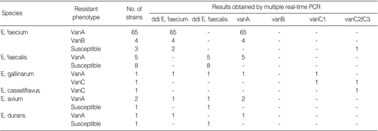
Resistant phenotype
No. of strains Species
Results obtained by multiple real-time PCR
ddl E. faecium ddl E. faecalis vanA vanB vanC1 vanC2/C3
E. faecium VanA 65 65 - 65 - - -
VanB 4 4 - 4 - - -
Susceptible 3 2 - - - - 1
E. faecalis VanA 5 - 5 5 - - -
Susceptible 8 - 8 - - - -
E. gallinarum VanA 1 1 1 1 - 1 -
VanC 1 - - - - 1 1
E. casseliflavus VanC 1 - - - 1
E. avium VanA 2 1 1 2 - - -
Susceptible 1 - 1 - - - -
E. durans VanA 1 1 - 1 - - -
Susceptible 1 - 1 - - - -
Table 2. Genotype analysis of 93 enterococcal isolates with different glycopeptides-resistant phenotypes by using multiplex real-time PCR and melting curve analysis
dF/dT
0.8
0.6
0.4
0.2
0.0
70 75 80 85
Deg.
dF/dT
1.50 1.25 1.00 0.75 0.50 0.25
70 75 80 85
Deg.
dF/dT
1.75 1.50 1.25 1.00 0.75 0.50 0.25
70 75 80 85
A Deg. B
C
Fig. 2. Melting curve analysis for the identification of mixed strains.
(A) Melting curve for DNA extracted from black colonies growing on the Enterococcal agar plate. VanA, ddl Enterococcus faecalis, and ddl Enterococcus faecium peaks are shown. (B) Melting curve for DNA extracted from pure colonies of E. faecium with phenotype VanA growing on the blood agar plate. Peaks for E. faecium with VanA and ddl genes. (C) Melting curve for DNA extracted from pure colonies of E. faecalis with phenotype VanA growing on the blood agar plate.
Peaks for E. faecalis with VanA and ddl genes.
와 일치하지 않았다.
한편, 한 검체에 2종 이상의 장구균이 섞여 있는 경우도 있었 다. 장구균 93주 중 VanA형E. faecium1주와 VanA형E.
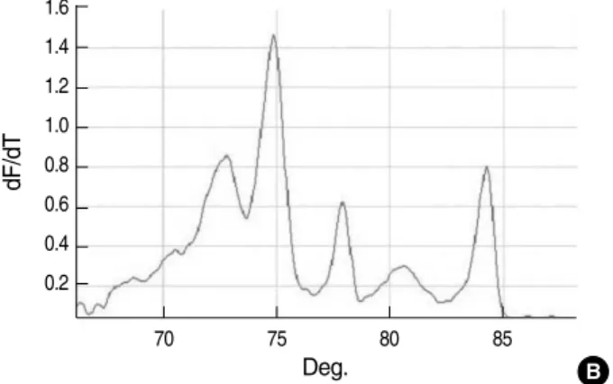
faecalis1주는 한 검체에서 분리된 균주로 EA 선택배지에서 자란 집락에서vanA E. faecium과vanA E. faecalis가 검출 되었고 혈액한천배지에 계대배양했을 때 두 종류의 순수 집락 으로 구분되어 분리되었다(Fig. 2). 그러나 VanA형E. avium 1주, VanA형E. gallinarum1주 및 VanC형E. gallinarum1 주는 2종 이상의 균주가 혼합된 것처럼 VanA형E. avium1주 는vanA유전자와E. faecium과E. faecalis의ddl유전자가 동시에 검출되었으며 VanA형E. gallinarum1주는vanC1유 전자 및vanA유전자와E. faecium과E. faecalis의ddl유전 자가 동시에 검출되었고 VanC형E. gallinarum1주는vanC1 유전자와vanC2/C3유전자가 함께 검출되었으나 EA 선택배 지에 자란 집락을 계대배양하였을 때 순수 집락으로 분리할 수 없었다(Fig. 3).
고 찰
최근 국내에 실시간-중합효소연쇄반응 장비를 도입하는 검
사실이 증가하는 추세이다. 실시간-중합효소연쇄반응 장비는 고가의 기기이지만 중합효소연쇄반응보다 더 민감하고 특이적 인 검사법의 개발이 가능하다. 염기서열에 특이한 소식자를 사 용하고 융해곡선분석으로 특이도가 높다[16]. 융해곡선분석은 1997년 처음 소개되었다. 융해는 두 가닥 DNA가 가온되면 한 가닥 DNA로 분리되는 현상을 말한다. 두 가닥 DNA에 비특이 적으로 결합하는 형광색소(fluorescent dye)인 SYBR Green 을 사용하여 융해과정이 실시간으로 측정된다. 반응산물의 염 기서열, 크기 및 GC 비율에 따라 특징적인 융해곡선을 보여 전 기영동 없이도 융해곡선분석만으로 반응산물을 확인할 수 있었 다[17]. 널리 사용되는 SYBR Green은 고농도에서 중합효소연 쇄반응을 저하시키고 융해과정에서 형광색소의 재분포가 일어 나 유전자형결정(genotyping)까지 가능한 고해상 융해곡선 (high-resolution melting curve)분석에는 적합하지 않았다.
최근 개발된 SYTO 9, EvaGreen, LCGreen 등의 형광색소는 이러한 단점이 개선되어 고해상 융해곡선분석에 사용되고 있다 [16, 18]. 본 연구에 사용된 Type-it HRM PCR 키트의 master mix에는 EvaGreen이 들어 있다.
다중 중합효소연쇄반응법은 두 쌍 이상의 시발체를 사용하여 두 가지 이상의 검출부위를 동시에 검출할 수 있게 고안된 중합
dF/dT
1.4 1.2 1.0 0.8 0.6 0.4 0.2
70 75 80 85
Deg.
dF/dT
1.50 1.25 1.00 0.75 0.50 0.25 0.00
70 75 80 85
Deg.
dF/dT
1.6 1.4 1.2 1.0 0.8 0.6 0.4 0.2
70 75 80 85
A Deg. B
C
Fig. 3. Melting curve analysis of amplicons obtained from 3 mixed strains of 2 or more enterococci by using multiplex real-time PCR.
Melting curves for Enterococcus faecalis and Enterococcus faecium with vanA gene (A); E. faecalis and E. faecium with vanA gene and Enterococcus gallinarum with vanC1 gene (B); and Enterococcus casseliflavus/Enterococcus flavescens with vanC2/C3 gene and E.
gallinarum with vanC1 gene (C).
효소연쇄반응법이다. 일회 반응으로 여러 대상을 검출할 수 있 어 검사 시간과 시약 비용을 줄일 수 있다. 실시간-중합효소연 쇄반응 장비에서도 소식자의 형광색상과 반응산물의 Tm 차이 를 이용하여 다중검사가 가능하다[19-21]. 융해곡선분석을 이 용하는 다중 실시간-중합효소연쇄반응법은 다중 중합효소연쇄 반응법과 달리 반응산물의 크기보다 반응산물의 Tm에 차이가 나도록 시발체를 설계한다. 다중반응은 dNTP, Taq polymerase 등 반응 시약들의 농도가 한정된 조건하에 놓이게 되어 시발체 간에 경합 반응이 일어난다. 상대적으로 약한 반응이 일어나는 시발체는 단일반응 때보다 민감도가 저하될 가능성이 있다. Sa- take 등[9]은 다중 중합효소연쇄반응으로 VRE 유전자를 검출 하는 경우 단일반응 때보다 검출률이 저하되는 것을 보고하였 다. 저자들은 EA 선택배지에 자란 집락에서 DNA를 추출하도 록 고안하였기에 주형 DNA의 농도를 민감도 이상으로 유지할 수 있어 다중반응에서 민감도가 저하되는 문제는 유의하지 않 다고 추정하였다. 본 검사법은 DNA를 추출할 때 증류수 50 mL 를 사용하면 EA 선별배지에 자란 집락 한 개로 내성 유전자형 결정과 균 동정이 가능하였다. 과량의 주형 DNA를 사용하면 융해곡선이 오른쪽으로 이동되며 Tm 차이가 적은vanB와 vanC1반응산물 간의 구분이 어렵게 되었다. 그래서 중합효소 연쇄반응에 사용되는 주형DNA 농도를 20-90 ng으로 조정하 여 반응산물이 검출되지 않거나 Tm값이 증가되는 현상이 일어 나지 않도록 하였다.
신속하고 정확한 VRE의 검출은 적절한 감염관리를 시행하 여 전파를 차단하는데 매우 중요하다[13]. 더욱이vanA유전자 를 가진 반코마이신 내성Staphylococcus aureus의 출현으로 VRE 감염관리의 중요성이 강조되고 있다[22]. 중합효소연쇄반 응법으로 VRE 유전자를 검출하는 방법은 가장 특이도가 높은 검사법이고 저도내성을 갖는 VRE 검출에도 유용하다[6]. 실시 간-중합효소연쇄반응 기기의 등장으로 중합효소연쇄반응보다 신속하게 내성 유전자의 검출이 가능하게 되었다[12]. 지금까지 여러 방식의 내성 유전자 검출법이 보고되었으나[7-13], 융해 곡선분석을 이용한 방법은 없었다. 이에 저자들은 염기서열에 특이한 소식자를 사용하지 않고 융해곡선분석을 이용하여van 유전자의 검출과 장구균의 동정이 동시에 가능한 다중 실시간- 중합효소연쇄반응법을 개발하게 되었다. VRE는 7종류(VanA, VanB, VanC, VanD, VanE, VanG, VanL)의 표현형이 있으 며 해당 유전자형에 의해 발현된다[6, 23]. 주로 VanA, VanB, VanC형이 임상검체에서 검출되며 균종으로는E. faecium과 E. faecalis가 대부분을 차지하고 있다[4, 24-27]. vanA와 vanB유전자와 달리vanC유전자는 내인성의 균종 특이성이
있으며 유전자형의 결정만으로 균 동정이 가능하다. vanC1은 E. gallinarum, vanC2는E. casseliflavus, vanC3는E. fla- vescens에 특이적이지만vanC2와vanC3유전자는 염기서열 이 98.3%가 동일하여 중합효소연쇄반응으로 구분하기 어렵다 [28]. 저자들은 임상검체에서 주로 검출되는 VanA, VanB, VanC 형의 VRE가 갖고 있는vanA, vanB, vanC1, vanC2/C3유전 자를 검출하고E. faecium, E. faecalis, E. gallinarum, E.
casseliflavus/E. flavescens의 동정이 가능하도록 시발체를 설계하였다.
유용성 평가에 사용된 장구균 93주는 2000년 2월 이후 강릉 아산병원 진단검사의학과에서 분리되어 -70℃에 보관된 균주 들과 2009년 5월과 6월 두 달 동안 반코마이신 6 mg/mL가 첨 가된 EA 선택배지에 의뢰된 대변 및 직장주변도말 검체를 배양 하여 분리한 균주였다. 신속하게 VRE를 검출하는 방법으로 대 변이나 직장주변도말 검체에서 직접 DNA를 추출하거나[9, 13, 29-31], 선택배지로 장구균을 선별하는 방법들이 연구되었다 [32-34]. 검체에서 직접 DNA를 추출하는 방법은 억제제로 인하 여 VRE 검출률이 저하된다[9, 13, 34]. 이에 저자들은 6 mg/mL 가 첨가된 EA 선택배지로 장구균을 선별하여 중탕 가열법으로 DNA를 추출하는 방법을 사용하였다. 이 방법으로 중합효소연 쇄반응의 저하 없이 EA 선별배지에 검은색 집락을 형성한 당일 에 내성 유전자형 결정과 동정이 가능하였다. 생화학적 균 동정 과 MIC의 측정은 EA 선별배지에 자란 검은 집락을 혈액한천배 지에 계대배양하여 실시하였으므로 실시간-중합효소연쇄반응 법을 이용한 VRE 검출보다 2일 이상이 더 소요되었다.
감시배양에서는 한 검체에 두 종 이상의 장구균이 섞여 있는 경우도 있었다. EA 선택배지에 검은 집락을 형성하는 장구균들 은 집락의 형태 감별만으로 2종 이상의 장구균이 섞여 있는지 알 수가 없었고 순수 집락을 분리하는데 2차 계대배양이 필요하 였다. 대상 균주 중 3주는 2차 계대배양에서도 순수집락으로 분 리할 수 없어 2차 계대배양의 집락으로 균 동정과 MIC를 측정 하여 VanA형E. gallinarum, VanA형E. avium, VanC형E.
gallinarum으로 판정하였다. 본 검사법으로는 이런 경우에도 EA 선택배지에 집락을 형성한 당일에 정확하게 유전자형의 결 정과 균 동정을 할 수 있었다. VanA형E. gallinarum은vanA E. faecium, vanA E. faecalis, E. gallinarum가 혼합된 경우 였고, VanA형E. avium은vanA E. faecium과vanA E. fae- calis가 혼합된 경우였으며 VanC형E. gallinarum은E. cas- seliflavus/E. flavescens와E. gallinarum이 혼합된 경우였다.
대상 균주 중 4주는 생화학적 동정결과와 분자생물학적 동정 결과가 불일치하였다. E. faecium1주, E. avium1주, E. dur-
ans2주는 실시간-중합효소연쇄반응법으로 각각E. casseli- flavus/E. flavescens, E. faecalis, vanA형E. faecium, E.
faecalis로 동정되었다. 상용화된 생화학적 동정 키트만으로 장 구균을 정확하게 동정하는데 한계가 있는 것 같다. Angeletti 등[35]은 임상검체에서 분리한 장구균 279주 중에 26주에서 중 합효소연쇄반응법과 16S 리보소옴 DNA의 염기순서분석법을 사용한 분자생물학적 동정결과와 생화학적 동정결과가 불일치 함을 보고하였다. Leuconostoc spp, Aerococcus spp, Strep- tococcus bovis등을 장구균으로 잘못 동정하였거나 장구균의 종명에 차이를 보였다. 한편, glycopeptides 내성유전자형과 표현형이 일치하지 않는 VRE 균주도 검출되었다. VanB형E.
faecium4주는vanA유전자가 검출되었다. 최근 국내병원에 서vanA유전자를 지닌 VanB형E. faecium이 높은 비율로 검 출되고 있는데 임상적 의미는 아직 명확하게 밝혀지지 않고 있 다[36, 37]. 이런 VRE는 생체 내에서 타이코프라닌에 내성을 보일 가능성이 있어 병원 검사실에서는 내성 유전자검사의 실 시가 필요하다. 결론적으로 저자들의 융해곡선분석을 이용한 다중 실시간-중합효소연쇄반응법은 EA 선택배지에서 검은 집 락을 형성한 당일에 유전자형의 결정과 균 동정이 가능할 뿐만 아니라 여러van유전자를 갖는 경우나 2종 이상의 장구균이 섞여 있는 경우에도 정확하고 신속하게 검출할 수 있어 VRE 감 시배양에 유용할 것이다.
요 약
배경 : 저자들은 융해곡선(melting curve)분석을 이용하여 반코마이신 내성 장구균(VRE)의 유전자형 결정과 임상에서 흔 히 검출되는 장구균의 동정이 동시에 가능한 다중 실시간-중합 효소연쇄반응법(multiplex real-time PCR)을 개발하여 그 유 용성을 평가하였다.
방법 : 검사법의 특이도는 반코마이신 내성 장구균 4개 표준 균주와 반코마이신에 감수성이 있는 장구균 2개 표준균주로 평 가하였고, 다양한 항균제감수성 양상을 갖는 임상 검체에서 분리 된 장구균 93 균주에 대한 내성유전자와 균 동정을 실시하였다.
결과 : Enterococcus faecium, Enterococcus faecalis, vanA형E. faecium, vanB형E. faecalis, Enterococcus gal- linarum및Enterococcus casseliflavus의 대표적인 융해곡 선을 얻었다. 장구균 93주 중 82주는 유전자형과 표현형이 모 두 일치하였다. 4주는 glycopeptides 내성양상이 불일치하였 고 다른 4주는 균 동정에 불일치를 보였다. 그리고 3주에서는 순수집락을 분리할 수 없어 glycopeptides 내성양상과 균 동정
에 불일치를 보였다.
결론 : 융해곡선분석을 이용한 다중 실시간-중합효소연쇄반 응법으로 VRE 유전자 검사와 장구균 동정검사를 신속하고 정 확하게 실시할 수 있었다.
참고문헌
1. Leclercq R, Derlot E, Duval J, Courvalin P. Plasmid-mediated resis- tance to vancomycin and teicoplanin in Enterococcus faecium. N Engl J Med 1988;319:157-61.
2. Willems RJ, Top J, van Santen M, Robinson DA, Coque TM, Baque- ro F, et al. Global spread of vancomycin-resistant Enterococcus fae- cium from distinct nosocomial genetic complex. Emerg Infect Dis 2005;11:821-8.
3. Park JW, Kim YR, Shin WS, Kang MW, Han KJ, Shim SI. Suscepti- bility tests of vancomycin-resistant enterococci. Korean J Infect Dis 1992;24:133-7. (박지원, 김양리, 신완식, 강문원, 한경원, 심상인. Van- comycin 내성 enterococci에대한감수성검사. 감염 1992;24:133-7.) 4. Lee WG, Jung MK, Kwak YS. Vancomycin-resistant enterococci:
incidence, antimicrobial susceptibility, and resistance genotypes.
Korean J Clin Pathol 1998;18:51-6. (이위교, 정민권, 곽연식. Vanco- mycin 내성 장구균의 분리율, 항균제 감수성 및 내성형에 관한 연구. 대 한임상병리학회지 1998;18:51-6.)
5. Korean Society for Nosocomial Infection Control. Konis web-based reports & analysis program. http://konis.cdc.go.kr/sub/reports_
icu.htm (Updated on Jun 2009).
6. Leven M. Molecular methods for the detection of antibacterial re- sistance genes. In: Lorian V, ed. Antibiotics in laboratory medicine.
5th ed. Philadelphia, PA: Lippincott Williams & Wilkins, 2005:509-31.
7. Clark NC, Cooksey RC, Hill BC, Swenson JM, Tenover FC. Charac- terization of glycopeptide-resistant enterococci from U.S. hospitals.
Antimicrob Agents Chemother 1993;37:2311-7.
8. Dutka-Malen S, Evers S, Courvalin P. Detection of glycopeptide resistance genotypes and identification to the species level of clini- cally relevant enterococci by PCR. J Clin Microbiol 1995;33:24-7.
9. Satake S, Clark N, Rimland D, Nolte FS, Tenover FC. Detection of vancomycin-resistant enterococci in fecal samples by PCR. J Clin Microbiol 1997;35:2325-30.
10. Kariyama R, Mitsuhata R, Chow JW, Clewell DB, Kumon H. Sim- ple and reliable multiplex PCR assay for surveillance isolates of van- comycin-resistant enterococci. J Clin Microbiol 2000;38:3092-5.
11. Perez-Hernandez X, Mendez-Alvarez S, Claverie-Martin F. A PCR assay for rapid detection of vancomycin-resistant enterococci. Diagn Microbiol Infect Dis 2002;42:273-7.
12. Palladino S, Kay ID, Costa AM, Lambert EJ, Flexman JP. Real-time PCR for the rapid detection of vanA and vanB genes. Diagn Micro- biol Infect Dis 2003;45:81-4.
13. Palladino S, Kay ID, Flexman JP, Boehm I, Costa AM, Lambert EJ, et al. Rapid detection of vanA and vanB genes directly from clini- cal specimens and enrichment broths by real-time multiplex PCR assay. J Clin Microbiol 2003;41:2483-6.
14. Clinical and Laboratory Standards Institute. Performance standards for antimicrobial susceptibility testing: nineteenth informational supplement. Wayne, PA: Clinical and Laboratory Standards Insti- tute, 2009.
15. Rozen S and Skaletsky HJ. Primer3 on the WWW for general users and for biologist programmers. In: Misener S and Krawetz SA, eds.
Bioinformatics methods and protocols. Totowa, NJ: Humana Press, 2000:365-86.
16. Shipley GL. An introduction to real-time PCR. In: Dorak MT, ed.
Real-time PCR. Abingdon, Oxfordshire: Taylor & Francis, 2006:1-26.
17. Ririe KM, Rasmussen RP, Wittwer CT. Product differentiation by analysis of DNA melting curves during the polymerase chain reac- tion. Anal Biochem 1997;245:154-60.
18. Wittwer CT, Reed GH, Gundry CN, Vandersteen JG, Pryor RJ. High- resolution genotyping by amplicon melting analysis using LCGreen.
Clin Chem 2003;49:853-60.
19. Henegariu O, Heerema NA, Dlouhy SR, Vance GH, Vogt PH. Mul- tiplex PCR: critical parameters and step-by-step protocol. Biotech- niques 1997;23:504-11.
20. Markoulatos P, Siafakas N, Moncany M. Multiplex polymerase chain reaction: a practical approach. J Clin Lab Anal 2002;16:47-51.
21. Wittwer CT, Herrmann MG, Gundry CN, Elenitoba-Johnson KS.
Real-time multiplex PCR assays. Methods 2001;25:430-42.
22. Chang S, Sievert DM, Hageman JC, Boulton ML, Tenover FC, Dow- nes FP, et al. Infection with vancomycin-resistant Staphylococcus aureus containing the vanA resistance gene. N Engl J Med 2003;348:
1342-7.
23. Boyd DA, Willey BM, Fawcett D, Gillani N, Mulvey MR. Molecu- lar characterization of Enterococcus faecalis N06-0364 with low-level vancomycin resistance harboring a novel D-Ala-D-Ser gene cluster, vanL. Antimicrob Agents Chemother 2008;52:2667-72.
24. Hong KS, Kang ES, Lee MA. Investigation of prevalence of vanco- mycin-resistant enterococci and genotypes of glycopeptide resis- tance using polymerase chain reaction. Korean J Clin Pathol 1998;18:
372-8. (홍기숙, 강은숙, 이미애. Vancomycin 내성 Enterococci의 빈도 조사 및 중합효소연쇄반응을 이용한 유전자형의 분석. 대한임상병리학 회지 1998;18:372-8.)
25. Lee SY, Park JH, Park HS, Lee MA, Kang ES, Hong KS. Compari- son of antimicrobial susceptibility testing methods to detect glyco- peptide resistance in enterococci: E-test, Vitek, disk diffusion and agar dilution method. Korean J Clin Pathol 2000;20:301-7. (이수연, 박진희, 박향숙, 이미애, 강은숙, 홍기숙. Glycopeptide 내성장구균의항 균제 감수성 검사법 비교: E-test, Vitek, 디스크 확산법 및 한천 희석법.
대한임상병리학회지 2000;20:301-7.)
26. Park JH, Lee SY, Lee MA, Chung WS. Investigation of risk factors for vancomycin-resistant enterococci (VRE) infection and coloniza- tion. Korean J Clin Pathol 2000;20:308-13. (박진희, 이수연, 이미애, 정 화순. 반코마이신 내성 장구균 장내 군집 및 감염의 위험인자 분석. 대한 임상병리학회지 2000;20:308-13.)
27. Kwon OG, Uh Y, Jang IH, Lee MK, Yoon KJ, Kim HY. Trend of iso- lation and genotypes of vancomycin-resistant enterococci isolated from tertiary care hospital in Wonju area. Korean J Clin Pathol 2000;
20:486-93. (권오건, 어영, 장인호, 이미경, 윤갑준, 김효열. 원주지역 3차 병원에서 분리된 vancomycin 내성 장구균의 분리 추이와 내성 유전자 형. 대한임상병리학회지 2000;20:486-93.)
28. Navarro F and Courvalin P. Analysis of genes encoding D-alanine- D-alanine ligase-related enzymes in Enterococcus casseliflavus and Enterococcus flavescens. Antimicrob Agents Chemother 1994;38:
1788-93.
29. Paule SM, Trick WE, Tenover FC, Lankford M, Cunningham S, Sto- sor V, et al. Comparison of PCR assay to culture for surveillance de- tection of vancomycin-resistant enterococci. J Clin Microbiol 2003;
41:4805-7.
30. Sloan LM, Uhl JR, Vetter EA, Schleck CD, Harmsen WS, Manahan J, et al. Comparison of the Roche LightCycler vanA/vanB detection assay and culture for detection of vancomycin-resistant enterococci from perianal swabs. J Clin Microbiol 2004;42:2636-43.
31. Stamper PD, Cai M, Lema C, Eskey K, Carroll KC. Comparison of the BD GeneOhm VanR assay to culture for identification of van- comycin-resistant enterococci in rectal and stool specimens. J Clin Microbiol 2007;45:3360-5.
32. Sahm DF, Free L, Smith C, Eveland M, Mundy LM. Rapid charac-
terization schemes for surveillance isolates of vancomycin-resistant enterococci. J Clin Microbiol 1997;35:2026-30.
33. Petrich A, Luinstra K, Page B, Callery S, Stevens D, Gafni A, et al.
Effect of routine use of a multiplex PCR for detection of vanA- and vanB- mediated enterococcal resistance on accuracy, costs and ear- lier reporting. Diagn Microbiol Infect Dis 2001;41:215-20.
34. Drews SJ, Johnson G, Gharabaghi F, Roscoe M, Matlow A, Tellier R, et al. A 24-hour screening protocol for identification of vancomy- cin-resistant Enterococcus faecium. J Clin Microbiol 2006;44:1578-80.
35. Angeletti S, Lorino G, Gherardi G, Battistoni F, De Cesaris M, Dic-
uonzo G. Routine molecular identification of enterococci by gene- specific PCR and 16S ribosomal DNA sequencing. J Clin Microbiol 2001;39:794-7.
36. Song JH, Ko KS, Oh WS, Park S, Heo ST, Kwon KT, et al. High fre- quency of vancomycin-resistant Enterococcus faecium isolates with VanB phenotype and vanA genotype in Korean hospitals. Diagn Microbiol Infect Dis 2006;56:401-6.
37. Park IJ, Lee WG, Shin JH, Lee KW, Woo GJ. VanB phenotype-vanA genotype Enterococcus faecium with heterogeneous expression of teicoplanin resistance. J Clin Microbiol 2008;46:3091-3.