http://dx.doi.org/10.5090/kjtcs.2014.47.3.201 ISSN: 2233-601X (Print) ISSN: 2093-6516 (Online)
1
Division of Thoracic Surgery, Department of Surgical, Medical, Molecular, and Critical Area Pathology, University of Pisa,
2Multidisciplinary Robotic Surgery Centre, Cisanello University Hospital
Received: March 24, 2014, Revised: March 31, 2014, Accepted: March 31, 2014, Published online: June 5, 2014
Corresponding author: Marcello C. Ambrogi, Division of Thoracic Surgery, Department of Surgical, Medical, Molecular, and Critical Area Pathology, University of Pisa, Via Paradisa 2, 56124 Pisa, Italy
(Tel) 39-050-995228 (Fax) 39-050-995352 (E-mail) [email protected]
C
The Korean Society for Thoracic and Cardiovascular Surgery. 2014. All right reserved.
CC
This is an open access article distributed under the terms of the Creative Commons Attribution Non-Commercial License (http://creative- commons.org/licenses/by-nc/3.0) which permits unrestricted non-commercial use, distribution, and reproduction in any medium, provided the original work is properly cited.
Robotic Surgery for Lung Cancer
Marcello C. Ambrogi, M.D., Ph.D.
1, Olivia Fanucchi, M.D.
1, Franco Melfi, M.D.
1,2, Alfredo Mussi, M.D.
1During the last decade the role of minimally invasive surgery has been increased, especially with the introduction of the robotic system in the surgical field. The most important advantages of robotic system are represented by the wristed instrumentation and the depth perception, which can overcome the limitation of traditional thoracoscopy.
However, some data still exist in literature with regard to robotic lobectomy. The majority of papers are focused on its safety and feasibility, but further studies with long follow-ups are necessary in order to assess the oncologic outcomes. We reviewed the literature on robotic lobectomy, with the main aim to better define the role of robotic system in the clinical practice.
Key words: 1. Robotocs 2. Lung neoplasms
3. Carcinoma, non-small-cell lung 4. Minimally invasive surgery
INTRODUCTION
Lobectomy with lymph node sampling or dissection re- mains the cornerstone of treatment for early stage non-small cell lung cancer (NSCLC) since the only randomised pro- spective study of the Lung Cancer Study Group [1]. Over the years, changes have happened in the thoracic approach with the intent has been to make surgery less invasive. In 1992, Lewis et al. [2] firstly reported the utilisation of video as- sisted thoracic surgery (VATS) to perform 40 lobectomies.
Many advantages were obtained by using VATS: less trauma and pain [3], short chest drainage duration, short hospital stay [4,5], and preservation of pulmonary function [6]. Although there are clear benefits, VATS has also some disadvantages for the surgeon. Long instruments placed through fixed entry points creating a fulcrum effect, with the surgical field
viewed on a bi-dimensional screen and with the camera under an assistant’s control, creating an unnatural environment where the surgeon can lose orientation, the eye–hand–target axis, and visual depth perception.
In order to overcome these limitations, some robotic sys- tems were developed during the last decades. For the pur- poses of this document, we define robotic surgery as a surgi- cal procedure that comprehends a computer technology en- hanced device, which is under the direct control of the sur- geon, during the interaction between surgeon and patient. The Automated Endoscopic System for Optimal Positioning, was the first robotic arm approved by the US Food and Drug Administration (FDA) to be used in laparoscopic surgery [7].
Subsequently, the same company (Computer Motion Inc.,
Goleta, CA, USA) developed the ZEUS system to assist sur-
geons in minimally invasive surgery [8,9]. At the same time,
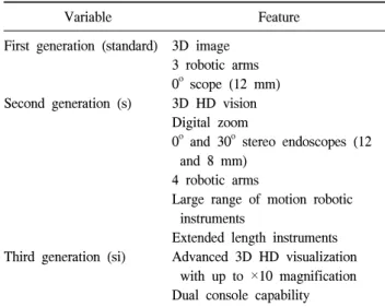
Table 1. Main features of the three generations of robotic system
Variable Feature
First generation (standard)
Second generation (s)
Third generation (si)
3D image 3 robotic arms 0
oscope (12 mm) 3D HD vision Digital zoom
0
oand 30
ostereo endoscopes (12 and 8 mm)
4 robotic arms
Large range of motion robotic instruments
Extended length instruments Advanced 3D HD visualization
with up to ×10 magnification Dual console capability 3D, 3-dimensional.
the da Vinci Surgical System was developed by Intuitive Surgical (Sunnyvale, CA, USA) and cleared by the FDA for laparoscopy, thoracoscopy and intracardiac mitral valve repair.
Currently, the da Vinci Robotic system is the only complete surgical system applied in a wide range of surgical procedures.
The aim of this review is to evaluate the application of ro- botic system in the field of thoracic surgery, in particular, of robotic lobectomy, analysing the perioperative and long-term outcomes. A systematic review of the literature was per- formed by accessing the MEDLINE database for entries from 1990 through January 2014. We selected and reviewed rele- vant original articles and case reports published in English language, excluding abstracts, and the reference lists from those sources were searched for additional trials. Data inves- tigation included different techniques, patients’ selection cri- teria, operative time, conversion rate, mortality, morbidity, postoperative stay, and oncologic results. Additionally, we fo- cused on the costs management of the robotic system.
ROBOTIC SYSTEM
The robotic system consists in a master remote console, in a computer controller, and in a manipulator with fixed remote centre kinematics connected via electrical cables and optic fibres. The camera used in the system provides a true stereo-
scopic picture (3-dimensional) transmitted to a surgeon's console. The master console is connected to the surgical ma- nipulator with the camera arm and three instrumental arms.
The surgeon manipulates two master handles and the move- ments are transmitted to the tips of the instruments thanks to trigger highly sensitive motion sensor, which is able to filter up to 6 Hz of the surgeon hands tremor. The surgical arm cart provides three degrees of freedom (pitch, yaw, and in- sertion), while the tip of the instrument is characterized by a mechanical cable-driven wrist (EndoWrist), providing four more degrees of freedom (internal pitch, internal yaw, rota- tion, and grip). These seven degrees of freedom of the in- strumentation allow to replicate the human wrist inside the chest cavity. Three generation of robotic systems (standard, s, and si) have been developed during the last decade (Table 1).
Recently the first generation was discontinued.
TECHNIQUES
Different surgical techniques were described over these years. The number of ports used in each study, as well as the size of the access port/additional incision varied between in- stitutions and sometimes within an institution in different time periods. Herewith we briefly report the different techni- ques used by several authors. Gharagozloo et al. [10] de- scribed a hybrid technique: three robotic arms are positioned at the 8th (camera), 6th, and 5th intercostal space for the dis- section of hilar structures. After the dissection phase, the ro- bot is removed, and the surgeon returns to the operating table for vascular, bronchial and parenchymal division [11,12].
Ninan and Dylewski [13] described a robotic lobectomy with
three arms: the robotic camera port is placed in the 5th or
6th intercostal space, directly over the mid-fissure area. The
two other ports are placed in the same intercostal space ante-
riorly and posteriorly, in order to avoid multiple intercostal
neurovascular bundles. An utility port is inserted over the
11th rib and bluntly tunneled over the 9th rib, entering in the
chest cavity through the 8th intercostal space [13]. Also Jang
et al. [14] used a 3 arms-technique with the camera placed in
the 7th intercostal space along the midaxillary line, another
arm placed in the 6th or 7th intercostal space along the pos-
terior axillary line and the other one positioned through a
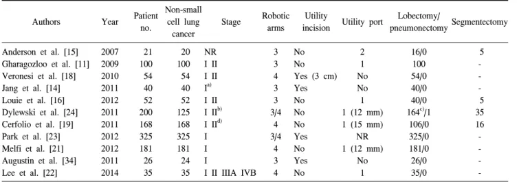
Table 2. Pantient’ features and surgical details of robotic procedures
Authors Year Patient
no.
Non-small cell lung
cancer
Stage Robotic arms
Utility
incision Utility port Lobectomy/
pneumonectomy Segmentectomy
Anderson et al. [15]
Gharagozloo et al. [11]
Veronesi et al. [18]
Jang et al. [14]
Louie et al. [16]
Dylewski et al. [24]
Cerfolio et al. [19]
Park et al. [23]
Melfi et al. [21]
Augustin et al. [34]
Lee et al. [22]
2007 2009 2010 2011 2012 2011 2011 2012 2012 2011 2014
21 100 54 40 52 200 168 325 181 26 35
20 100 54 40 52 125 168 325 181 24 35
NR I II I II I
a)I II I II
b)I II
d)I I I
I II IIIA IVB 3 3 4 3 3 3/4
4 3/4
4 3 4
No No Yes (3 cm) Yes No No No Yes No Yes No
2 1 No No 1 1 (12 mm) 1 (15 mm)
NR 1 (12 mm)
No 1
16/0 100 54/0 40/0 40/0 164
c)/1
106/0 325/0 181/0 26/0 35/0
5 - - - 5 35 16 - - - - NR, not reported.
a)
Three patients with solitary recurrence after chemo-radiotherapy included.
b)
Five patients underwent neoadjuvant chemotherapy for locally advanced IIIA disease.
c)
Including 4 bilobectomy, 3 sleeve lobectomies, and 3 en bloc lobectomies.
d)