진구성 척추 골다공증성 압박 골절과 경피적 척추 성형술 후 인접분절 척추 압박골절의 비교 분석 - 예비보고 -
원광대학교 의과대학 정형외과학교실, 마취통증의학과교실1, 응급의학과교실2
채수욱․김영진․양정환․이지완․추지웅․최덕화1․위대한2
Comparison of Adjacent Spinal Compression Fracture after Old Osteoporotic Spinal Compression Fracture and Percutaneous Vertebroplasty - A Preliminary Report -
Soo Uk Chae, Yeung Jin Kim, Jung Hwan Yang, Ji Wan Lee, Ji Woong Choo, Deok Hwa Choi1, Wi Dae Han2
Department of Orthopedic Surgery, Department of Anesthsiology and Pain Medicine1, Department of Emergency medicine2, School of Medicine, Wonkwang University, Iksan, Korea
Objectives: New adjacent spinal compression fractures occur not only in patients with osteoporotic spinal compression fractures after vertebroplasty but also in untreated old spinal compression fractures. The objective was to analyze and compare the characteristics in a group who had untreated old osteoporotic spinal compression fractures with those who had adjacent spinal compression fractures, which occurred after a vertebroplasty.
Materials & Methods: From April 2006 to April 2009, 103 patients who had undergone vertebroplasty were evaluated. Eighteen patients (22 cases) who had untreated old osteoporotic spinal compression fractures were in group I, and 12 patients (13 cases) who had adjacent spinal compression fractures, which occurred after a vertebroplasty were in group II. In each group, we measured age and gender, body mass index (BMI), lumbar bone marrow density (BMD), sagittal index, and compression rate at the initial fracture site.
Results: The mean age of group I was 76.7–years-old (males/females: 5/13), and that of group II was 78.7–years-old (males/females: 6/6). The most common fracture site was T12 (36%) in group I, and the most common site was L1 (54%) in group II. The mean BMI of group I was 23.3 and that in group II was 19.8. The mean t-score of group I was -4.59 and that of group II was -3.99. The mean compression rate and sagittal index of group I was 30.7% and 24.5°, respectively and those in group II were 32.1 % and 26°, respectively. No differences in sagittal index or compression rate were observed between the two groups.
Conclusions: Patients require continuous education, exercise, and osteoporotic medication after an osteoporotic spinal compression fracture. In particular, after a vertebroplasty, patients must wear an orthosis, and in elderly patients with a low BMD, it is important to continually observe for an adjacent spinal compression fracture.
Key Words: Adjacent fracture, Osteoporosis, Compression fracture, Vertebroplasty, Bone marrow density
Received: August 24, 2010 Revised: September 10, 2010 Accepted: October 25, 2010
Corresponding Author: Jung Hwan Yang, Department of Orthopeadic Surgery, School of Medicine, Wonkwang University, 344-2, Shinyong-dong, Iksan, 570-711, Korea
Tel: +82-63-472-5100, Fax: +82-63-472-5688, E-mail: [email protected]
* 본 논문은 2010학년도 원광대학교의 교비지원에 의해서 수행됨.
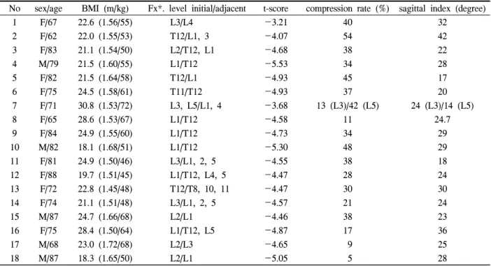
No sex/age BMI (m/kg) Fx*. level initial/adjacent t-score compression rate (%) sagittal index (degree)
1 F/67 22.6 (1.56/55) L3/L4 -3.21 40 32
2 F/62 22.0 (1.55/53) T12/L1, 3 -4.07 54 42
3 F/83 21.1 (1.54/50) L2/T12, L1 -4.68 38 22
4 M/79 21.5 (1.60/55) L1/T12 -5.53 34 28
5 F/82 21.5 (1.64/58) T12/L1 -4.93 45 17
6 F/75 24.5 (1.58/61) T11/T12 -4.93 37 20
7 F/71 30.8 (1.53/72) L3, L5/L1, 4 -3.68 13 (L3)/42 (L5) 24 (L3)/14 (L5)
8 F/65 28.6 (1.53/67) L1/T12 -4.58 11 24.7
9 F/84 24.9 (1.55/60) L1/T12 -4.73 34 29
10 M/82 18.1 (1.68/51) L1/T12 -5.30 48 29
11 F/81 24.9 (1.50/46) L3/L1, 2, 5 -4.55 38 18
12 F/88 19.7 (1.51/45) L1/T12, L4, 5 -4.47 28 24
13 F/72 22.8 (1.45/48) T12/T8, 10, 11 -4.47 30 30
14 F/74 21.1 (1.51/48) L3/L1, 2, 5 -4.57 21 24
15 M/87 24.7 (1.66/68) L2/L1 -4.46 38 23
16 F/75 28.4 (1.50/64) L1/T12, L5 -4.87 17 36
17 M/68 23.0 (1.72/68) L2/L3 -4.65 9 25
18 M/87 18.3 (1.65/50) L2/L1 -5.05 5 28
* Fx: fracture.
Table 1. Patients developed adjacent new compression fracture has previous old compression fractures who not undergone vertebroplasty
골다공증성 척추 압박골절의 치료로 침상 안정 및 골다공증 치료 약물을 포함한 약물 치료와 흉요천추 보조기 등의 일반적인 보존적 치료를 시행하나, 1990년대 초부터 최소 침습적인 방법으로 경피적 척 추 성형술이 골다공증성 척추 압박골절에 적용되면 서 치료에 한 방법으로 시행되고 있으며, 2000년도 초부터 풍선 성형술이 도입되어 시행되고 있다. 통 증의 감소와 조기 보행을 가능하게 하며, 골절 척추 추체의 높이 회복과 척추 시상면을 회복시켜 후만 변형을 방지하는 등의 여러 가지 장점이 있으나 시 술에 관련된 합병증으로 시멘트 유출과 관련된 신경 손상, 정맥 내 시멘트 유출에 의한 색전증, 감염이나 인접 추체의 새로운 골절 등의 단점이 보고되고 있 다. 특히 풍선 성형술 후 인접분절의 골절은 3~29%
의 발생률이 보고되고 있고, 인접분절 골절 발생 시 질환의 유병 기간이 길어져 이에 대한 예방과 치료 에 대해서 많은 논란이 되고 있다.1-3 이러한 골다공 증성 척추 압박골절의 경피적 척추 성형술 후 새로 운 인접분절 골절이 합병증으로 발생하여 관심이 높 아지고 있으나, 시술하지 않은 진구성 척추 골다공
증성 압박 골절 후에도 새로운 인접분절 골절이 발 생하기에 이 두 군을 골다공증 검사 결과와 함께 각 각의 특성 및 발생 인자에 대해서 비교 통계 분석하 여 예비 보고하고자 한다.
대상 및 방법
2006년 04월부터 2009년 04월까지 본원에 내원한 흉요추부 골절 환자 538명중 풍선 성형술을 포함한 경피적 척추 성형술을 시행한 103명의 환자를 대상으 로 하였다. 이중 골다공증 척추 압박골절 후 새로운 골절이 발생한 환자 중에서 인접분절이 아닌 떨어진 (remote) 골절 환자를 제외한 환자 30명 중, 척추 성형 술 등의 시술을 시행한 적 없는 진구성 골다공증 척 추 압박골절 후에 발생한 인접분절 골절 환자 18명, 22례를 I군으로(Table 1, Fig. 1), 경피적 척추 성형술 후 발생한 인접분절 골절 환자 12명, 13례를 II군으 로 분류하였다(Table 2, Fig. 2). 각 군의 전 환자들의 나이와 신장, 체중을 이용하여 신체 질량지수(BMI:
kg/m2)를 측정하여 골밀도 수치와 상관관계를 보정하
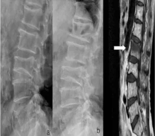
Fig. 1. The 78 years old women who had previous old L3 compression fracture (a) developed new L2 compression fracture (b) in lumbar spine lateral x-ray view and MRI (T1-weighted image) showed compression fracture of L2 with loss of height (white arrow).
No. sex/age BMI (m/kg) Fx*. level initial/adjacent t-score compression rate (%) sagittal index (degree)
1 M/84 21.5 (1.60/58) L1/T12, L2 -2.92 40 26
2 F/72 14.9 (1.64/40) L2/L1 -2.25 62 23
3 F/87 16.9 (1.53/38) L2/T11 -4.19 27 17
4 M/83 20.4 (1.73/61) T12/L1 -3.30 30 29
5 M/79 18.4 (1.65/50) T12/L1 -5.53 34 28
6 M/87 14.9 (1.64/40) L1/L2 -5.05 38 23
7 F/88 22.9 (1.45/48) T11/L1 -4.47 28 24
8 F/71 16.9 (1.50/38) T12/L1 -4.39 48 29
9 F/66 28.4 (1.50/64) L1/L2 -4.27 20 29
10 M/78 23.2 (1.70/67) L2/L1 -5.82 16 33
11 F/72 20.0 (1.50/45) L1/T12 -4.14 21 28
12 M/78 18.9 (1.51/43) L2/L3 -5.82 21 23
* Fx: fracture.
Table 2. Patients developed adjacent new compression fracture who undergone previous undergone vetebroplast Fig. 2. The 72 years old women who had previous
undergone kyphoplasty on L1 (a) developed adjacent new compression fracture T12 (b) in lumbar plain lateral x-ray view and MRI (T1- weighted image) showed compression fracture of T12 with loss of height (white arrow).
였고, 과거력상 당뇨병, 호흡기 질환, 고혈압 등의 심 장질환 등의 전신 질환 유무와 흡연이나 스테로이드 계 약물 투여 등의 과거력을 검토하였다. 요추부 골 다공증 검사(BMD)는 P-QCT (sensation 16-channels, SIEMENS, Germany)를 이용하여 흉추 12번부터 요추 5번까지에서 진구성 골다공증성 압박골절이 있는 분 절이나 척추체 성형술을 시행한 부위를 제외하여, 척 추 분절 4부위의 각각 망상골과 피질골의 골밀도 측
정값을 평균으로 하여 골밀도와 함께 t-점수(20세 기 준), z-점수를 시행하였다. 요추부 측면 기립 단순 방 사선 상으로 최초 골절부를 중심으로 Cobbs 방법을 이용하여 시상면 지수(sagittal index, degree angle)와 골절의 압박률(compression ratio, %)을 2명의 다른 측 정자가 2번씩 독립적으로 반복 측정하였다. 풍선 성 형술의 시술 방법으로 환자를 복와위로 취한 후 C-형 영상 증폭장치를 이용하여 양측 척추경 도달법으로
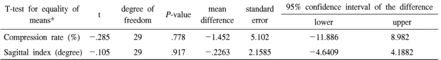
T-test for equality of
means* t degree of
freedom P-value mean difference
standard error
95% confidence interval of the difference
lower upper
Compression rate (%) -.285 29 .778 -1.452 5.102 -11.886 8.982
Sagittal index (degree) -.105 29 .917 -.2263 2.1585 -4.6409 4.1882
* Indepentent sample test with levene’s test for equality of variance.
Table 3. Correlation with compression rate and sagittal index between two groups
골시멘트를 주로 척추체 상판 부위 골절부에 집중하 여 약 3~6 cc 가량을 주입하였다. 술 후 4시간 후부 터 흉요천추 보조기를 착용하여 보행하였다.
통계분석 방법으로 각각의 측정된 평균값을 각 군의 차이의 비교를 Levene의 등분산 검정(SPSS version 18.0 for Windows)으로 독립 표본 t-검정으로 각 군의 유의성을 분석하였으며, Pearson 상관 계수를 이용하 여 상관관계를 분석하였다.
결 과
I군의 평균 연령은 76.7세(남자 5명, 여자 13명)이 며, II군의 평균 연령은 78.7세(남자 6명, 여자 6명)이 다. 대상 환자 중에서 I군의 골절 발생률은 17.4%이 며, II군은 11.8%였으며, 경미한 충격이나 낙상 등의 외상에 의해 발생하였다. 인접분절 골절의 발생 시 기는 수주부터 수개월까지 다양하게 보였다. I군의 최초 골절 요추 1번(7례, 37%)으로 가장 많았고, II군 의 골절은 요추 1번과 요추 2번이 각각 4례(33%)였 다. I군의 인접분절 골절 부위는 흉추 12번(36%)이 가장 많았고, 요추 1번(32%) 순이었다. II군의 인접 분절 골절은 요추 1번(54%), 요추 2번(23%) 순이었 다. 대상 환자 모두 척추 성형술을 시행 후 VAS 점 수 평균 4 이상의 증상 호전이 보였다. 신체 질량지 수의 평균은 I군 23.3, II군 19.8이었다. 요추부 골다 공증 점수(T-score) 평균은 각각 -4.59, -3.99이며 모두 중증의 골다공증 소견이었으며, BMI와 Pearson 상관계수에 의한 분석 상 유의한 상관관계를 보이지 않았다. 골절 압박률 및 시상면 지수는 I군에서 30.7
%, 24.5도이며, II군에선 32.1%, 26도로, Levene의 등 분산 검정에서 P-값이 각각 0.616, 0.213으로 각 군 간에 통계적 유의한 차이를 보이지 않았다(Table 3).
고 찰
골다공증성 척추 골절은 골다공증에 의한 병적 골 절로서 가벼운 충격에 의해서 쉽게 골절이 일어날 수 있는 상태를 의미한다. 골다공증에 의하여 대퇴 골 근위부, 요골 원위부, 상완 근위부 등 여러 부위 골절이 발생할 수 있고 그 중 척추 골절이 차지하는 비율이 전체 골절의 50% 이상을 차지할 정도로 그 의의가 매우 높다. 그 치료로 침상 안정, 보조기 치 료, 약물 치료 등 보존적 요법과 풍선 성형술을 포함 한 경피적 척추 성형술, 기기 고정 및 척추 유합술 등 수술적 치료 등이 행해지고 있다. 특히 수술적 치 료 방법 중 풍선 추체 확장 성형술은 풍선을 이용하 여 무너진 척추체를 회복하여 후만각과 시상면을 유 지하며, 통증을 감소시키고, 조기 보행을 가능하게 하여 현재 널리 사용되고 있는 치료 방법이다.4,5 그 러나 시멘트가 척추체외로 유출되어 혈관 색전증을 유발하거나 신경 손상 문제를 야기할 수 있고, 인접 분절 추체의 추가 골절을 야기하는 단점이 있다. 인 접 척추의 압박골절의 발생은 척추 성형술 후 52%
까지 발생률이 보고되며, 풍선 성형술 후 약 3~29%
까지 다양하게 보고하고 있다.6-9 Lindsay 등10은 폐경 후 골다공증 여성에서 1년 이내에 새로운 골절이 일 어날 가능성이 381명 중 19.2%가 일어난다 하였으 며, 현재까지의 문헌에서 대부분의 풍선 성형술 후 새로운 골절이 2개월 이내에 발생하는 것으로, 이는 척추 성형술 후 골 시멘트로 강화된 분절의 강도가 커지면서 기둥효과에 의해 인접 상하 분절로 더 큰 응력이 집중으로 인해 발생한다 하였다.7-9 본 연구 에서도 풍선 성형술 후 인접 상하 분절의 새로운 압 박골절이 발생하여 다시 척추 성형술을 시행한 경우 가 11.8%였다. 그러나 인접 분절 압박 골절의 발생
은 척추 성형술등의 시술을 시행한 적 없는 진구성 골다공증성 척추 압박 골절을 가진 환자에서도 새롭 게 인접 추체에 압박 골절이 야기되기도 한다. 위험 인자로 폐경 후 고령환자, 낮은 골밀도, 성별, 인종 이 있다. 특히 낮은 골밀도를 가진 골다공증성 척추 압박 골절 환자에서는 새로운 척추 압박 골절이 발 생할 확률이 그렇지 않은 환자보다 위험도가 4배 이 상된다고 보고되기도 하였다.11-15 Lindsay 등10은 골 다공증 검사 상 골다공증으로 진단된 여성 환자 중 10년간 추적 관찰 중 55%가 골다공증성 척추 골절 이 발생하였으며, Cauley 등12은 척추 골절이 없는 환자 중 15년간 추적 관찰 중 23.3%에서 28.3%가 척 추 골절이 발생하였다 하였다.
여러 보고에서 척추 성형술 후 새로운 인접 분절 의 골절의 발생률 및 영향을 주는 요소 등에 대하여 다양한 보고들이 있지만,2,6,8,9 인접분절의 골다공증 성 척추 압박 골절환자에서 척추 성형술 후와 척추 성형술을 시행한 적 없는 진구성 압박 골절의 주요 인자 및 차이에 관한 연구에 대하여 알려져 있지 않 다는 점에서 본 연구가 의의가 있다 하겠다. 새로운 인접 분절 골절을 야기하는 데 있어서 이전 척추 성 형술 유무가 위험인자로 작용하지만 그 중요도와 기 여도에서 있어서는 진구성 압박 골절을 가진 환자에 서의 새로운 인접 분절 발생률과 큰 차이를 보이지 않았다. 발생 연령 및 T-score, 평균 압박률과 시상면 지수에서도 통계학적 차이를 보이지 않았으며, 이전 척추 성형술 유무가 인접 분절 압박골절의 발생 시 기에 영향을 미칠 수 있으며, 진구성 압박 골절과 마 찬가지로 새로운 인접 분절 골절의 위험 요소가 될 수는 있으나 중요도 및 발생률에 있어서는 더 큰 의 미를 지닌다고 할 수는 없다는 것이다.
본 연구가 후향적 연구이며, 환자의 수가 많지 않 다는 점, 골절이 발생하여 병원에 내원한 환자 중에 서 추체 성형술 및 풍선 성형술을 시행했던 환자를 대상으로 하였고, 추적 기간 동안 병원에 내원하여 순차적으로 시술을 받은 환자를 대상으로 하였기에 이러한 요소들이 인접 분절 골절률의 조사에 영향을 주었을 가능성이 있고, 고령 환자가 많기에 정기적 추적 검사가 용이하지 않았다는 점과 사망 등으로 인하여 추적 검사 중단 등이 있었다는 점은 이 연구
의 제한점이라 할 수 있다. 전향적으로 지속적인 대 상군을 늘려 포괄적인 연구가 필요하다.
본 연구에서는 골다공증성 압박 골절을 가진 환자 들 중에서, 척추 성형술을 시행한 환자에서 여러 원인 들에 의해 인접 분절에 새롭게 척추 압박 골절이 발생 한 경우와 척추 성형술을 시행한 적 없는 진구성 압박 골절 환자에서 인접 분절에 새롭게 척추 압박 골절이 발생한 경우를 비교 분석하였다. 두 군에서 인접분절 골절 발생에 대한 통계학적으로 의미 있는 차이가 관 찰되진 않지만, 골다공증성 척추 압박 골절 후 지속적 인 환자 교육과 운동 및 골다공증 치료가 요하며, 특히 척추 성형술 후 보조기 착용과 고령 및 골밀도가 낮은 경우 인접분절 압박골절 발생에 대해 지속적인 관찰 및 예방이 필요할 것으로 사료된다.
참 고 문 헌
1. Ahn DK, Lee S, Choi DJ, Park HS, Kim KS, Kim TW. The efficacy of kyphoplasty on osteoporotic vertebral compression fracture–A 1-year follow-up study-. J Korean Soc Spine Surg 2009;16:79-88.
2. Fribourg D, Tang C, Sra P, Delamarter R, Bae H.
Incidence of subsequent vertebral fracture after kyphoplasty. Spine 2004;29:2270-6.
3. Kim CH, Choi YJ, Hwang JK, Kim KH, Lee JH, Song JS. Long term outcome of vertebroplasty in the treatment of osteoporotic compression fracture.
J Korean Soc Spine Surg 2005;12:69-74.
4. Bouza C, Lopez T, Magro A, navalpotro L, Amate JM. Efficacy and safety of ballon kyphoplasty in the treatment of vertebral compression fractures:
systemic review. Eur spine J 2006;15:1050-67.
5. Chan JH, Peh WC, Tsui EY, Chau LF, Cheung KK, Chan KB, et al. Acute vertebral body compre- ssion fractures: discrimination between benign and malignant causes using apparent diffusion coeffi- cients. Br J Radiol 2002;75:207-14.
6. Berlemann U, Ferguson SJ, Nolte LP, Heini PF.
Adjacent vertebral failure after vertebroplasty. A biomechanical investigation. J Bone Joint Surg
2002;84B:748-52.
7. Harrop JS, Prpa B, Reinhardt MK, Lieberman I.
Primary and secondary osteoporosis incidence of subsequent vertebral compression fractures after kyphoplasty. Spine 2004;29:2120-5.
8. Tseng YY, Yang TC, Tu PH, Lo YL, Yang ST.
Repeated and multiple new vertebral compression fractures after percutaneous transpedicular vertebro- plasty. Spine 2009;34:1917-22.
9. Tanigawa N, Komemushi A, Kariya S, Kojima H, Shomura Y, Omura N, et al. Relationship between cement distribution pattern and new compression fracture after percutaneous vertebroplasty. AJR 2007;189:348-52.
10. Lindsay R, Stuart LS, Cooper C, Hanley DA, Barton I, Broy SB, et al. Risk of new vertebral fracture in the year following a fracture. JAMA 2001;17:320-3.
11. Cauley JA, Palermo L, Vogt M, Ensrud KE, Ewing
S, Hochberg M, et al. Prevalent vertebral fractures in black women and white women. J Bone Miner Res 2008;23:1458-67.
12. Cauley JA, Hochberg MC, Lui LY, Palermo L, Ensrud KE, Hillier TA, et al. Long-term risk of incident vertebral fractures. JAMA 2007;298:2761-7.
13. Melton LJ lll, Atkinson EJ, Cooper C, O’Fallon WM, Riggs BL. Vertebral fractures predict subsequent fractures. Osteoprosis Int 1999;10:214-21.
14. Ross PD, Davis JW, Epstein RS, Wasnich RD.
Preexisting fractures and bone mass predict vertebral fracture incidence in women. Ann intern Med 1991;114:919-23.
15. Klotzbuecher CM, Ross PD, Landsman PB, Abbott TA, Berger M. Patients with prior fractures have an increased risk of future fractures: a summary of the literature and statistical synthesis. J Bone Miner Res 2000;15:721-39.