The Journal of the Korean Society for Surgery of the Hand VOLUME 7, NUMBER 2, DECEMBER 2002
External Fixation with Distraction for Distal Radius Fracture
Myung-Hwan Son, M.D., Byung-Cheol Kim, M.D., Nam-Wook Kang, M.D., Dae-Cheol Jeon, M.D.
Department of Orthopaedic Surgery, Busan Medical Center, Busan, Korea
P u r p o s e : To evaluate the clinical and radiologic results in the using of external fixation with distraction for distal radius fractures.
Materials and Methods : We retrospectively analysed 25 patients who have taken operative treatment with external fixation for distal radius from 1995 to 2000.
They had been followed for average of 21 months (range, 12 to 38 months). Average age was 55.84 years (range, 24 to 84 years). The patients were evaluated by Frykman classification for fracture type, and by Demerit point sys- tem for clinical results. We also analysed the amount of distraction by carpal height ratio. Radiologic results were obtained by anteroposterior and lateral view of preopera- tive, postoperative, and final result.
Results : The clinical results were excellent in 5 cases, good in 15 cases, fair in 3 cases, and poor in 2 cases. The radiologic results were the averaged radial length, 10.1 mm (range, 8 to 12 mm), the averaged radial inclination, 19.4 degree (range, 14 to 28 degree), the averaged volar
inclination, 6.1 degree (range, 0 to 12 degree), and the averaged step-off, 0.9 mm (range, 0 to 4 mm). The degree of distraction was averaged 0.59 (range, 0.53 to o.67). No significant correlation was found between amount of distraction and final result. A significant cor- relation was seen between severity of fracture and final result.
C o n c l u s i o n s : External fixation with distraction for distal radius fracture is a satisfactory method of treating severe fractures.
Key Words : Distal radius, Fracture, External fixation, Distraction
서 론
요골 원위부 골절은 성인에서 흔히 발생하는 골절로서 특히 노년기에는 낙상으로, 중년층에서는 고에너지 손상 이 주요한 원인이다. 과거에는 치료방법에 관계없이 비 교적 좋은 예후를 보이는 것으로 받아들였으나1 , 4 , 1 2 , 1 3 ), 생활구조의 복잡화, 교통사고 및 산업재해 등으로 인한 고에너지 손상의 빈도가 증가하여 분쇄골절 등 불안정성 골절이 증가하고 있어 단순한 보존적인 차원을 넘어 적 극적인 치료가 필요하게 되었다.
이에 저자들은 요골 원위부 골절에 있어서, 외고정 장 치로 신연에 의한 치료를 수행하고 이에 대한 결과를 임 상적 및 방사선학적으로 분석하고자 하였다.
연구대상 및 방법
1 9 9 5년 1월부터 2 0 0 0년 1 2월까지 5년간 부산의료 원 정형외과에서 요골 원위부 골절로 치료했던 환자중 외고정 장치를 이용하여 수술을 시행받은 3 1례에서 1년
요골 원위부 골절의 신연에 의한 외고정적 치료
지방공사 부산의료원 정형외과 손명환・김병철・강남욱・전대철
통신저자 : 손 명 환
부산광역시 연제구 거제2동 1330 지방공사 부산의료원 정형외과
TEL : 051-607-2862, FAX : 051-507-3001 E-mail : osmhwon@hotmail.com
본 논문의 요지는 대한정형외과학회 부산, 울산, 경남지회 월례집담회에서 구연되었음
이상 추시가 가능하였던 2 5례( 2 5명)을 대상으로 하였 으며, 추시기간은 최단 1년에서 최고 3년 2개월이었고 평균 2 1개월이었다. 성별은 남자가 1 1례, 여자가 1 4례 이었으며, 연령은 2 4세부터 8 4세까지 평균 5 5 . 8 4세였 다. 수상원인은 낙상 1 1례, 추락사고7례, 교통사고 5 례, 압궤손상 2례로 고에너지 손상이 많은 부분을 차지 하였다. 동반손상은 1 4례( 5 6 % )에서 발생하였으며, 척 추의 동반 골절이 6례로 가장 빈도가 높았다.
골절의 분류는 F r y k m a n에 의한 분류법을 이용하였으 며, 전체 2 5례중 7형 1 0례( 4 0 % )로 가장 많았으며, 8형 6례(24%), 4,5형이 각각 3례(12%) 등의 순서였다.
수술은 도수 정복 후 방사선 사진상 정복의 만족성에 있어 2 mm 이상의 요골 단축이 있거나, 2 mm 이상의 관절면의 전위가 있는 경우 혹은 2 0도 이상 후방 경사가 있는 불안정 골절과 심한 분쇄상 골절 및 심한 골다공증 등에 외고정 장치로 신연을 이용한 인대 정복( l i g a- m e n t o t a x i s )에 의한 골절 정복을 시도하였으며, 영상 증폭장치로 골절부를 확인하여 관절면 정복이 만족스럽 지 못한 경우 필요에 따라 K 강선을 이용하여 제한된 내 고정술을 시행하였다. 신연의 정도는 전후면 방사선 상 에서 측정한 수근 높이 비(carpal height ratio)1 6 )를 이용하였다.
임상적 결과는 Demerit point system6 , 1 4 )을 이용하 여 우수(0,1,or 2 demerit-points), 양호(3~8), 보 통(9~20), 불량(21 이상)으로 평가하였다. 방사선적 인 계측 결과는 술전, 술후 및 최종 추시시의 수근 관절
며, K 강선은 8주에 제거하였다.
결 과
임상적으로는 우수 5례, 양호 1 5례, 보통 3례, 불량 2례로 80% 차지하는 2 0례에서 양호 이상의 결과를 보 였으며 Frykman 분류상 손상의 정도가 심할수록 결과 또한 좋지 못하였다(Table 1). 방사선학적인 계측 결과 는 평균 요골 길이의 감소가 0.9 mm, 평균 요측 경사 의 감소가 3 . 6°, 평균 전방 경사의 감소는 4 . 9°, 관절면 의 불일치는 0.9 mm로 나타났다. 또한 신연의 정도는 수근 높이 비가 평균 0 . 5 9 ( 0 . 5 3 - 0 . 6 7 )로 측정되었으 며 정상 범위인 0 . 5 4±0 . 0 3에 포함되는 경우, 우수 2 례, 양호 4례, 보통 1례로, 정상 범위 이상인 경우, 우수 3례, 양호 1 1례, 보통 2례, 불량 2례로 각각 나타났다 (Table 2). 통계처리를 위하여, 우수와 양호를 G r o u p 1, 보통과 불량을 Group 2로 나누고, 수근 높이 비가 정상과 그 이상으로 하여 χ2- t e s t로 분석하였다. 그 결과 통계학적으로 두 G r o u p간의 차이는 없었다 . (χ
2=0.656, p=0.564)
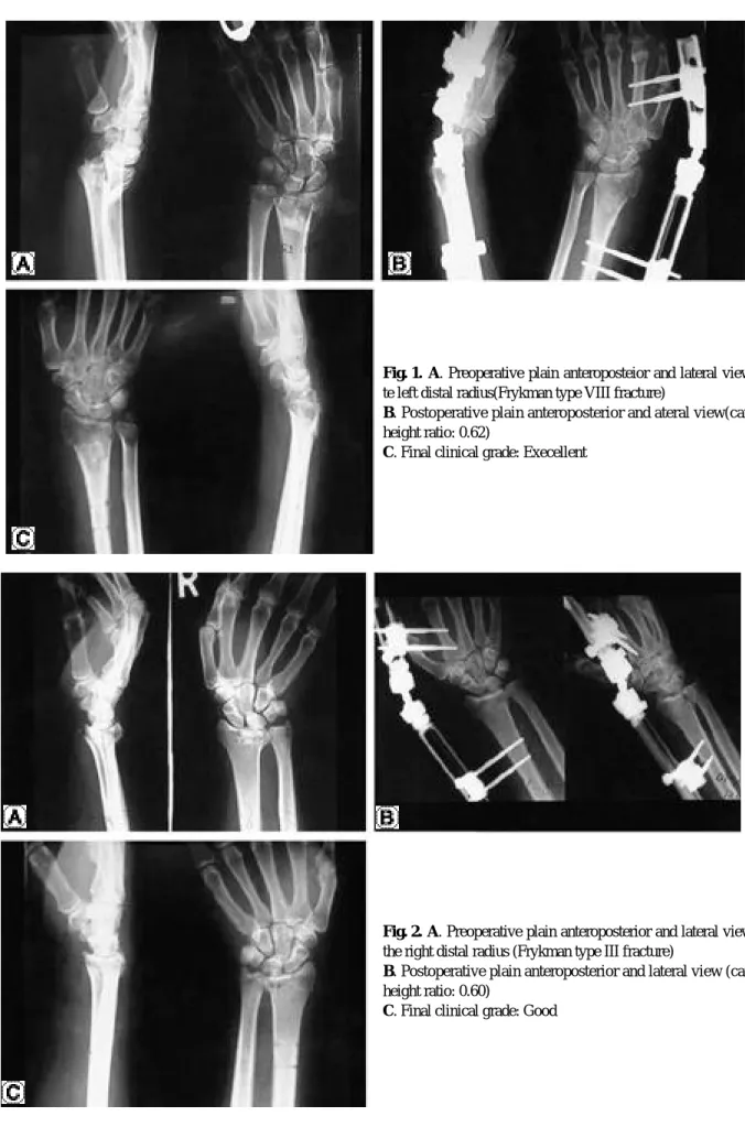
증례 1
4 7세 여자환자로 실족손상으로 Frykman 제8형이 며, 술후 수근 높이 비가 0 . 6 2로 나타났고 최종 추시 판 정시 우수의 결과를 보였다(Fig. 1).
증례 2
5 5세 남자환자로 낙상손상으로 Frykman 제3형이 며, 술후 수근 높이 비가 0 . 6 0으로 나타났고 최종 추시 판정시 양호의 결과를 보였다(Fig. 2).
고 찰
Table 1. Evaluation of Results with the modification, by Sarmiento et al., of the demerit-point scale of Gartland and Werley.
Fracture type III IV V VI VII VIII Total(%)
Excellent 1 2 1 1 5(20)
Good 1 2 1 1 7 3 15(60)
Fair 2 1 3(12)
Poor 1 1 2(8)
Total 1 3 3 2 10 6 25
Table 2. Comparison of carpal height ratio and results of the demerit-point scale.
Carpal height ratio normal (0.54±0.03) >0.57
Excellent 2(29) 3(17)
Good 4(57) 11(61)
Fair 1(14) 2(11)
Poor 2(11)
Total 7 18
(x2=0.656, p=0.564)
Fig. 1. A. Preoperative plain anteroposteior and lateral view of te left distal radius(Frykman type VIII fracture)
B. Postoperative plain anteroposterior and ateral view(carpal height ratio: 0.62)
C. Final clinical grade: Execellent
Fig. 2. A. Preoperative plain anteroposterior and lateral view of the right distal radius (Frykman type III fracture)
B. Postoperative plain anteroposterior and lateral view (carpal height ratio: 0.60)
C. Final clinical grade: Good
손상이 경미한 경우는 도수 정복 및 부목 고정이 좋은 치 료법으로 알려져 있다2 , 8 ). 그러나 요골 원위부 골절의 전 이된 골절은 전통적인 비수술적 방법에 의해서 성공적으 로 치료하기가 어렵게 되었다. 그래서 치료의 목표를 최 대한의 기능 복원, 강도의 유지, 외상후 관절염 발생의 최소화, 합병증 방지에 두고, 골절이 치유될 때까지 정복 을 획득하고 유지하여 수근관절의 재활을 수행하여 운동 과 강도를 복원하는 것이다. 이를 위해서 C o o n e y등3 ) 은, 방사선 소견상 후방 경사가 2 0도 이상, 심한 후방 골 피질의 분쇄, 10 mm 이상의 요골 단축이 있는 경우 를 불안정 골절로 보았고, Lafontaine 등1 0 )도 골절 정 복 후 불안정성에 관여하는 다섯 가지 인자로 2 0도 이상 의 후방 경사, 후방 골간단 분쇄(metaphyseal com- minution), 관절 내 손상, 동반된 척골 손상, 60세 이
으로 주장하였다. 불안정한 요골 원위부 골절의 정복 유 지는 석고 고정만으로는 어렵다는 것이 밝혀짐에 따라, 많은 저자들에 의해 경피적 핀 삽입, 핀 삽입과 석고를 이용한 고정, 외고정 장치의 이용, 관혈적 정복 및 내고 정, 관혈적 정복 및 내고정과 자가 장골 이식 그리고 외 고정 장치를 이용한 병합 요법 등이 시행되어 왔다.
요골원위부의 심각한 골절 치료에 외고정 장치의 사용 은 광범위하게 수용되고 있으며, 만족할만한 결과를 보 고하고 있다2 , 7 , 1 1 , 1 5 ). 하지만, 외고정 장치를 이용한 요골 원위부 골절의 치료에 있어서 신연의 정도를 명확히 제 시한 연구는 그리 많지 않다. Kaempffe 등9 )에 의하면, 신연의 증가는 골절 정복을 향상시켜서 방사선적 결과상 좋게 보이지만, 기능과 동통, 운동 범위, 악력 등에는 유 해하다고 하였으나 통계적 유의성을 나타내지는 못하였 Total Case
Case Gender Follow up
Type R.S (mm) decreased decreased
S.O (mm) C.H.R Result
/Age (months) R.I (degree) V.I (degree)
1 M/43 13 IV 0 5 1 0 0.56 Excellent
2 M/38 16 V 0 0 1 1 0.6 Excellent
3 M/52 18 VI 1 8 4 1 0.53 Good
4 M/27 12 VIII 2 3 7 1.5 0.6 Fair
5 M/51 17 VII 2 5 6 1.5 0.63 Fair
6 M/67 20 IV 1 3 4 1 0.59 Good
7 M/24 15 VIII 2 3 7 0.5 0.62 Good
8 M/36 18 VII 1 2 8 1 0.57 Good
9 M/52 12 VI 0 0 1 0.5 0.59 Excellent
10 M/55 20 III 1 2 4 1 0.6 Good
11 M/54 26 VIII 1 3 5 0.5 0.62 Good
12 F/70 32 VII 2 9 11 2 0.61 Poor
13 F/36 14 VII 1 5 8 1.5 0.57 Fair
14 F/47 38 VIII 0 1 1 0 0.62 Excellent
15 F/65 22 VII 1 3 4 0.5 0.6 Good
16 F/62 21 V 0 1 0 0 0.57 Excellent
17 F/80 13 VII 1 7 6 1 0.59 Good
18 F/76 23 VII 1 5 6 1.5 0.55 Good
19 F/58 28 VII 1 3 5 1 0.59 Good
20 F/72 29 IV 0 4 5 0.5 0.62 Good
21 F/68 21 VII 1 3 6 1 0.58 Good
22 F/64 34 VIII 2 5 9 3 0.64 Poor
23 F/84 12 VIII 1 3 4 0 0.58 Good
24 F/59 24 VII 0 3 5 0.5 0.54 Good
25 F/55 27 V 0 4 5 0.5 0.58 Good
* R.S Radial shortening
* R.I Radial inclination
* V.I Volar inclination
* S.O Step-off
* C.H.R Carpal-height ratio
본 연구에서도 요골 원위부 골절의 수술적 치료에 있 어서 신연의 정도와 최종 추시 결과 사이에는 그 결과에 차이가 없었다.
결 론
요골 원위부 골절에서 신연을 이용한 외고정술을 시행 하여 8 0 %에서 양호 이상의 결과를 얻었으며, 신연의 정 도와 최종 추시 결과 사이에는 그 결과에 차이가 없는 것 으로 나타났으며, 골절의 심각성과 오히려 깊은 관련성 이 있을 것으로 보이며, 제한된 례로 보다 많은 연구가 필요할 것으로 사료된다.
REFERENCES
01) Colles A : The classic: On the fracture of the carpal extremity of the radius.(Reprinted from the original 1814 article). Clin Orthop,71-A:839-847,1972.
02) Cooney WP : External fixation of distal radial fractures.
Clin Orthop,180:44-49,1983.
03) Cooney WP, Linscheid RL and Dobyns JH : External pin fixation for unstable Colles` fracture. J. Bone and Joint Surg.,61-A:840-845,1979.
04) Dupytren B : On the injuries and disease of bones. The Sydenham Society, London, 1847.
05) Fernandez DL and Palmer AK : Fractures of the distal radius. In Green DP (ed.: Operative hand surgery,vol1.
New York,Churchill-Livingstone,pp809-864,1999.
06) Garland JJ and Werley CW : Evaluation of healed
Colles’ fractures. J. Bone and Joint Surg.,33-A:895-907, 1951.
07) Jenkins NH, Jones DG, Johnson SR, Mintowt-Czyz WJ : External fixation of Colles’fractures. J Bone Joint Surg.,69-B:207-211,1987.
08) Jun-O Yoon and Ji-Chul Kim : Surgical treatment of the distal radius fracture. J Korean Orthop Assoc,30(5):1423- 1432,1995.
09) Kaempffe FA and Walker KM : External fixation for distal radius fractures-effect of distraction on outcome.
Clin Orthop.,380:220-225 ,2000.
10) Lafontaine M, Hardy D and Delince P : Stability assess- ment of distal radius fractures. Injury,20:208-210, 1989.
11) Leung KS, Shen WY, Leung PC, Kinninmonth AW, Chang JC and Chan GP : Ligamentotaxis and bone grafting for comminuted fractures of the distal radius. J.
Bone Joint Surg.,71B:838-842,1989.
12) Peltier LF : Eponymic fractures: Abraham Colles and Colles` fracture. Surgery, 15:322-328,1954.
13) Peltier LF : Eponymic fractures: Fractures of the distal radius. An historical account. Clin Orthop, 187:18-22, 1984.
14) Sarmiento A, Pratt GW, Berry NC and Sinclair WF : Colles` fractures. Functional bracing in supination. J. Bone and Joint Surg.,57-A:311-317,1975.
15) Seitz WH, Froimson AI, Leb R, Shapiro JD : Augmented external fixation of unstable distal radius frac- tures. Orthop Rev 20:169-177,1991.
16) Youm Y, McMurty RY, Flatt AE and Gillespie TE : Kinematics of the wrist. J Bone Joint Surg.,60-A:423- 431,1978.