- 104 -
Toxicity of Polylactic Acid Polymer in the Treatment of Paranasal Sinusitis*
Yang-Gi Min, M.D.
1, Young-Ki Kim, M.D.
2, Sea-Young Jeon, M.D.
3,,,,Ki-Sang Rha, M.D.
4and Seo-Young Jeong, Ph.D.
5 ABSTRACTThe authors, after inducing acute sinusitis in rabbits, compared cases where we opened the natural ostium and a general dose of administered antibiotics and cases where we opened the natural ostium and locally administered dose of antibiotics using polymer, and found that the application of local antibiotics using polymer is the superior of the two in treating sinusitis. To be able to develop a treatment for sinusitis using antibiotics incorporating polymer, we first need to examine the toxicity of polymer.
The purpose of this study was to examine the toxicity of polymer through a general toxicity test as well as a special toxicity test in experiments using animals. As a result of this study, we discovered that poly-L-lactic acid (PLA) polymer works as an in- traperitoneal foreign body and causes adhesion of viscera, but nethier acute nor subacute toxicity of PLA polymer was detected, and no negative effect on reproductive function was observed. It was also observed to induce neither immune reaction such as hypersensitivity nor local toxicity to the maxillary sinus mucosa of rabbit. We conclude that the results of this study can provide basic information for developing antibiotics-incorporating PLA polymer for the treatment of sinusitis and for clinical experiments involving such antibiotics.
KEY WORDS:Polylactic Acid Polymer·Treatment·Sinusitis.
INTRODUCTION
In the sinus, because of its limited blood flow1) and its co- vered with bone, much doubt is raised as to whether antibio- tics could infiltrate by secretion into the sinus when systemic antibiotics are administered.2) It is possible that treatment with systemic antibiotics could cause side effects. Therefore, local administration could be a more effective way of medical tre- atment. The conventional liquid medicine, however, only has a short period of contact with mucosa and does not stay in the nasal cavity and sinus area long enough. Moreover, medicine
that uses lipids or an organic ingredient as its solvent does not mix with mucus. It deteriorates ciliary movement of epithelial cells of nasal mucosa and has very low intratissue permeabi- lity.3) There are documented reports that treatment drugs-inc- orporating polymer are locally administered in the vagina,4) the central nervous system5) and the subcutaneous tissue6) for a relatively long period of time without side effects, and we had also already proved that the treatment of local antibiotics using polymer is superior in treating sinusitis through exper- imentally induced acute sinusitis in rabbits by comparing cases where we opened the natural ostium and aministered systemic antibiotics and cases where we opened the natural ostium and administered antibiotics locally using polymer.7)8) Henceforth, it is first necessary to examine the toxicity of polymer in order to develop a new treatment of sinusitis using antibiotics-inc- orporating polymer. In this study, we attempted to examine the toxicity of polymer through general toxicity tests as well as special toxicity tests by experiments in animals.
MATERIALS AND METHODS Production of Poly-L-lactic acid polymer
Sonication of 700 mg PLA for 30 minutes after dissolving Span 80 in 5 ml of CH2Cl2 in organic phase produced an em-
*Supported by grant 96-0403-11-01-1 from the Korea Science and Engineering Foundation.
1Department of Otorhinolaryngology, College of Medicine, Seoul National University, Seoul, 2Department of Otorhinolar- yngology, Inje University, Seoul, 3Department of Otorhinolar- yngology, Gyeongsang National University, Chinju, 4Depart- ment of Otorhinolaryngology, College of Medicine, Chungnam National University, Taejon, 5Biochemical Research Center, Korea Institute of Science and Technology, Seoul, Korea Address correspondence and reprint requests to Yang-Gi Min, M.D., Department of Otorhinolaryngology, College of Medicine, Seoul National University, Chongno-ku, Yongon-dong 28, Seoul 110-744, Korea
Tel:82-2-760-2448, Fax:82-2-745-2387 Accepted for publication November 10, 1997
ulsion. Then, through resedimenting it with ethyl ether, we pr- oduced poly-L-lactic acid polymer in sponge form and used it in the experiments.
Acute toxicity experiment
Out of 90 SPF ICR male mice weighing 20-35 g, taking 10 as a control group and the remaining of 80 as the experime- ntal group, we made 8 experiment groups of 10 mice each. We did the same experiments with female mice with these same conditions. Having measured polymer by the amounts shown in Fig. 1, we inserted it into the peritoneal cavity of the ana- esthetized mice. For the mice in the control group, we instilled phosphate buffered saline (PBS) into the peritoneal cavity.
We observed the mice for 30 minutes daily over the two week experiment period and recorded clinical appearance:
abnormal behavior, reactions to stimuli, changes of respiration, activity, diarrhoea and so on. We also measured the weight of the mice before the experiment and 1 and 2 weeks after the ex- periment respectively, and recorded deviations as percentages.
When the experiment ended, or when a mouse died, we did au- topsy and made a general examination of gross findings in the lungs, liver, spleen, heart, urogenital tract, gastrointestinal tract and so on. We recorded any abnormal findings and did a his- topathological examination of abnormal parts. We also made light microscopic examination after having stained them with Hematoxylin and Eosin (H&E). We included damage to hair, injury to ears and injury to the tail as subjects of general exa- mination.
Subacute toxicity experiment
For the subacute toxicity experiment, we used 64 SPF ICR male and female mice as animals for 13 weeks. To observe the whole body toxicity, we referred to the results of the acute toxicity experiment, and set the maximum amount of 1% sp- onge form polymer as 5 g/kg, and with a common ratio of 5, prescribed 1 g/kg and 200 mg/kg respectively for each of the three stages. In each experiment, having taken 16 male and fe- male mice, we divided them into four groups, i.e. three groups to which we administered polymer and one group to which we administered PBS. Three dosed groups were divided into the first group to which we administered 200 mg/kg of PLA po- lymer, the second to which we administered 1 g/kg of PLA polymer and the third to which we administered 5 g/kg of PLA polymer. Prescription of the amount of the medicine was based on the weight of the animal as measured before treatment, and after intraperitoneal instillation of the sponge form of polymer, we observed the animal for 13 weeks. During the period of the experiment, we checked the mice more than once a day to see
whether general symptoms, toxicity symptoms or death had occurred and recorded the results of observation. The weight of each experiment animal was measured immediately before the start of treatment and again once a week afterward. On the day when we measured the weight after treatment, we also me- asured the daily food intake of the animal. At the 3rd, 6th, 9th and 13th week after treatment we did a complete blood count and blood chemistry test, and sacrificed 4 male and female mice in order to make a general examination of them for ab- normal findings and recorded the results. We did a multiple organ biopsy of their lungs, kidneys, livers, brains, and peri- toneum, and fixing them with formalin, embedded them in paraffin. In addition, we fixed their tissue in 4% glutaraldeh- yde for examination under transmission electron microscopy to see whether polymer had accumulated inside the organ.
General fertility and reproduction
We took 10 normal male and female mice per group with 60 in total, to form experiment groups, and 10 normal male and female mice per group with 20 in total, as a control group.
Dosage amounts were based on 5 g/kg maximum amounts and we made an intraperitoneal instillation of antibiotic polymer of 200 mg/kg in the first group, 1 g/kg in the second group and 5 g/kg in the third group. As per dosage timing, we adminis- tered the polymer 60 days before mating in the male mice, and, in the female mice, by experiment group, we intraperitoneally instilled the amounts prescribed in three stages as in the ch- ronic toxicity experiment. For the control group, we used 10 male and female mice each, to which we administered only PBS. Then, we made a comparative examination of the ex- periment group with the control group, focusing on survival, general symptoms, mating rate, implantation rate, pregnancy, death of fetus, birth weight of fetus. In addition, for the sur- viving fetuses, we observed growth, hearing, reactions to vi- sual stimuli as well as general symptoms, and recorded the results.
Teratogenicity
For the experiment group, we took 30 pregnant rabbits and divided them into three groups of 10 each, and, for the control group, we took 10 pregnant rabbits. Based on the maximum amount of dosage prescribed in the acute toxicity experiment, we made an intraperitoneal insertion of PLA polymer in three respective stages:200 mg/kg in the first group, 1 g/kg in the second group, and 5 g/kg in the third group on the sixth day after conception.
We examined the survival rate, general symptoms, weight, and daily food intake of all the rabbits. On the 28th day of
pregnancy, 1/2 of each group was sacrificed for dissection in order to make a comparative examination for pregnancy, nu- mber of corpus lutea, death of fetus, weight of survived fetus, sex and the malformation of external and internal organs, the musculoskeletal system, and other viscus in comparison with the control group. We left the other 1/2 for natural parturition and fostered the fetus, and examined the abnormalities in the same way.
Perinatal and postnatal effects
For the experiment group, we took 30 pregnant rats and divided them into three groups with 10 per group, and for the control group, we took 10 pregnant rats. We set the experiment period from the 15th day of pregnancy to the 21st day after the beginning of breast feeding of the fetus. We did an intraperi- toneal insertion of the prescribed polymer on the experiment animals. For the control group, we instilled PBS only on the 15th day of pregnancy. Polymer was inserted, in the following stages:200 mg/kg in the first group, 1 g/kg in the second gr- oup, and 5 g/kg in the third group. After dosing, we watched for survival rates, general symptoms, weight, and the daily food intake of all the rats. We fostered all the groups till delivery, and examined for pregnancy period, death of fetus, death of the newborn, weight of the newborn, abnormality of external or- gans, the skeletal system of those stillborn, growth of the fetus, development of the fetus, abnormal symptoms of the fetus, the reproductive organs of the fetus, breast feeding of the mo- ther, and effects on the nurturing instinct of the mother rat. Then, we sacrificed them to do a thorough autopsy. The weight, su- rvival rate, and abnormal behavior of fetuses born in the ex- periment group were comparatively examined with those of fetuses born in the control group.
Local irritation
To evaluate local irritation by polymer in the maxillary si- nus, we used the maxillary sinus of rabbit. We divided 57 ra- bbits that did not appear to have sinusitis into two groups:
in the first group of 24 rabbits, we inserted 10 mg of polymer and in the second group of 24 rabbits, 25 mg of polymer was inserted:the control group consisted of 9 rabbits. Having re- moved the anterior bony wall of the maxillary sinus of the rabbits and confirming the normality of the maxillary sinus, we inserted sponge-form polymer that had been produced, th- rough the opened anterior bony wall. In the control group, we did not insert polymer, just resuturing cheek flaps instead. We sacrificed 8 experiment animals per group, with respective in- tervals of 2 weeks, 4 weeks, and 8 weeks, taking 3 from the control group as well. We examined for secretions in the ma-
xillary sinus and for general findings, and cut a sample of mucosa from the medial wall of the maxillary sinus for histo- pathological examination. We divided the appearances of the maxillary sinus, based on gross examination, into 5 stages;and recorded them on a primary mucosal irritation index (Table 2).
We recorded histologic findings from light microscopic exa- mination with focusing on loss of cilia, permeation of infla- mmatory cells, epithelial ulceration, edema and congestion and from transmission electron microscopic examination.
We recorded the changes in clinical symptoms daily throu- ghout the examination period to examine systemic side effects that could be caused by absorption of polymer through mucosa.
We took a blood sample from each experiment animal through the marginal vein located in the posterior surface of the auricle on the day of PLA insertion and after 2 weeks, 4 weeks, and 8 weeks. We did various blood tests:complete blood count, glucose test, kidney function test, liver function test, lipid ba- ttery test, and electrolyte test.
Immunotoxicity
Twenty-eight normal Hartley guinea pigs were divided into two groups, with 5 of them taken as a control group and 20 as an experiment group. Using 1 ml tuberculin syringe, we in- jected 0.1 ml of 1% polylactic acid polymer solution at the dorsum of 20 guinea pigs. Two weeks later, we again injected the same amount of PLA polymer at the same spot of the do- rsum to each group. Then, we observed for signs of swelling, inflammation, anaphylaxis and shock at the injection spot. The
Table 1. Groups of mice for acute toxicity experiments
Group No Dosage
Group 1 20 4 mg/kg
Group 2 20 10 mg/kg
Group 3 20 40 mg/kg
Group 4 20 100 mg/kg
Group 5 20 400 mg/kg
Group 6 20 1 g/kg
Group 7 20 4 g/kg
Group 8 20 6 g/kg
Control 20 PBS
Table 2. Grading of primary mucosal irritation index for local irritation experiment
Grade Findings 0 No inflammation, redness or petechia
1 Slighely redness, barely perceptible
2 Distinct redness with apparent vessels and a few petechia
3 Marked redness with traces of hemorrhage and/or marked edema
4 Severe injury with tissue erosion and necrosis
control group was injected with PBS and monitored for any changes.
Carcinogenicity
Four groups, two each of 60 male and 60 female mice and rats (i.e. 240 in total) between 1 and 2 months old, were taken as a control group. The 240 additional young mice and rats with the same conditions were taken as the experiment group.
Based on 5 g/kg of the maximum dosage we intraperitoneally inserted PLA polymer of 200 mg/kg in the first group of 120 mice and rats, and 1 g/kg in the second group of 120. We int- raperitoneally inserted PLA polymer in each experiment ani- mal after 2 months. The same amount of PBS was injected in the control group. While we fostered the experiment animals for a period of 4 months, upon the death of the animal, we took a sample of the main organs, and did an H&E stain on them.
We examined their carcinogenicity under light microscopy. Af- ter 4 months, we sacrificed all the surviving experimental an- imals and sampled the main organs.
RESULTS Acute toxicity experiment
In all the mice, no abnormal behavior, reaction to stimuli, change in respiration nor diarrhoea was observed. Also, all of them displayed a normal level of activity. However, 2 mice from the 7th group to which 4 g/kg of PLA polymer was in- serted died 7 days after the insertion, and from the 8th group to which 6 g/kg of PLA polymer was instilled, 2 mice died 5 days after the insertion, and 1 mice 9 days after insertion. We dissected the deceased mouse and did a autopsy of the body.
The autopsy showed obstructive ileus and peritonitis caused by intestinal adhesion of PLA polymer, but no abnormal fin- dings were shown in other organs. An increase in weight was observed in all groups.
As a result of gross examination of internal organs, we fo- und a small pale yellow patch on the surface of the liver in 2 cases from the 3rd group, 1 case from the 4th group, 2 cases from the 5th group, 2 cases from the 7th group, 1 case from the 8th group, and 4 cases from the control group. In addition, in 1 case from the 5th group, 4 cases from the 6th group, 5 cases from the 7th group, 9 cases from the 8th group, we observed adhesion of PLA polymer to the liver or the large intestine.
With light microscopic examination using Hematoxylin and Eosin staining, we observed omentum with frequent foreign body giant cells around the part where polymer had adhered in 2 cases from the 6th group (Fig. 1), but the rest of the gr- oups and the control group were normal. In the liver tissue of some of the mice in the experiment group, we observed evid-
ence of toxic hepatitis, but these findings were also observed in the control group. The pancreas, spleen, and kidney were normal in all groups. We observed the adhesion of polymer onto peritoneum in a case from the group to which a large dose of polymer was administered.
Subacute toxicity experiment
At the 3rd week, the 6th week, the 9th week and the 13th week after administering the dosage, we could not observe any abnormal apparent findings in any mice, and the weight of all animals increased. However, 1 mouse from the 3rd week gr- oup, 3 mice from the 6th week group, 3 mice from the 9th week group, and 2 mice from the 13th week group died of adhesive ileus. There were no abnormal findings from the complete bl- ood count, but in the liver function test, we observed a slight increase in OT/PT values in all groups, including the control group. All groups turned out normal in the lipid battery test.
In light microscopic examination after H & E staining, 2
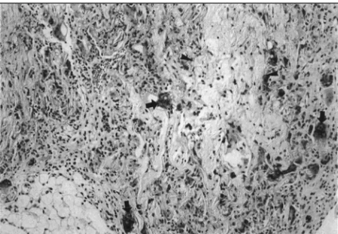
Fig. 2. Maxillary sinus mucosa of the rabbit showing mild muc- osal thickening (arrow) 8 weeks after 25mg of PLA polymer insertion.
Fig. 1. Photomicrograph showing many giant cells (arrows) and intermingled PLA polymer (arrowheads) in omentum (H & E,
×100).
cases from the 3rd week group, 4 cases from 6th week group, 1 case from the 9th week group, 5 cases from the 13th week group and 7 cases from the control group showed findings of chronic hepatitis. Neither the experiment group nor the con- trol group exhibited accumulation of polymer in any organs in light microscopic examination.
General fertility and reproduction
One male mouse and 1 female mouse from the second group to which 1 g/kg of polymer had been intraperitoneally inserted for general fertility and reproduction experiment died from obstructive ileus and peritonitis. Two male mice and 3 female mice out of the third group to which 5 g/kg of polymer had been inserted also died. However, we failed to observe any abnormal apparent symptoms in surviving F0 mice. Mating was successful in each group and the number of fetuses born per mouse did not show significant difference between the co- ntrol and experiment groups. The increase of weight in preg- nant mice also did not show significant difference between the two groups. The experiment group also did not show any abnormality in mating rate and implantation rates. No new- borns died in any of the groups. Birthweights did not show significant difference, and the reactions of surviving fetuses toward auditory and visual stimuli were normal as well. Fin- ally, no significant difference was observed in the sex of fetus.
Teratogenicity
No rabbits died after administration of polymer. No abnor- mal behavior nor symptoms were observed in any rabbits, and the weight of all rabbits increased. However, there was no pr- oof that weight increase slowed in the experiment group as compared with the control group. The daily food intake of pr- egnant rabbits was 160 g per day. In a rabbit sacrificed on the 28th day after fertilization, we observed no significant differ- ence in pregnancy nor in the number of corpus lutea in either the experiment groups or the control group. No external ma- lformation nor abnormality of the internal organs of fetuses was observed. The weight of surviving fetuses did not show significant difference between the experiment groups and the control group, nor was there any difference in the number of fetuses. No difference was found in the sex of fetuses either.
Neither the experiment group nor the control group exhibited malformation of external or internal organs, the musculoske- letal system and other viscera.
Perinatal and postnatal effects
No F0 female and male rats died after intraperitoneal instil- lation of polymer, or showed abnormal symptoms. There was
no significant difference between the weights of F0 female rats from the control group and from the experiment groups both before and after the insertion of polymer. The daily food intake was 1.2 g. No abnormal symptoms were observed in F0
female and male rats after the insertion of polymer. The period of pregnancy was 22.1±1.45 days in the 1st group, 21.3±
1.01 days in the 2nd group, 22.5±1.73 days in the 3rd group and 20. 7±1.15 days in the control group, and there was no significant difference between the control and experiment gr- oups. There were no cases of death of fetuses, or new-borns.
No external malformation, abnormality of the skeletal system, or significant difference was observed in the weight of ne- wborns in each group, nor in the stillborn. The growth of the fetuses in all groups including the control group was normal, and no abnormality was observed during development. The re- productive function of the fetus was normal, and the breast feeding ability of F0 female rats was observed to be normal in all groups.
Local irritation
We examined the rabbits 2 weeks after insertion of polymer into the maxillary sinus, observing that no abnormal secretion in the maxillary sinus had accumulated and that the maxillary sinus mucosa showed a slight flare in the control group, but the other groups were observed to be normal. However, no se- cretion was observed in the first group, in which 10 mg of po- lymer was inserted in the maxillary sinus, however, 2 cases exhibited a slight flare. No secretion was observed in the se- cond group, in which 25 mg of polymer was inserted to the maxillay sinus, the mucosa showed slight flared in 3 cases.
In the complete blood count, glucose test, kidney function test, liver function test, lipid battery test and electrolyte test, there was no abnormality observed in comparison with the control group. In an examination done after 4 weeks, no secretion was observed in the maxillary sinus of all experiment groups, but the mucosae of 2 cases from the control group, 1 case from the first group and 3 cases from the second group were slightly flared, with the rest of the groups normal. In the meantime, the complete blood count, glucose test, kidney function test, liver function test, lipid battery and electrolyte failed to show any abnormality in the experiment group. In the examination of the maxillary sinus mucosa of rabbits after 8 weeks, 1 case from the second group showed mild mucosal thickening (Fig.
2), and the mucosae of 2 cases from the second group, 1 case from the first group and 1 case from the control group showed mild erythema. The maxillary sinus mucosa of the rest of the rabbits was normal. No abnormal findings, however, were ob- served upon the hematological examination of all rabbits.
The findings of light microscopic examination after H & E staining revealed that the first group, the second group, and
the control group, all observed in the second week, and only the first group and control group in the 4th week were obser- ved to be of normal maxillary sinus mucosa, but the infiltration of PMN into the maxillary sinus mucosa was observed in 2 cases from the second group in the 4th week. In the examina- tion of the maxillary sinus mucosa of the rabbits in the 8th week, 3 cases from the third group, to which 25 mg of pol- ymer was inserted, showed infiltration of PMN mixed with lymphocyte, and 1 case among them demonstrated thickening of the mucosal ephithelium as well. In electron microscopic examination of the maxillary sinus mucosa after 2 weeks, 1 case from the control group, 2 cases from the first group, and 3 cases from the second group-and after 4 weeks, 1 case from the control group, 1 case from the first group, and 3 cases from the second group-showed compound cilia, but no other abn- ormality of ciliary epithelial cell was observed.
Immunotoxicity
We observed no swelling, inflammation, anaphylaxis, or sh- ock at the injection spot in all guinea pigs.
Carcinogenecity
There was no findings of cancer generation observed after intraperitoneal insertion of PLA polymer. The death of 4 mice from the first group, and of 8 mice and 2 rats from the second group was, however, observed;but there were no carcinoge- nicity finding in necropsies of these cases, and the cause of death, we assumed, was aspyxia related to anesthesia. Those rats and mice that died in the 1st and 2nd week of the experi- ment displayed severe intraperitoneal polymer adhesion to the large and small intestines, and we supposed that they died of the obstructive ileus, accompanied with the resulting perito- nitis.
DISCUSSION
In total, there were 5 cases of death in the acute toxicity experiment and 9 cases of death in the subacute toxicity ex- periment. We assumed all of these cases died of whole body septic shock as the inserted PLA polymer heavily adhered onto the small or large intestines causing obstructive ileus and pe- ritonitis. Apparently these cases of intestinal adhesion were normal reactions to polymer as a foreign body, rather than by the chemical toxicity of PLA polymer itself.9) Intestinal adh- esion was severe particularly in the animals when a large qu- antity of polymer were inserted. Therefore, it is postulated that these cases did not have a direct cause to the toxicity of PLA polymer.
We observed abnormal findings of liver tissue in many mice
under light microscopic examination. The abnormal findings in liver tissue were various indications of chronic hepatitis:
infiltration of inflammatory cells, microvacuolation of liver cell, granulomatous inflammation, septal fibrosis, and sinus- oidal congestion. These findings are frequently observed in the liver of mice, and we suppose them to be evidence of he- patitis mainly caused by the MHV virus of the Coronaviridae family.10) The large number of abnormal findings in the 9th group which was part of the control group may support this opinion.
During general fertility and reproduction experiment, no si- gnificant difference between the control and experiment gr- oups was observed in the birth and growth of the fetus during pregnancy, and it is more likely that polymer doesn’t deterio- rate the fertility or reproductive capability of mice. No differ- ence was observed between the experiment and control groups in terms of survival of the fetus upon the administering of PLA polymer, nor in implantation, the number of surviving fetuses, and the sex. These results indicate that no malformation or side effects are caused by polymer in the delivery period and after birth.
In local irritation experiment, we could not observe any si- gnificant difference between the experiment and control groups on the maxillary sinus mucosa by the polymer. This finding could be regarded as conflicting with the finding that intrape- ritoneally inserted polymer adhered with nearby viscera. Ho- wever, assuming that intraperitoneal viscera could have kept rubbing with the PLA polymer due to the movement of the large and small intestines, we suppose that the maxillary sinus mucosa does not adhere or adhere slowly with PLA polymer as it is not apparently active. We did not examine whether ab- normal mucosa as in case of sinusitis, has a local mucosal change with PLA polymer. However, as the adhesion of po- lymer with maxillary mucosa had not been observed in the authors’ earlier experiment where we treated acute maxillary sinusitis of rabbits with PLA polymer,7)8) it is speculated that there should not be any local toxicity of PLA polymer to the mucosa of sinusitis. In the meantime, experiment using more than 25 mg of PLA polymer were impossible to do in our st- udy because volumes of PLA polymer greater than 25 mg were more than those of the maxillary sinuses of rabbits and inser- tion was impossible.
We assume, from the experiment using guinea pigs, that PLA polymer does not have immunotoxicity, and that this is more likely because lactic acid is a biological material made in the body11) and does not result in immunotoxicity in a living body.
In the carcinogenecity experiment, we did not observe, in any mice or rats, either development of cancer or death caused by cancer in a period of 4 months. This result indicates that poly-L-lactic acid polymer is not carcinogenic.
We conclude that PLA polymer is not toxic. We expect this
study to provide basic data for future clinical experiments us- ing antibiotics-incorporated PLA polymer.
REFERENCES
1) Kumlien J, Schiratzki H. Blood flow in the rabbit sinus mucosa during experimentally induced chronic sinusitis. Measurement with a diffusible and with and non-diffusible tracer. Acta Otolaryngol (Stockh) 1985;99:630-6.
2) Axelsson A, Brorson JE. The concentration of antibiotics in sinus secretions. Ampicillin, cephradine and erythromycinestolate. Ann Otol Rhinol Laryngol 1974;83:323-31.
3) Piskunov S, Piskunov G, Razinkov S, et al. The prolongation of drug action in the treatment of diseases of the nose and paranasal sinuses. Rhinology 1993;31:33-6.
4) Taylor AV, Boland J, McKenzie IZ. The concorrent in vitro and in vivo release of PGE2 from a controlled release hydrogel polymer pessary for cervical ripening. Prostaglandins 1990;40:89-90.
5) Becker JB, Robinson TE, Barton P, et al. Sustained behavioral recovery from unilateral nigrostriatal damage produced by the co-
ntrolled release of dopamine from a silicone polymer pellet placed into the denervated striatum. Brain Res 1990;508:60-4.
6) Gangadharam PR, Ashtekar DR, Farhi DC, et al. Sustained release of isoniazid in vivo from a single implant of biodegradable poly- mer. Tubercle 1991;72:115-22.
7) Yang-Gi Min, Young-Ki Kim, Young-Seok Choi, Jin-Sung Shin.
Mucociliary activity and histopathology of sinus mucosa in expe- rimental maxillary sinusitis: A comparison of systemic administr- ation of antibiotic and antibiotic delivery by polylactic acid poly- mer. Laryngoscope 1995;105:835-42.
8) Yang-Gi Min, Young-Ki Kim, Seo-Young Jeong. Application of polylactic acid polymer in the treatment of acute maxillary sinus- itis in rabbits. Acta Otolaryngol (Stockh) 1995;115:548-52.
9) Gourlay SJ, Rice RM, Hegyeli AF, et al. Biocompatibility testing of poly-mers: In vivo implantation studies. J Biomed Mater Res 1978;12:219-32.
10) Lee YS. Comparative biology. In: Laboratory animal medicine, 1st ed, Seoul, Seoul National Universtiy Publishing, 1992:151-278.
11) Visscher GE, Robison RL, Maulding HV, Fong JW, Pearson JE, Argentieri GJ. Note: Biodegradation of and tissue reaction to poly (DL-lactide) microcapsules. J Biomed Mater Res 1986;20:667-76.