20 Copyright © 2016 Korean Dementia Association
INTRODUCTION
Neuromyelitis optica (NMO) is an inflammatory disease of the central nervous system mainly characterized by optic neuritis and extensive myelitis where the optic nerve and spi- nal cord have an inflammatory disease of the central nervous system caused by an autoimmune mechanism.
Its clinical symptoms are similar to multiple sclerosis (MS), which has made it difficult to distinguish between them and has caused confusion in their diagnosis. However, autoanti- bodies to the water channels protein aquaporin-4 (AQP4) of the central nervous system was found since 2004, the patho- genesis of NMO and many facts about the clinical character-
istics had been known.1
The typical characteristics of cognitive functional disorders that were exhibited in patients with MS through the previous study, include deterioration of concentration and information processing speed, decreased memories and frontal lobe dys- functions and decreased flexibility of thinking.2 The authors report a case that had shown improvement of frontal lobe dys- functions in NMO after treatment.
CASE REPORT
A 68-year-old right-handed man with an educational back- ground of a high school diploma had visited the hospital due to weakness in the bilateral upper limbs and lower limbs, de- creased sensation of lower extremities and dysuria occurred 5 days ago as main complaints. Due to decreased vision of the left eye, which had occurred seven years ago, and decreased
Improvement of Frontal Lobe Dysfunctions in Neuromyelitis Optica after Treatment: A Case Report
JaeJeong Joo,1 Sul-Ki Lee,2 In Ha Hwang,2 Kyum-Il Kwon,1 Byoung June Ahn,1 YoungSoon Yang2
1Department of Neurology, Soonchunhyang University Gumi Hospital, Gumi, Korea
2Department of Neurology, Veterans Health Service Medical Center, Seoul, Korea
Background Neuromyelitis optica (NMO) is characterized by optic neuritis and longitudinally extensive transverse myelitis. Generally, the brain had been considered healthy in NMO patients, though recent studies have demonstrated that T2-weighted abnormalities may be ob- served in various brain regions. Logically, NMO brain lesions are localized at sites of high aquaporin-4 expression.
Case Report A 68-year-old right-handed man with dysuria, weakness in the bilateral upper and lower limbs, and decreased sensation of the lower extremities, was diagnosed with neuromyelitis optica. The patient was gradually speaking less, was showing reduced interest in hobbies, and had undergone changes in character and behavior. An examination was performed using the Seoul Neuropsychological Screening Battery (SNSB), which revealed that the profile of frontal lobe dysfunctions was prominent as compared with other cognitive domains. The patient was treated with prednisolone and azathioprine for about 1 year without recurrence, and showed prognostic improvement according to further SNSB testing.
Conclusions Further studies are considered necessary in order to find the most effective medication regimen for improving cognitive func- tions in those accurately diagnosed with NMO, and to develop systematic treatment using even more diversified immune-related agents.
Key Words neuromyelitis optica, frontal function, treatment.
Received: March 4, 2016 Revised: March 14, 2016 Accepted: March 14, 2016
Correspondence: YoungSoon Yang, MD, Department of Neurology, Verterans Health Service Medical Center, 53 Jinhwangdo-ro 61-gil, Gangdong-gu, Seoul 05368, Korea
Tel: +82-2-2225-4106, Fax: +82-2-2225-4105, E-mail: astro76@naver.com
cc This is an Open Access article distributed under the terms of the Cre- ative Commons Attribution Non-Commercial License (http://creative- commons.org/licenses/by-nc/3.0) which permits unrestricted non-com- mercial use, distribution, and reproduction in any medium, provided the ori- ginal work is properly cited.
DND
Print ISSN 1738-1495 / On-line ISSN 2384-0757
Dement Neurocogn Disord 2016;15(1):20-23 / http://dx.doi.org/10.12779/dnd.2016.15.1.20
CASE REPORT
www.dnd.or.kr 21
DND
vision of the right eye occurring 2 months before visiting the hospital, he was diagnosed as optic neuritis and received a steroid treatment from other hospital. He had discontinued the medication and was under prognostic observation. The vision in both of his eyes at the time of visiting the hospital were decreased but still he was able to distinguish shapes or colors of all objects.
His consciousness was clear, and eye movements, facial symmetry and sensory functions were normal other than the existing sight disorder. The muscular strength of extremities showed MRC grade III in the right upper limb, grade IV in the left upper limb, and grade IV in both lower limbs. When conducting the sensory test, there were slightly degraded pain senses, vibration senses, tactile senses and location senses be- low T4 skin fragments. He showed a positive response to Lher- mitte’s sign where the patient has sensations of electrical cur- rent flowing through his bilateral arms when suddenly flexing his head forward. The deep tendon reflexes of extremities were all normal, and both sides showed negative responses to the Babinski reflex. As abdominal distention was observed, 1000 cc of urine was drained after insertion of a urinary catheter.
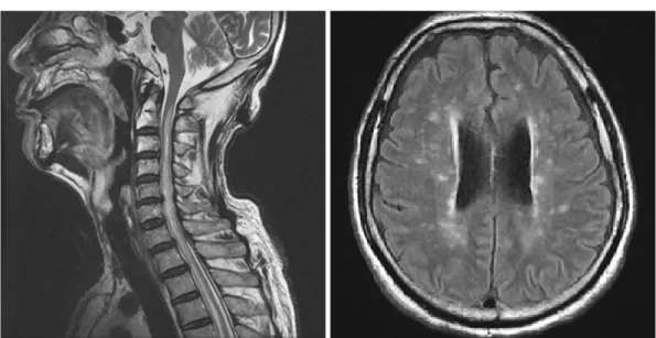
During the spinal magnetic resonance imaging (spine MRI), a high intensity signal of T2 was shown from C1-T8 spine (Fig. 1) and the findings from the brain MRI were normal. The number of cells, the chemical values and IgG index were nor- mal in the cerebrospinal fluid tests, whereas the oligoclonal band was negative also.
The visual evoked potential test showed poor waveforms from both sides, whereas the left posterior tibial nerve sensory evoked a potential test among the somatosensory evoked po- tential test showing an abnormality reasonable for central
nerve conduction disorder, and the auditory evoked potential test results were normal. Basic blood tests, urinalysis, vasculi- tis test and thyroid function tests were all normal and from the autoimmune antibody test, it showed positive response to the AQP4 antibody.
As the guardian commented that the patient is gradually speaking less, has a reduction of interest in hobby activities, and has character and behavioral changes such as being un- able to control his appetite and becoming furious to insignif- icant matter, we conducted the Seoul Neuropsychological Screening Battery for additional evaluation.3 The patient’s edu- cation level was graduation from high school; his score on the Korean Version of the Mini-Mental State Examination was 22 points whereas the clinical dementia rating was 1 point (Table 1). In the case of this patient, the profile of frontal lobe dysfunctions was prominent compared to other cognitive domains.
The patient was diagnosed as NMO and treated with meth- ylprednisolone 1g via IV administration once daily for 5 days.
After the treatment, the muscle power of the bilateral upper limbs was observed as MRC grade IV, and the muscle power of his lower limbs was the same as measured at the time of ad- mission. Since the patient had no large scale change in the conditions, he is under prognostic observation as being treat- ed with prednisolone and azathioprine from the out-patient department currently without recurrence for about 1 year.
After 1 year of time lapse, we conducted the Neuropsychologi- cal Screening Battery (NSB) again for cognitive function eval- uation, and frontal lobe functions showing an improving trend but there were no large changes in other domains compared to the conditions before NSB (Table 1).
Fig. 1. Sagittal T2-weighted image of spine shows diffuse enlargement of cervical and thoracic spinal cord and continuous high signal in- tensity in the central gray matter from C1 level. Axial fluid-attenuated inversion recovery image of brain shows high signal intensity lesions in the cerebral periventricular and deep white matter.
JaeJeong Joo et al.
NMO and Frontal Lobe Function
22 Dement Neurocogn Disord 2016;15(1):20-23
DISCUSSION
NMO is a disease that shows clinical findings differentiated from MS and can measure auto-antibodies associated with the disease onset mechanism. Currently, MS occurs as sensi- tized T-lymphocytes passing through the Blood-Brain Barrier whereas NMO is considered to be an inflammation that oc- curs through an auto-antibody. In the past, it has been known that NMO did not invade into the brain, and even if any brain
lesions were found from MRI images, they were known as asymptomatic. But according to a recent study, brain lesions are found in ≥70% of patients with anti-AQP4 positive anti- bodies and associated symptoms are expressed in a substantial number of patients.4 In addition, as a case is being identified in whom the brain lesion is expressed as the initial symptom before the optic neuritis and myelitis are displayed, the clini- cal spectrum of NMO is gradually widening.
Notwithstanding there is a debate on whether anti-AQP4 Table 1. Initial and follow-up (F/U) neuropsychological test
Neuropsychological test Initial F/U
K-MMSE
Orientation to time 3 5
Orientation to place 3 5
Registration 3 3
Attention and calculation 2 2
Recall 2 2
Language 8 8
Drawing 1 1
Total score 22 26
Attention
Digit span, forward 3 6
Digit span, backward 3 4
Language & related funtions
K-BNT 58/60 57/60
Rt-Lt orientation Normal Normal
Calculation (+/-/×/÷) Normal (3/3/3/3) Normal (3/3/3/3)
Praxis Normal Normal
Visuospatial function and memory
Reye complex figure, copy 34/36 35/36
Immediate recall 16.5/36 16/36
Delayed recall 11/36 14/36
Recogntion score 16 18
Korean verbal learning test
Immediate recall 4-5-7:16 5-5-9:19
Delayed recall 0 0
Recognition score 16 19
Frontal executive funtions
Contrasting program Ab NL
Go-no-go Ab NL
Fist-edge-palm Ab NL
Alternating hand movement Ab NL
Alternating square & triangle Ab NL
Luria loop Ab NL
Phonemic word fluency (animal/supermarket) 9/7 12/11
Semantic word fluency (ㄱ/ㅇ/ㅅ/total) 3/3/3/9 8/4/3/15
Stroop test, word reading (correct/error) 112/0 112/0
Stroop test, color reading (correct/error) 86/3 84/3
K-BNT: Korean-Boston Naming Test, K-MMSE : Korean Version of the Mini-Mental State Examination.
www.dnd.or.kr 23
DND
antibody is just a simple disease specific indicator or if it plays an important role in etiology, but currently it is accepted that anti-AQP4 antibody binds to AQP4 in the central nervous system and through activation of complementary body and immuno-reaction, it plays the role of etiology of NMO, ac- cording to the results of several studies. Therefore, in case of NMO that causes the functional damage of the central ner- vous system, it can be seen as it is natural to have occurrence of dysfunction in cognitive function acting in integrated man- ner of several sites.
From the previous study, the patients with MS showed the most abnormalities in the information processing speed, mem- ory and execution function and in the patients with NMO, it is known that they showed cognitive dysfunctions with simi- lar frequency.5,6 In addition, a study result was presented that suggests the decreased cognitive function in NMO is associat- ed with the volumetric reduction of the brain white matters.7
In MS, the immunomodulatory medication and the immu- nosuppressive medications were reported as effective for treat- ment of cognitive dysfunctions,5 thereby it is likely to presume that they may present the similar effectiveness after treatment in NMO as well. From the initial NSB actually performed in this patient, frontal lobe dysfunctions were observed notably among cognitive functions and the patient showed prognos- tic improvement in the NSB performed again after using the medication for treatment of NMO.
This study aimed to differentiate a diagnosis of NMO from MS and other auto-immune diseases invading the central ner- vous system at an early stage. And in addition to the studies
in respect to the mechanism that can improve cognitive func- tions after medication treatment based on accurate diagnosis in the future, further studies are considered as necessary for the most effective medication regimen in improving cogni- tive functions through systematic treatment using even more diversified immune-related agents.
Conflicts of Interest
The authors have no financial conflicts of interest.
Acknowledgements
This study was supported by a VHS Medical Center Research Grant, Re- public of Korea (grant number: VHSMC 15012).
REFERENCES
1. Lee S, Kim BJ. Diagnosis and treatment of neuromyelitis optica. J Neurocrit Care 2011;4 Suppl 1:S38-S41.
2. Lee CN, Park KW. Cognitive impairment in multiple sclerosis. J Mult Scler 2013;4:29-35.
3. Ahn HJ, Chin J, Park A, Lee BH, Suh MK, Seo SW, et al. Seoul Neu- ropsychological Screening Battery-dementia version (SNSB-D): a use- ful tool for assessing and monitoring cognitive impairments in de- mentia patients. J Korean Med Sci 2010;25:1071-1076.
4. Pittock SJ, Lennon VA, Krecke K, Wingerchuk DM, Lucchinetti CF, Weinshenker BG. Brain abnormalities in neuromyelitis optica. Arch Neurol 2006;63:390-396.
5. Blanc F, Zéphir H, Lebrun C, Labauge P, Castelnovo G, Fleury M, et al.
Cognitive functions in neuromyelitis optica. Arch Neurol 2008;65:84-88.
6. Saji E, Toyoshima Y, Yanagawa K, Nishizawa M, Kawachi I. Neuro- psychiatric presentation of neuromyelitis optica spectrum disorders.
Neurol 2010;74:A169.
7. Blanc F, Noblet V, Jung B, Rousseau F, Renard F, Bourre B, et al.
White matter atrophy and cognitive dysfunctions in neuromyelitis op- tica. PLoS One 2012;7:e33878.