Disturbed Osteoblastic Differentiation of Fibrous Hamartoma Cell from Congenital Pseudarthrosis of the Tibia Associated with
Neurofi bromatosis Type I
Dong Yeon Lee, MD, Tae-Joon Cho, MD, Hye Ran Lee, MS, Kang Lee, MD, Hyuk Joo Moon, MD, Moon Seok Park, MD, Won Joon Yoo, MD, Chin Youb Chung, MD, In Ho Choi, MD
Department of Orthopedic Surgery, Seoul National University College of Medicine, Seoul, Korea
Copyright © 2011 by Th e Korean Orthopaedic Association
Th is is an Open Access article distributed under the terms of the Creative Commons Attribution Non-Commercial License (http://creativecommons.org/licenses/by-nc/3.0) which permits unrestricted non-commercial use, distribution, and reproduction in any medium, provided the original work is properly cited.
Clinics in Orthopedic Surgery • pISSN 2005-291X eISSN 2005-4408 Received February 7, 2011; Accepted March 2, 2011
Correspondence to: Tae-Joon Cho, MD
Division of Pediatric Orthopedics, Seoul National University Children’s Hospital, 101 Daehak-ro, Jongno-gu, Seoul 110-744, Korea Tel: +82-2-2072-2878, Fax: +82-2-745-3367, E-mail: [email protected]
Background: Fibrous hamartoma is the key pathology of congenital pseudarthrosis of the tibia (CPT), which was shown to have low osteogenicity and high osteoclastogenicity. This study further investigated the mechanism of impaired osteoblastic differen- tiation of fi brous hamartoma cells.
Methods: Fibroblast-like cells were obtained from enzymatically dissociated fi brous hamartomas of 11 patients with CPT as- sociated with neurofi bromatosis type I (NF1). Periosteal cells were also obtained from the distal tibial periosteum of 3 patients without CPT or NF1 as control. The mRNA levels of Wnt ligands and their canonical receptors, such as Lrp5 and β-catenin, were assayed using reverse transcriptase PCR (RT-PCR). Changes in mRNA expression of osteoblast marker genes by rhBMP2 treatment were assayed using quantitative real time RT-PCR. Changes in mRNA expression of transcription factors specifi cally involved in osteoblastic differentiation by rhBMP2 treatment was also assayed using quantitative real-time RT-PCR.
Results: Wnt1 and Wnt3a mRNA expression was lower in fi brous hamartoma than in tibial periosteal cells, but their canonical receptors did not show signifi cant difference. Response of osteoblastic marker gene expression to rhBMP2 treatment showed patient-to-patient variability. Col1a1 mRNA expression was up-regulated in most fi brous hamartoma tissues, osteocalcin was up- regulated in a small number of patients, and ALP expression was down-regulated in most fi brous hamartoma tissues. Changes in mRNA expression of the transcription factors in response to rhBMP2 also showed factor-to-factor and patient-to-patient variabil- ity. Dlx5 was consistently up-regulated by rhBMP2 treatment in all fi brous hamartoma tissues tested. Msx2 expression was down- regulated by rhBMP2 in most cases but by lesser extent than control tissue. Runx2 expression was up-regulated in 8 out of 18 fi brous hamartoma tissues tested. Osterix expression was up-regulated in 2 and down-regulated in 3 fi brous hamartoma tissues.
Conclusions: Congenital pseudarthrosis of the tibia appears to be caused by fi brous hamartoma originating from aberrant growth of Nf1 haploinsuffi cient periosteal cells, which failed in terminal osteoblastic differentiation and arrested at a certain stage of this process. This pathomechanism of CPT should be targeted in the development of novel therapeutic biologic intervention.
Keywords: Congenital, Pseudarthrosis, Osteoblastic differentiation
Congenital pseudarthrosis of the tibia (CPT) is a very rare but specifi c condition characterized by anterolateral bow- ing at birth, and recurrent pathologic fractures of the tibia during early childhood. Fracture usually occurs spontane- ously or aft er minor trauma and rarely heals without ap- propriate surgical intervention, and easily results in pseud- arthrosis.
Fibrous hamartoma tissue is regarded as the key pathology of CPT,1) which occupies the site of pseudar- throsis, and is continuous with abnormally thickened peri- osteum in the adjacent bone segment.
CPT frequently develops in patients with neurofi- bromatosis type 1 (NF1). It was reported that association with NF1 was found in 40 to 60 per cent of CPT patients,2) and CPT develops in 1 to 4 percent of patients with NF1.3) NF1 is one of the most common autosomal dominant disorders, aff ecting approximately 1 in 3500 births.4) It is characterized by neurofibromas, cafe-au-lait spots, Lisch nodules and numerous skeletal manifestations, such as macrocephaly, short stature, kyphoscoliosis, sphenoid wing dysplasia, congenital bowing and congenital pseud- arthrosis of the tibia.
NF1 is caused by a heterozygous mutation in the Nf1 gene located in chromosome 17q11.2.5) Th is gene en-
codes neurofi bromin, which is expressed in a wide range of cells and tissues,6) including maturing chondrocytes, hypertrophic chondrocytes, osteoblasts, osteocytes, and osteoclasts.7) It is also expressed in tissues around CPT.8) Neurofibromin negatively regulates the activity of an in- tracellular signaling molecule p21ras (Ras), by functioning as a GTPase activating protein (Ras-GAP).9,10) Haploinsuf- ficiency or complete deficiency in Nf1 gene function re- sults in a dose-dependent elevation in Ras activity, which can activate the mitogen-activated protein kinase (MAPK) pathway and the phosphatidylinositol-3-phosphate kinase (PI-3K) pathway.11)
The Nf1 gene is classified as a tumor suppressor gene. Heterozygous Nf1+/– mice experienced tumors and premature death.12) Chimeric mice containing moderate levels of Nf1–/– defi cient cells went on to form numerous neurofi bromas, which progressed to malignancy with loss of the p53 tumor suppressor gene.13) Furthermore, the loss of heterozygosity (LOH) is seen in benign and malignant tumors, including neurofibroma, malignant peripheral nerve sheath tumor (MPNST) and astrocytoma.14,15)
Previous studies demonstrated that the phenotype of the fi brous hamartoma cells is consistent with the im- munophenotype of mesenchymal lineage cells as that
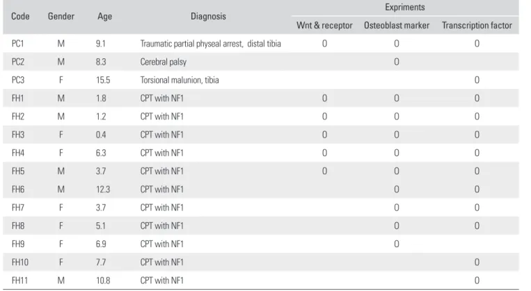
Table 1. Tissue Samples Used in This Study
Code Gender Age Diagnosis
Expriments
Wnt & receptor Osteoblast marker Transcription factor
PC1 M 9.1 Traumatic partial physeal arrest, distal tibia O O O
PC2 M 8.3 Cerebral palsy O
PC3 F 15.5 Torsional malunion, tibia O
FH1 M 1.8 CPT with NF1 O O O
FH2 M 1.2 CPT with NF1 O O O
FH3 F 0.4 CPT with NF1 O O O
FH4 F 6.3 CPT with NF1 O O O
FH5 M 3.7 CPT with NF1 O O O
FH6 M 12.3 CPT with NF1 O O
FH7 F 3.7 CPT with NF1 O O
FH8 F 5.1 CPT with NF1 O O
FH9 F 6.9 CPT with NF1 O
FH10 F 7.7 CPT with NF1 O
FH11 M 10.8 CPT with NF1 O
PC: periosteal cell, FH: fi brous hamartoma, CPT: congenital pseudarthrosis of the thibia, NF1: neurofi bromatosis type 1.
of tibial periosteal cells.16,17) However, these cells do not undergo osteoblastic differentiation in response to bone morphogenetic protein (BMP), and they are more osteo- clastogenic than the tibial periosteal cells.16) In this study, we investigated further steps of osteoblastic diff erentiation in cells isolated from fi brous hamartoma, in order to de- lineate the mechanism of impaired osteoblastic diff erentia- tion of fi brous hamartoma cells.
METHODS
Primary Cell Culture
Th is study was approved by the institutional review board of Seoul National University Hospital. Harvested human tissues were investigated in experiments with informed consent. Fibrous hamartoma tissue was harvested from 11 patients (5 male and 6 female) with an atrophic type of CPT associated with NF1, during surgical procedures for osteosynthesis. Control tissue was obtained from the distal tibial periosteum. A strip of about 2 × 5 mm distal tibial periosteum was harvested during supramalleolar tibial derotation osteotomy from 3 patients without CPT or NF1 (Table 1). Th ey underwent tissue culture. Fibrous hamar- toma or tibial periosteum was enzymatically dissociated to obtain fi broblast-like cells, which were plated on a 100- mm dish, and cultured in DMEM containing 10% FBS and antibiotics at 37oC in 5% CO2. When the second pas- sage cells reached confl uence, they were trypsinized, fro- zen, and stored in liquid nitrogen until required. Th e cells from fi brous hamartoma were denoted by the labels listed in Table 1 as FH1 through FH11, and cells from the tibial periosteum as PC1 through PC3. A vial of cells from one confl uent dish were thawed and plated on 100-mm dish.
When they reached confl uence again, they were re-plated in 1:3 ratio, and rhBMP2 (R&D Systems, Minneapolis, MN, USA) treatment was commenced 48 hours aft er plat- ing. As the amount of tissue harvested from patients was limited, not all tissues were used in the experiments (Table 1).
Wnt Ligands and Receptors Expression
Total RNA was extracted from plated cells using Trizol reagent (Invitrogen, Carlsbad, CA, USA). The mRNA expressions of Wnt1, Wnt3a, LRP-5 and β-catenin were as- sayed by reverse transcription-polymerase chain reaction (Table 2), to investigate for endogenous Wnt signaling.
Glyceraldehyde-3-phosphate dehydrogenase (GAPDH) was used as internal control. Regular PCR was performed using AccuPower® HotStart PCR Premix (Bioneer, Dae- jeon, Korea) with primers for each gene (Table 2). Quanti-
tative real-time PCR was performed for Wnt1 and Wnt3a as they showed different band intensity in regular PCR.
After reverse transcription using the SuperScript First- strand Synthesis System for RT-PCR (Invitrogen), real- time PCR was performed in TaqMan Universal PCR Master Mix (Applied Biosystems, Foster City, CA, USA) using an ABI Prism 7000 Sequence Detection System (Applied Biosystems). To compare diff erent gene expres- sion levels, threshold cycle (CT) values were compared for relative gene expression analysis. Th e expression levels of target genes were presented as ratio to that of GAPDH. All samples were amplifi ed four times.
Changes in mRNA Expression by rhBMP Treatment Th e plated cells were grown in osteogenic media (DMEM containing 10% FBS, 100 nM dexamethasone, 10 mM sodium-glycerophosphate, and 0.05 mM ascorbic acid-2- phosphate). Th ey were divided into two groups. Group I was grown solely in osteogenic media. Group II was treat- ed with 100 ng/mL rhBMP2 (R&D Systems) for 2 weeks.
Total RNA was extracted following 2 weeks of treatment.
Th e mRNA levels of osteoblast markers, such as col1a1, os-
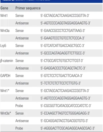
Table 2. Custom-made Primers List
Gene Primer sequence
Wnt1 Sense 5’-GCTAGCACTCAAGACCCGGTTA-3’
Antisense 5’-AGTCCCCAGGTAGGAGGAAGTG-3’
Wnt3a Sense 5’-GAACCGCCCTCCTGATTAAG-3’
Antisense 5’-GAAGTCCCTGTCCTCTCCCA-3’
Lrp5 Sense 5’-GTCATCATTGACCAGCTGCC-3’
Antisense 5’-GCCCAGTAGAGGTTCTTGCC-3’
β-catenin Sense 5’-CTGCCATCTGTGCTCTTCGT-3’
Antisense 5’-GAGGACCCCTGCAGCTACTC-3’
GAPDH Sense 5’-GTCTCCTCTGACTTCAACA-3’
Antisense 5’-TCTCTCTCTTCCTCTTGTG-3’
Wnt1* Sense 5’-GCTAGCACTCAAGACCCGGTTA-3’
Antisense 5’-AGTCCCCAGGTAGGAGGAAGTG-3’
Probe 5’-CGCGGTTCATACGCATCCCATCTC-3’
Wnt3a* Sense 5’-CCAAGCTTAGTCCTGGGAGAGG-3’
Antisense 5’-GCAGGAGTACCTGACGGTGTG-3’
Probe 5’-AGGGACTTCGCAGAGGCAAGCGAC-3’
*Primers and probe sequences for quantitative real-time RT-PCR.
teocalcin, and alkaline phosphatase, liver/bone/kidney type (ALPL), were quantitatively analyzed by real-time RT-PCR using ready-made primers. Th e mRNA levels of transcrip- tion factors involved in osteoblastic diff erentiation, such as Dlx5, Msx2, Runx2, and Osterix, were also quantitatively analyzed by real-time RT-PCR using ready-made primers (Table 3). Real-time PCR and quantifi cation method was identical to that described previously. Th e expression level of rhBMP2 treated cell was divided by that of untreated cell to calculate the extent of up-regulation or down-regu- lation of the expression.
RESULTS
Wnt Ligands and Receptors Expression
Control tissue (PC1) and fi brous hamartoma tissues from five patients (FH1-5) underwent quantitative real-time RT-PCR for Wnt1 and Wnt3a, and qualitative RT-PCR for Lrp-5 and β-catenin. In the fi brous hamartoma from all 5 CPT patients, the mRNA levels of Wnt ligands were lower than that of control tissue. Expression level of these ligands varied from patient to patient. The mRNA level of Wnt1 in the fibrous hamartoma ranged from 3.3% to 34.2% of that of normal control tissue, while Wnt3a mRNA expres- sion level ranged from 0.14% to 13.5%. All tissues showed strong mRNA expression of both Lrp-5 and β-catenin in RT-PCR (Fig. 1).
Osteoblast Marker Gene Expression Change by BMP Treatment
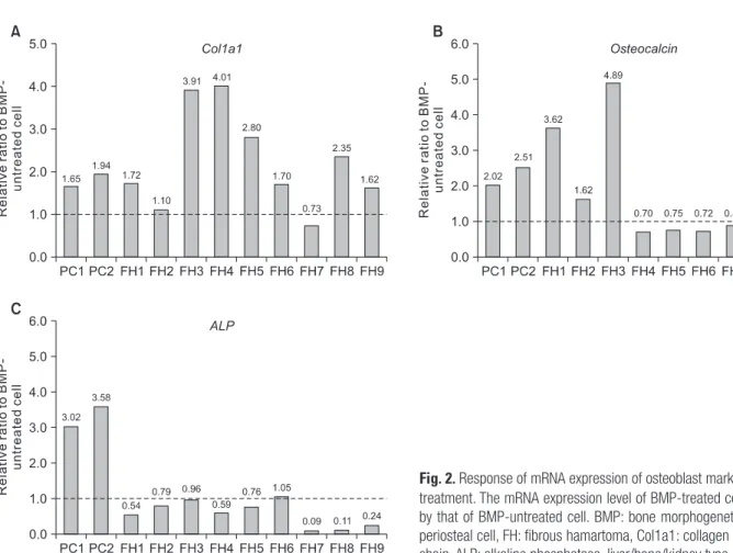
Th e mRNA level of osteoblast markers, including col1a1, osteocalcin, and ALPL were analyzed in 2 control tissues (PC1, PC2) and 9 fi brous hamartoma tissues (FH1-FH9).
Th e control tissues (periosteal cells) showed up-regulation of mRNA expression of col1a1, osteocalcin and ALPL in response to rhBMP-2 treatment. Col1a1 mRNA level increased by 1.65- or 1.94-fold, osteocalcin by more than 2.0-fold, and ALPL by more than 3.0-fold. However, the response of fi brous hamartoma remained inconsistent. In 7 of 9 FH’s, the mRNA expression of col1a1 increased by more than 10% of non-treatment control, while in 2 FH’s, it increased by 10% or decreased by 21%, respectively (Fig.
2A). Osteocalcin mRNA expression was up-regulated by rhBMP in fewer FH’s than col1a1. Only 2 FH’s showed up-regulation of osteocalcin gene expression, and 6 FH’s showed down-regulation of osteocalcin gene expression by rhBMP2 treatment (Fig. 2B). ALP gene expression was down-regulated by rhBPM2 treatment in 7 FH’s, and that of the remaining 2 FH’s remained within 10% of that of untreated control. Th is was in contrast to more than 3-fold up-regulation in the periosteal cells (PC1 and PC2) (Fig.
2C).
Fig. 1. The mRNA expression of Wnt ligands and their canonical receptors. (A) When the mRNA level of periosteal cell (PC1) was set as 1.0, the relative levels of mRNA expression of FH1-5 were less than 0.4 in both Wnt1 and Wnt3a, which are the most important factors in canonical signal pathway involved in osteoblastic differentiation. (B) PCR showed abundant expression of Lrp-5 and β-catenin mRNA in all tissues tested. PC: periosteal cell, FH: fibrous hamartoma, Lrp: low density lipoprotein receptor-related protein, Wnt: wingless-type MMTV intergration site.
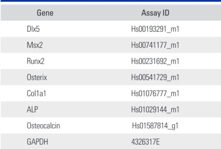
Table 3. Ready-made Primer Set List for Real-time PCR*
Gene Assay ID
Dlx5 Hs00193291_m1
Msx2 Hs00741177_m1
Runx2 Hs00231692_m1
Osterix Hs00541729_m1
Col1a1 Hs01076777_m1
ALP Hs01029144_m1
Osteocalcin Hs01587814_g1
GAPDH 4326317E
*TaqMan® Gene Expression Assay.
Change in the mRNA Expression of Transcription Factors Involved in Osteoblastogenesis by BMP Treatment (Fig. 3)
Transcription factors involved in osteoblastic diff erentia- tion, such as Dlx5, Msx2, Runx2, and Osterix, were ana- lyzed in 10 FH’s (FH1-FH8, FH10, FH11) and 2 periosteal cells. Periosteal cells showed responses compatible with osteoblastic differentiation. Dlx5, Runx2, and Osterix mRNA expressions were up-regulated with rhBMP2 treat- ment by 52%, 59, and 20%, respectively, and that of Msx2 was down-regulated by 48%. In FH’s, changes in mRNA expression of these transcription factors were not consis- tent. Dlx5 expression was up-regulated in all FH’s tested.
Th e extent of up-regulation was similar to or larger than the two periosteal cells in 9 of 10 FH’s tested. FH2 showed mild up-regulation, which was smaller than that of ei- ther PC1 or PC3. Msx2 was down-regulated as periosteal cells in 9 of 10 FH’s tested. However, only one FH (FH10) showed down-regulation more than the PC’s. In 8 of 10 FH’s tested, the extent of down-regulation of Msx2 mRNA expression was less than 20% of the untreated cells, while that of PC’s were more than 40%. One FH (FH4) even showed up-regulation of Msx2 expression. Runx2 mRNA
expression was up-regulated in 8 FH’s, and down-regulat- ed in 2 FH’s. FH8 showed 8.7-fold up-regulation, which also showed the largest up-regulation of Dlx5 expression.
However, FH8 showed down-regulation of osteocalcin and ALPL mRNA expression. Osterix mRNA expression was diverse. Two HF’s showed up-regulation larger than PC1, while three showed down-regulation by rhBMP2 treat- ment.
DISCUSSION
This study was limited by the fact that mRNA expres- sions of fibrous hamartoma from CPT associated with NF1 were compared to that of periosteal cells from non- NF1 patients. The findings might reflect the difference between Nf1 haploinsufficient cells and wild-type cells, but it does not explain why fi brous hamartoma develops on distal tibia specifi cally in systemically Nf1 haploinsuf- fi cient individual. In order to address this question, fi brous hamartoma cells should be compared with periosteal cells from normal-looking contralateral distal tibia of the same patient. However, it would be unethical to harvest it only for research purpose.
Fig. 2. Response of mRNA expression of osteoblast markers to rhBMP2 treatment. The mRNA expression level of BMP-treated cell was divided by that of BMP-untreated cell. BMP: bone morphogenetic protein, PC:
periosteal cell, FH: fibrous hamartoma, Col1a1: collagen type 1 alpha 1 chain, ALP: alkaline phosphatase, liver/bone/kidney type.
We investigated several aspects of the osteoblastic differentiation process in primary cultured cells from fi brous hamartoma believed to be the main pathologic tis- sue in CPT associated with NF1. Although the data were not consistent throughout all cases investigated in this study, fibrous hamartoma cells showed deficient expres- sion of the Wnt ligands which are important factors in the regulation of bone mass, postnatal bone formation and fracture healing process.18) When stimulated by rhBMP2 treatment, most fibrous hamartoma cells up-regulated col1a1 expression. However, only a subset up-regulated osteocalcin expression, and all of them down-regulated ALP expression. These findings were consistent with a published report on a smaller number of cases.16)
Osteoblastogenesis is a complex process of cell differentiation from mesenchymal stem cell through os- teoprogenitor and preosteoblast to osteoblast. Wnts and BMPs are the most important signal molecules that regu-
late this process. In this study, we investigated mRNA ex- pression of the Wnt ligands (Wnt1 and Wnt3a), and their receptors of the canonical pathway (Lrp5 and β-catenin).
Wnts are a large family of growth factors involved in a va- riety of biological processes. Th e canonical pathway of Wnt signal mediated through β-catenin/Lrp5 modulates most aspects of osteoblastic physiology, including proliferation, differentiation, bone matrix formation/mineralization and apoptosis.19) We showed that the mRNA expression of Wnt1 and Wnt3a, which are known to be involved in os- teoblastogenesis, were signifi cantly low in fi brous hamar- toma cells, although the receptors’ mRNA expression was comparable to control tissue. Th is means that defi ciency of local Wnt ligands may contribute to disturbed osteoblastic diff erentiation in fi brous hamartoma in the pathogenesis of CPT. It appears worthwhile to further investigate Wnt signaling systematically in CPT associated with NF1.
In a previous study, BMP2, BMP4, BMPR1A, BM- Fig. 3. Response of mRNA expression of transcription factors involved in osteoblastic differentiation. The mRNA expression level of BMP-treated cell was divided by that of BMP-untreated cell. BMP: bone morphogenetic protein, PC: periosteal cell, FH: fibrous hamartoma, Dlx: distal-less homeobox, Runx: runt-related transcription factor, Msx: muscle segment homeobox, drosophila homolog, Osx: osterix.
PR1B, and BMPR2 were reported to express in fibrous hamartoma of CPT.16) BMP signals induce receptor- mediated signaling pathways involving Smads, and subse- quently transcriptions factors specifi c for osteoblastic dif- ferentiation. Runx 2 is the master osteogenic transcription factor, expresses in early skeletal development and persists through subsequent stages of bone formation.20,21) Dlx5 is a bone inducing homeodomain transcription factor, expressed in the later stages of osteoblastic diff erentiation, which is considered to be an upstream regulator of Runx2 and Osterix in the BMP-2 signaling pathway.22) Osterix is a zinc fi nger-containing transcription factor, specifi cally ex- pressed in all developing bones, which is considered to act mainly during the terminal diff erentiation of osteoblasts.21) Msx2 is a mammalian homologue of the Drosophila muscle segment homeobox gene, which appears to play a negative role in osteoblast diff erentiation, stimulating cell proliferation and suppressing osteogenic diff erentiation.21) Hence, BMP signaling appears to be transduced via Smads to Dlx5, and then to Runx2 for early diff erentiation, and to Osterix for terminal differentiation and Msx2 antago- nizes these pathways. In our control tissue - the periosteal cells - up-regulated Dlx5, Runx2 and Osterix were seen in response to BMP stimulation, whereas Msx2 was down- regulated. Hence, we attempted to delineate the response of these transcription factor expressions in fibrous ham- artoma cells. It is noteworthy that both Dlx5 and Runx2 responded in a way similar to the periosteal cells in most
1. Ippolito E, Corsi A, Grill F, Wientroub S, Bianco P. Patholo- gy of bone lesions associated with congenital pseudarthrosis of the leg. J Pediatr Orthop B. 2000;9(1):3-10.
2. Heft i F, Bollini G, Dungl P, et al. Congenital pseudarthrosis of the tibia: history, etiology, classification, and epidemio- logic data. J Pediatr Orthop B. 2000;9(1):11-5.
3. Crawford AH, Schorry EK. Neurofi bromatosis update. J Pe- diatr Orthop. 2006;26(3):413-23.
4. Friedman JM. Epidemiology of neurofibromatosis type 1.
Am J Med Genet. 1999;89(1):1-6.
5. Shen MH, Harper PS, Upadhyaya M. Molecular genetics of neurofi bromatosis type 1 (NF1). J Med Genet. 1996;33(1):2- 17.
6. Gutmann DH, Wood DL, Collins FS. Identification of the neurofi bromatosis type 1 gene product. Proc Natl Acad Sci U S A. 1991;88(21):9658-62.
REFERENCES
7. Kuorilehto T, Nissinen M, Koivunen J, Benson MD, Pel- tonen J. NF1 tumor suppressor protein and mRNA in skel- etal tissues of developing and adult normal mouse and NF1- defi cient embryos. J Bone Miner Res. 2004;19(6):983-9.
8. Leskela HV, Kuorilehto T, Risteli J, et al. Congenital pseud- arthrosis of neurofibromatosis type 1: impaired osteoblast differentiation and function and altered NF1 gene expres- sion. Bone. 2009;44(2):243-50.
9. Bollag G, Clapp DW, Shih S, et al. Loss of NF1 results in ac- tivation of the Ras signaling pathway and leads to aberrant growth in haematopoietic cells. Nat Genet. 1996;12(2):144-8.
10. Cichowski K, Jacks T. NF1 tumor suppressor gene function:
narrowing the GAP. Cell. 2001;104(4):593-604.
11. Schindeler A, Little DG. Recent insights into bone develop- ment, homeostasis, and repair in type 1 neurofi bromatosis (NF1). Bone. 2008;42(4):616-22.
12. Jacks T, Shih TS, Schmitt EM, Bronson RT, Bernards A, cases of fi brous hamartoma. On the other hand, Msx2 ex- pression was not suppressed by BMP stimulation as much as in the periosteal cells in most cases of fibrous hamar- toma. In addition, Osterix expression was down-regulated in 3 of 5 FH’s. Th ese fi ndings suggest that impairment of osteoblastic differentiation in fibrous hamartoma is the terminal step of the differentiation cascade and interfer- ence by Msx2 may work in this process. Another aspect of these fi ndings that we need to pay attention is that the behavior of FH’s varies from patient to patient.
In conclusion, the fi ndings in this study support the hypothesis that fi brous hamartoma originates from aber- rant growth of Nf1 haploinsuffi cient periosteal cells, which failed in terminal osteoblastic diff erentiation, arrested at a certain stage of this process. Th is pathomechanism of CPT should be targeted in the development of novel therapeu- tic biologic intervention.
CONFLICT OF INTEREST
No potential confl ict of interest relevant to this article was reported.
ACKNOWLEDGEMENTS
Th is work was supported by the Korea Research Founda- tion Grant funded by the Korean Government (KRF-2008- 313-E00352).
Weinberg RA. Tumour predisposition in mice heterozygous for a targeted mutation in Nf1. Nat Genet. 1994;7(3):353-61.
13. Cichowski K, Shih TS, Schmitt E, et al. Mouse models of tumor development in neurofibromatosis type 1. Science.
1999;286(5447):2172-6.
14. Upadhyaya M, Han S, Consoli C, et al. Characterization of the somatic mutational spectrum of the neurofi bromatosis type 1 (NF1) gene in neurofibromatosis patients with be- nign and malignant tumors. Hum Mutat. 2004;23(2):134- 46.
15. Li Y, Bollag G, Clark R, et al. Somatic mutations in the neuro- fi bromatosis 1 gene in human tumors. Cell. 1992;69(2):275- 81.
16. Cho TJ, Seo JB, Lee HR, Yoo WJ, Chung CY, Choi IH. Bio- logic characteristics of fi brous hamartoma from congenital pseudarthrosis of the tibia associated with neurofi bromato- sis type 1. J Bone Joint Surg Am. 2008;90(12):2735-44.
17. Mariaud-Schmidt RP, Rosales-Quintana S, Bitar E, et
al. Hamartoma involving the pseudarthrosis site in pa- tients with neurofibromatosis type 1. Pediatr Dev Pathol.
2005;8(2):190-6.
18. Chen Y, Alman BA. Wnt pathway, an essential role in bone regeneration. J Cell Biochem. 2009;106(3):353-62.
19. Bodine PV, Komm BS. Wnt signaling and osteoblastogen- esis. Rev Endocr Metab Disord. 2006;7(1-2):33-9.
20. Ducy P, Zhang R, Geoff roy V, Ridall AL, Karsenty G. Osf2/
Cbfa1: a transcriptional activator of osteoblast differentia- tion. Cell. 1997;89(5):747-54.
21. Ryoo HM, Lee MH, Kim YJ. Critical molecular switches involved in BMP-2-induced osteogenic differentiation of mesenchymal cells. Gene. 2006;366(1):51-7.
22. Ryoo HM, Hoffmann HM, Beumer T, et al. Stage-specific expression of Dlx-5 during osteoblast differentiation: in- volvement in regulation of osteocalcin gene expression. Mol Endocrinol. 1997;11(11):1681-94.