Received on January 6, 2016. Revised on March 16, 2016. Accepted on March 19, 2016.
CC This is an open access article distributed under the terms of the Creative Commons Attribution Non-Commercial License (http://creativecommons.org/licenses/by-nc/4.0) which permits unrestricted non-commercial use, distribution, and reproduction in any me- dium, provided the original work is properly cited.
*Corresponding Author. Soon-Wuk Jeong, Department of Veterinary Surgery, College of Veterinary Medicine, Konkuk University, 120 Neungdong-ro, Gwangjin-gu, Seoul 05029, Korea. Tel: 82-2-450-3670; Fax: 82-2-450-3037; E-mail: [email protected]
Abbreviations: CT, computed tomography; FNA, fine needle aspiration; ICD-0, international classification of diseases for oncology; OHE, ovar- iohysterectomy; PMR, proportional morbidity ratio
Clinical Outcomes of Surgically Managed Spontaneous Tumors in 114 Client-owned Dogs
Ji-Won Choi, Hun-Young Yoon and Soon-Wuk Jeong*
Department of Veterinary Surgery, College of Veterinary Medicine, Konkuk University, Seoul 05029, Korea
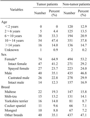
Medical records of 139 tumors from 114 dogs that under- went surgery from May 2010 through March 2015 were retrospectively reviewed. Among 114 dogs, females (64.9%) were significantly more common than males (35.1%) (p<0.05). Dogs aged 6 to 10 years were more presented than non-tumor patients, however, there was no significant difference. The mean age (±SD) was 10.3±3.0 years. Although we found no significant difference of breed predisposition, the most common breed was Maltese (19.3%), followed by Shih-Tzu (14.0%), and Yorkshire terrier (13.2%). Proportional morbidity ratios (PMRs) of mammary gland, oral cavity, and skin tumors were high in Poodles, Yorkshire terriers, and Golden retrievers, re- spectively. Mammary gland (36.0%) was the most com- mon site, followed by skin and soft tissues (12.2%), oral cavity (10.8%), and digestive organs (8.6%), but there was no significant difference. The objectives of surgery were curative surgery (86.2%), biopsy (4.9%), and palliative surgery (6.5%). In this study, 123 of 139 tumors had histo- pathological diagnoses. Adenocarcinoma was the most common type (n=24), followed by adenoma (n=17), soft tissue sarcoma (n=13), benign mixed tumor (n=5), and oth- ers (n=64). Recurrence or suspected metastasis was identi- fied in 26 dogs. Median survival times of malignant mam- mary gland tumors, skin and subcutaneous tumors, and splenic tumors were 1,563.0±1,201.7, 469, and 128 days, respectively.
[Immune Network 2016;16(2):116-125]
Keywords: Tumor, Surgical Outcome, Dog
INTRODUCTION
As the global burden of cancer continues to increase due to the population aging and adoption of cancer-causing life-styles, numerous reports describing cancer epidemiol- ogy not only the national aspect, but also the global aspect are published each year in human medicine (1,2). Since the small animal population is also aging like the human population, neoplasia is emerging as an important cause of death in the small animal (3,4).
Cancer epidemiology can help identify risk factors in carcinogenesis (5). Also, it is possible to identify fluctua- tion of tumor occurrence and to find out the association of environmental factor and tumor occurrence (1,6). Many cancer registries have yielded descriptive studies on the distribution of neoplastic diseases (7-11). However, a marked variation between countries and different breeds has been noted (6). Therefore, tumor distribution and rela- tive incidence rate depending on breed, age, and sex would be valuable data for veterinary medicine in Korea.
According to previous studies, tumor incidence increases with age (10). Possibly due to the high incidence of sex-
specific tumors, a higher incidence in females than in males has been reported (9,11). Although a significant dif- ference in breed-related incidence has been found in many studies, marked variations between the geographic areas where the studies were performed have been noted (6).
According to a previous retrospective investigation of skin and mammary tumors in Korea, Yorkshire terrier and Maltese were the most common breeds (12,13).
Tumors are usually classified by tumor site and histo- pathological diagnosis. The most common site of tumor development is also influenced by the region and the period. Although the most of studies reported that the skin and soft tissues and the mammary gland were most com- monly observed tumors (7,8,11,14), a decreased incidence of mammary gland tumors and an increased incidence of lymphoma have been observed in other study (9). With re- spect to histopathology, lipoma, adenoma, mast cell tu- mors, and histiocytoma have been reported as the common tumor types (8,11).
For surgeons and clients, it is difficult to make decision of tumor surgery, especially for the geriatric patients who have underlying diseases like heart failure which threaten patient’s life under the anesthesia. With the tumor easily visualized, fine needle aspiration (FNA) can be performed, and approximate differential diagnosis can be made. Using this information, surgeons can discuss with clients about suspected prognosis. However, according to anatomic sites, FNA may be hard to perform without anesthesia and can be a risky procedure with malignant tumor cell seeding.
Therefore, knowledge of cancer epidemiology can be val- uable information for surgeons to make surgical decision and discuss with client.
In Korea, there have been studies on the relative preva- lence and distribution of canine skin and mammary tumors (12,13). However, there have been no studies on sponta- neous tumors in client-owned dogs, especially surgically treated tumors, in Korea. Therefore, to identify the occur- rence pattern of tumor and association with the pattern and surgery, we provide an overview of the tumor epidemiol- ogy managed with surgery in Korea and its clinical outcomes.
MATERIALS AND METHODS
Case selection
The medical records of 139 tumors from 114 patients that
underwent surgery at the Veterinary Medical Teaching Hospital of Konkuk University from May 2010 through March 2015 were retrospectively reviewed.
Inclusion criteria
All 114 patients had tumors treated surgically. Data ob- tained from medical records included the followings: pa- tient history (Initial or recurrence of tumor), signalment (age, sex, breed), anatomical site of tumor, modality of treatment (surgery, chemotherapy), histopathological diag- nosis, survival time, and recurrences and metastasis.
Signalment and physical examination
The age, sex, reproductive status, and breed of patients were recorded. These data were compared with the data of other patients presented with non-tumor diseases.
Patients were divided into five non-overlapping age groups (<2 years, 2 to <6 years, 6 to <10 years, 10 to <14 years, and >14 years). Complete physical examinations (such as palpation of enlarged superficial lymph nodes, and tumor size, number, and location if visualized) were done in all patients.
Diagnostic imaging
Three-view thoracic radiographs and abdominal radio- graphs were obtained to evaluate distant metastasis. If nec- essary, additional investigations, such as computed tomog- raphy (CT) or magnetic resonance imaging, were done with internal organ tumors.
Surgical treatment
Surgical removal of the tumors was performed under gen- eral anesthesia and perioperative pain control carried out.
Client did informed consent to the surgery. Patients sub- mitted for tumor treatment were stabilized with supportive treatment if necessary. Surgical procedures were per- formed for various objectives (e.g., for diagnosis, cure, and palliation) depending on tumor size and site. Incisional or excisional biopsy was performed in case of diagnostic surgery. Depending on the resection of tumor margins, in- tracapsular, marginal, wide, and radical surgeries were categorized as curative surgeries. In the intact patients with sex-specific tumors, sterilization was also performed with tumor resection to prevent recurrence of tumor. Postopera- tively, according to histopathological diagnosis, chemo- therapy was performed as an adjuvant therapy.
Table I. The signalments of the patients with tumor
Variables
Tumor patients Non-tumor patients Number Percent
(%) Number Percent (%) Age
<2 years 0 0 120 12.9
2∼6 years 5 4.4 125 13.5
6∼10 years 38 33.3 194 20.9
10∼14 years 54 47.4 351 37.8
>14 years 16 14.0 136 14.7
Unknown 1 0.9 2 0.2
Sex
Female* 74 64.9 494 53.2
Intact female 47 41.2 271 29.2 Spayed female 27 23.7 223 24.0
Male 40 35.1 435 46.8
Castrated male 26 22.8 278 29.9 Intact male 14 12.3 157 16.9 Breed
Maltese 22 19.3 147 15.8
Shih-tzu 15 13.2 131 14.1
Yorkshire terrier 16 14.0 81 8.7
Cocker spaniel 11 9.6 66 7.1
Mongrel 10 8.8 67 7.2
Other breeds 40 35.1 437 47.1
Differences for analysis are considered to be significany at *p< 0.05.
Other breeds: Poodle, Schnauzer, Golden retriever, Alaskan malamute, Labrador retriever, Jindo, Doberman pinscher, Ame- rican pitbull terrier, Beagle, Chihuahua, Collie, Dachshund, Flat coated retriever, Miniature pinsher, Pug, Setter, Siberian husky.
Histopathological examination
The surgically removed tumors were submitted for histo- pathological examination to the Institute for Veterinary Pathology of Konkuk University, Seoul, Korea. Immunohis- tochemistry was also performed to characterize poorly differ- entiated neoplasm. Cancer cases were classified according to their primary site and recorded according to the International Classification of Diseases for Oncology 3rd edition (15) to facilitate comparisons with existing animal registries.
Survival
Follow-up evaluations were recommended every 1∼2 months in the initial 6 months or earlier if the owner ob- served clinical signs related to the tumor. Follow-up evalu- ations to detect local recurrence and metastasis of tumors were based on thoracic and abdominal radiography, ultra- sonography, and CT, if necessary. Survival time was calcu- lated from the date of surgical removal of the tumor to the date of death or the last date of follow-up. Survival analysis was done in patients performed the surgical resection of tumor except the patients undergone the chemotherapy.
Statistical analysis
The Pearson chi-square test was used to evaluate the rela- tive distribution of tumor incidence across age groups.
Within breeds, the Fisher exact test was applied to identify the relative incidence rates of all tumors. For all analyses, values less than 0.05 were considered significant.
Commercial statistical software (IBMⓇ SPSSⓇ Statistics ver. 21.1; IBM, USA) was used to complete all data analyses. To quantify cancer occurrence, the proportional morbidity ratio (PMR) was used. The PMR for a specific tumor type for each breed was calculated as the incidence rate of the specific tumor type within breed (A) divided by the incidence rate of the specific tumor type within all other breeds (B); that is:
= (Tumor type in breed)/(Total tumors in breed)
(Tumor type in all other breeds)/(Total tumors in all other breeds)
The Kaplan-Meier method of survival function estima- tion was used to calculate overall survival time. The data from the dogs that died of causes unrelated to their malig- nant tumors and from those that were alive at the end of
the study were considered as censored data. Differences in survival distribution were compared using the log-rank test.
RESULTS
Age
The mean age at diagnosis was 10.3 years (SD, 3.0; range 3∼18 years), and it was higher for malignant (10.6 years) than for benign (9.9 years) neoplasms (Table I). The age-specific tumor incidence rate was relatively low in younger animals, but it increased dramatically after the age of 6 to a peak in dogs aged 10 years and then decreased in animals older than this. Although we found no sig- nificant difference, tumor incidence rate was higher in the 6 to <10 years patients group and the 10 to <14 years
Figure 1. The distribution of purebred and crossbred in the malignant tumor patients.
Figure 2. The site of tumor development in the male and female patients.
Table II. The histopathologic diagnosis of the patients with tumor
Type of neoplasm No. %
Benign 36.7
Adenoma 17 33.3
Epithelioma 6 11.8
Benign mixed tumor 5 9.8
Others 23 45.1
Subtotal 51 100
Malignant 51.8
Adenocarcinoma 24 33.4
Soft tissue sarcoma 13 18.1
Mast cell tumor 5 6.9
Malignant melanoma 5 6.9
Osteosarcoma 5 6.9
Others 20 27.8
Subtotal 72 100
Not examined 16 11.5
Total 139 100
group of tumor patients than non-tumor patients.
Breed
Tumors were diagnosed in 22 breeds, including mixed- breed dogs. Maltese (19.3%) were most frequent, followed by Shih-Tzu (14.0%), Yorkshire terrier (13.2%), Cocker spaniel (9.6%), and Mixed-breed (8.8%) (Table I). However, there was no significant difference in breed distribution.
Other breeds (35.1%) consisted of the following: Poodle, Schnauzer, Golden retriever, Alaskan malamute, Labrador retriever, Jindo, Doberman pinscher, American Pitbull ter- rier, Beagle, Chihuahua, Collie, Dachshund, Flat-coated re- triever, Miniature pinscher, Setter, and Siberian husky.
Among the patients with malignant tumor, purebred con- stituted 91.4% of the tumor patients, while crossbred con- stituted 8.6% (Fig. 1).
Sex
In the sex distribution of the tumor patients, 64.9% were female and 35.1% were male (Table I). The female patient group had a significantly higher incidence than the male group (p<0.05). The ratio of male to female dogs in the study was 0.541; after the exclusion of sex-specific neo- plasms, the ratio was 0.95. In addition, while the incidence of all tumors was not highly influenced by neutering sta- tus, the incidence of sex-specific tumors was significantly influenced by neutering status (p<0.001).
Tumor distribution
Histopathological diagnosis of tumors: The histopatho-
logic diagnosis was done in 123 (88.5%) specimens of to- tal 139 tumor specimens. Seventy-two (51.8%) specimens were malignant with the benign tumors of 51 (36.7%) specimens (Table II). Adenoma was the most common tu- mor within benign tumors, followed by epithelioma and benign mixed tumors. In the case of malignant tumors, the most frequently observed tumor was adenocarcinoma, fol- lowed by soft tissue sarcoma, mast cell tumor, malignant
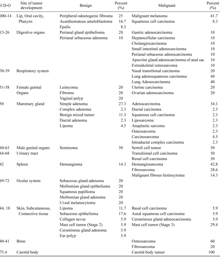
Table III. The histopathologic diagnosis classified by the sites of tumor development
ICD-O Site of tumor
development Benign Percent
(%) Malignant Percent
(%) 000-14 Lip, Oral cavity, Peripheral odontogenic fibroma 25 Malignant melanoma 41.7
Pharynx Acanthomatous ameloblastoma 16.7 Squamous cell carcinoma 8.3
Epulis 8.3
15-26 Digestive organs Perianal gland epithelioma 20 Gastric adenocarcinoma 10 Perianal sebaceous adenoma 10 Hepatocellular carcinoma 10
Cholangiocarcinoma 10
Small intestinal adenocarcinoma 10 Perianal sebaceous adenocarcinoma 10 Apocrine gland adenocarcinoma of anal sac 10
Extraskeletal osteosarcoma 10
30-39 Respiratory system Nasal transitional carcinoma 20
Lung adenosquamous carcinoma 40
Lung Adenocarcinoma 40
51-58 Female genital Leimyoma 20 Uterine carcinoma 20
Organs Fibroma 20 Ovarian adenocarcinoma 20
Vaginal polyp 20
50 Mammary gland Simple adenoma 27.3 Adenocarcinoma 34.1
Complex adenoma 2.3 Ductal carcinoma 2.3
Benign mixed tumor 11.3 Squamous cell carcinoma 2.3
Ductal adenoma 2.3 Liposarcoma 2.3
Lipoma 4.5 Anaplastic sarcoma 2.3
Osteosarcoma 2.3
Carcinosarcoma 4.5
Intraductal complex carcinoma 2.3
60-63 Male genital organs Seminoma 50 Sertoli cell tumor 50
64-68 Urinary tract Transitional cell carcinoma 50
Renal cell carcinoma 50
42 Spleen Hemangioma 14.3 Hemangiosarcoma 42.8
Fibrosarcoma 28.6
Malignant fibrous histiocytoma 14.3 69-72 Ocular system Sebaceous gland adenoma 20
Meibomian gland epithelioma 20
Squamous papilloma 20
Meibomian gland adenoma 20
Uveal melanocytoma 20
44, 18 Skin, Subcutaneous, Lipoma 11.7 Basal cell carcinoma 5.9
Connective tissue Sebaceous epithelioma 17.6 Aural squamous cell carcinoma 5.9
Collagen nevus 5.9 Ceruminous gland adenocarcinoma 5.9
Mast cell tumor (Stage 2) 5.9 Mast cell tumor (Stage 3) 29.4 Ceruminous gland adenoma 5.9
Ear polyp 5.9
40-41 Bone Osteosarcoma 60
Fibrosarcoma 20
75.4 Carotid body Carotid body tumor 100
Table III. Continued
ICD-O Site of tumor
development Benign Percent
(%) Malignant Percent
(%) 47 Peripheral nerves and
autonomic system
Hemangiopericytoma 100
45 Mesothelioma Mesothelioma 100
48 Retroperitoneum Hemangiosarcoma 100
49 Other soft tissue sarcoma Fibrosarcoma 50
Rhabdomyosarcoma 50
81-96 Malignant neoplasms of lymphoid, hematopoietic and related tissue
Lymphoma 100
ICD-O, International Classification of Diseases for Oncology.
Table IV. The Proportional Morbidity Ratio of breeds in specific tumor site
Breeds Mammary
gland tumor
Skin tumor
Oral cavity tumor
Maltese 1.111 0.870 1.111
Yorkshire Terrier 0.617 0.403 2.572
Shih-tzu 0.521 0.906 -
Cocker spaniel 1.389 1.610 -
Mongrel 0.833 1.449 1.852
Poodle 2.315 0.604 -
Alaskan malamute 0.556 - -
Golden Retriever - 5.435 -
melanoma, and osteosarcoma. While adenoma and benign mixed tumors were virtually exclusive to the mammary glands and skin and soft tissues, adenocarcinoma was found in the mammary glands, digestive organs, and respi- ratory systems.
Site of tumor development: The incidence rates of neo- plasia, categorized by site, are shown in Fig. 2. The ma- jority of the neoplasms in females were found in the mam- mary gland (51.5%) and in the skin (9.3%). Similarly, the majority of the neoplasms in males were found in the skin (19.0%), oral cavity (16.7%), and digestive organ (16.7%).
As shown in Table III, the histopathologic diagnosis of tu- mor was classified by the sites of tumor development. In all, 67 different histopathologic types of tumor were identified. While the majority of tumors in the digestive and respiratory system were malignant, the majority of tu- mors in the ocular system were benign. In PMRs of breeds, marked differences were found as shown in Table IV. In mammary gland tumor, Poodles had PMR slightly over 2 which indicating that this breed developed mam- mary gland tumors about double times than other breeds.
Likewise, PMR of Golden retrievers in skin tumor was more than 5 and Yorkshire terrier had PMR about 2.5.
Management of tumors
One-hundred twenty-three surgeries were performed in 114 patients with more than one tumor for the purposes of diagnosis, resection for cure, and palliation of symp- toms. Most patients (106, 86.2%) received surgery for cure. Six patients (4.9%) received biopsy for diagnosis, and eight patients (6.5%) received surgery for palliation of symptoms. Concurrent sterilization with tumor resection
was performed in 28 patients. After the surgery, chemo- therapy was performed in four patients (3.5%).
Surgery
The surgical procedures included biopsy for diagnosis, re- section for cure, and palliation of symptoms. Among the curative surgeries, wide resection (44, 41.5%) was the most common, followed by radical surgery (33, 31.1%), and marginal resection (29, 27.4%). Sterilization with tu- mor resection for intact patients was performed to prevent the recurrence of sex-specific tumor such as tumors of mammary gland, vagina, and perianal gland.
Chemotherapy
Four patients with three mast cell tumors and one fi- brosarcoma were treated by chemotherapy after the surgery. Three patients with mast cell tumors had received treatments, including surgery and the administration of
Table V. The recurrence or suspected metastasis of tumor in the patients
Site of tumor development Recurrence or suspected metastasis (%)
Alimentary tract tumor 33
Mammary gland tumor 26.5
Hematopoietic system tumor 25
Bone tumor 20
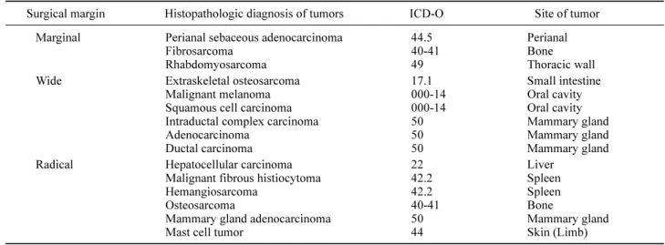
Table VII. The histopathologic diagnosis and site of tumors classified according to the surgical margin
Surgical margin Histopathologic diagnosis of tumors ICD-O Site of tumor Marginal Perianal sebaceous adenocarcinoma
Fibrosarcoma Rhabdomyosarcoma
44.5 40-41 49
Perianal Bone Thoracic wall
Wide Extraskeletal osteosarcoma
Malignant melanoma Squamous cell carcinoma Intraductal complex carcinoma Adenocarcinoma
Ductal carcinoma
17.1 000-14 000-14 50 50 50
Small intestine Oral cavity Oral cavity Mammary gland Mammary gland Mammary gland Radical Hepatocellular carcinoma
Malignant fibrous histiocytoma Hemangiosarcoma
Osteosarcoma
Mammary gland adenocarcinoma Mast cell tumor
22 42.2 42.2 40-41 50 44
Liver Spleen Spleen Bone
Mammary gland Skin (Limb) ICD-O, International Classification of Diseases for Oncology.
Table VI. The survival rates of 6, 9, 12 months and local recurrence or suspected metastasis rate according to the surgical margin
Surgical margin No. 6 months
survival rate (%)
9 months survival rate (%)
12 months survival rate (%)
Local recurrence or suspected metastasis rate (%)
Marginal 3 100 100 100 67
Wide 11 72.7 63.6 45.5 45.5
Radical 7 50 50 33 67
vinblastine and imatinib mesylate, and these patients re- ceived excisional biopsy, incisional biopsy, and radical limb excision, respectively. One patient with intrapelvic fi- brosarcoma received carboplatin, ifosfamide, cyclo- phosphamide, and doxorubicin after palliative surgery.
Survival
Of all patients, 54% follow-up evaluations were not available. Twenty-two percent of patients had died because of malignant tumors, including squamous cell carcinoma, uterine carcinoma, osteosarcoma, hepatocellular carcinoma, nasal transitional cell carcinoma, malignant melanoma, and malignant fibrous histiocytoma. Roughly 5% died because of causes unrelated to the tumor, including chronic renal failure and postoperative shock, while 19% were alive at the end of the study. Recurrence or suspected metastasis was identified in 26 dogs. The recurrence or metastasis rate was identified in alimentary tract tumor (33%), followed by mammary gland tumor (26.5%), tumors of hema- topoietic system (25%), and bone tumor (20%) (Table V).
The survival rate and recurrence or suspected metastasis rate of patients undergone curative surgery was assessed (Table VI). The patients lost during follow-ups were ex- cluded, and the survival rate was calculated from the date of surgery. While the survival rate of patients with wide surgical margin was decreased to 45.5 percent, it was main- tained 100 percent in the patients with marginal surgical margin. The half of the patients with radical surgical margin died at 6 months after the surgery. To find out factors that influence the survival rate, the site and histopathological di-
Figure 3. Survival curve of the malignant mammary tumor.
OHE, Ovariohysterectomy.
agnosis of tumor were classified as shown in Table VII.
Data regarding the survival of dogs with mammary gland tumors after the surgery are presented in Fig. 3.
Median survival times of malignant mammary gland tu- mor, skin and subcutaneous tumor, and splenic tumor were 1,563.0±1,201.7, 469, and 128 days, respectively.
DISCUSSION
The findings about the tumor incidence between the sexes were similar to those of previous studies (9,11,16). It was significantly higher in female dogs than male dogs (p< 0.01). The fact that the male: female ratio was close to 1 after the sex-specific tumors were excluded may have been because of the sex-specific neoplasms, such as mammary gland tumors. The association between the incidence rate of mammary gland tumors and neutering was significantly high (p<0.01), while there was no significant difference in the association with the incidence of overall tumors. This finding was consistent with the previous studies (14).
Although making comparisons with the earlier studies performed around the world can be risky because of the different age structure of the population, age distribution was similar to that of previous studies (9,11). The mean age at diagnosis for benign tumors was lower than that for malignant tumors. This might be because a large pro-
portion of the tumors were mammary gland tumors, which the malignancy of tumor has been reported to increase with aging (17).
The most frequently presented breeds were Maltese, Shih-Tzu, and Yorkshire terrier. These breeds were also commonly observed breeds in another study in Korea, and this may be mainly because these are the major pop- ulations in Korea (13). Because the study was conducted at a teaching hospital, these data did not represent the en- tire canine population in Korea. Therefore, to quantify can- cer occurrence, the PMRs were used. Higher PMRs for mammary gland tumor, skin tumor, and oral cavity tumor were found in Poodles, Golden retrievers, and Yorkshire terriers, respectively.
The predisposition to mammary gland tumors have been found in several breeds, including Poodles, Chihuahuas, Dachshunds, Yorkshire terriers, Maltese, and Cocker span- iels (7,18,19). Like BRCA1 gene and BRCA2 genes in women breast cancer, studies on the English springer span- iels have proved that the same genes influence mammary cancer development (20). As the findings of studies per- formed in the U.S. and Europe are consistent with our own, the high occurrence of mammary gland tumors in Poodles may not be because of the different population structure but because of the fact that Poodles are influ- enced by genetic factors.
Similar to the studies from northern Italy, Tulsa (US), and California (US), the two most frequent tumor locations in female patients were mammary glands and skin and subcutaneous tissues (7,10,14). For male patients, skin, or- al cavity, and digestive tissues were the most common tu- mor sites. Because the tumors of mammary gland, genital organs, and skin and subcutaneous tissues are easier to rec- ognize by physical examination than the tumors of other internal organs that require specific examinations, these tu- mors may be easily found and overrepresented (10).
According to the recent studies, skin tumors are more prevalent than mammary gland tumors when compared with the past studies (8,11). This may be attributable to the increasing frequency of spaying at a young age.
Adenoma and epithelioma comprised a large proportion of benign tumors in the present study. This finding with adenoma is similar to earlier observations (8,11), but dif- ferent frequencies for the lipoma and the cutaneous histio- cytoma were found, which have been cited as commonly observed tumors in many studies (8,11-13). The cutaneous
histiocytoma is a benign tumor that undergoes spontaneous regression and, with respect to lipoma, marginal resection is recommended only when tumors interfere with normal function (21,22). Because only cases verified using histol- ogy were included in this study, and lipoma and cutaneous histiocytoma generally do not require surgery, they might have been underrepresented.
The most commonly observed malignant tumor types were adenocarcinoma and soft tissue sarcomas. The major proportional trends in terms of tumor types were quite sim- ilar to previous studies except for lymphosarcoma (11,16).
Most patients with lymphosarcoma have multicentric diseases. Therefore they require systemic chemotherapy to effectively treat their disease (23). Because only surgically managed patients were involved in the study, lympho- sarcoma might have been underrepresented.
Due to the small size of patients with complete fol- low-up, only tendency of outcomes from curative surgeries was investigated. According to the results of the study, no propensity of the recurrence rate classified by the surgery type was identified, and the wider the margin of surgery, the lower the survival rate of 6 and 12 months. While be- havior of tumor excised by marginal surgery tends to prog- ress slowly and present relatively low rate of metastasis, most of the tumors undertaken radical and wide surgery were highly malignant with aggressive biological behavior and rapid and widespread metastasis such as hemangio- sarcoma, osteosarcoma, and splenic fibrous histiocytoma (24-27). Generally, surgical margin for curative surgery is determined by the propensity for local recurrence and mi- croscopic metastasis (28). Considering that, the results of our study may be explained by tendency to choose sur- geries of extensive margins with aggressive tumors. Also, there are a lot of reports describing micrometastasis (29-31). The intra-abdominal tumors can metastasize through the vessels, lymphatics, and peritoneal surface (30). Because of this, metastasis can occur in patients un- dergone tumor resection for cure (31). In human medicine, many reports describing shedding of malignant tumor cells into the blood according to surgical stress especially color- ectal tumors were published, but there are still con- troversies (29,31,32). By contrast, animal studies have re- ported a lot of evidence about the tumor cells in the blood (33-35). Micrometastasis may more often develop with the tumors within the abdomen because of the relatively richer blood supply and lymphatics than external organs such as
skin. Since the majority of tumors undertaken wide or radi- cal surgery were tumors within the abdomen, the results of study may be influenced by stress from surgical manip- ulation and anatomic condition.
According to the previous studies, OHE before or at the time of mastectomy is not a prognostic factor in dogs with established mammary gland tumors (36,37). Contrary to these studies, a recent study suggested that OHE up to two years before mastectomy significantly influences survival time (38). Our findings also support that OHE may influ- ence the survival time like the recent study.
Identifying the incidence rate and patterns of specific breeds, ages, and sex groups within specific geographic areas may help veterinary clinicians because the data are highly influenced by geographic areas (6). Although in- cidence is the most useful statistic for occurrence, the ac- curately calculated population at risk is needed to find out valid incidence (22). In this respect, this study has some limitations because the study was conducted in a teaching hospital, so the population might have been biased.
Therefore, further investigations based on larger pop- ulation will be needed to increase the accuracy of the pop- ulation size assessment.
In this study, we focused on canine spontaneous tumors treated surgically and investigated the outcome from clin- ical perspective. The data obtained from this study may be also very useful for the translation study in human can- cer field in future.
CONFLICTS OF INTEREST
The authors have no financial conflict of interest.
REFERENCES
1. Jemal, A., F. Bray, M. Melissa, J. Ferlay, E Ward, and D. Forman.
2011. Global cancer statistics. CA Cancer J. Clin. 61: 69-90.
2. Siegel, R., J. Ma, Z. Zou, and A. Jemal. 2014. Cancer statistics, 2014. CA Cancer J. Clin. 64: 9-29.
3. Fleming, J. M., K. E. Creevy, and D. E. Promislow. 2011.
Mortality in North American dogs from 1984 to 2004: An inves- tigation into age-, size-, and breed-related causes of death. J. Vet.
Intern. Med. 25: 187-198.
4. Inoue, M., A. Hasegawa, Y. Hosoi, and K. Sugiura. 2015. A cur- rent life table and causes of death for insured dogs in Japan. Prev.
Vet. Med. 120: 210-218.
5. Misdorp, W. 1996. Veterinary cancer epidemiology. Vet. Q. 18:
32-36.
6. Bronden, L. B., A. Flagstad, and A. T. Kristensen. 2007. Veterinary
cancer registries in companion animal cancer: a review. Vet.
Comp. Oncol. 5: 133-144.
7. MacVean, D. W., A. W. Monlux, P. S. Jr. Anderson, S. L. Silberg, and J. F. Roszel. 1978. Frequency of canine and feline tumors in a defined population. Vet. Pathol. 15: 700-715.
8. Dobson, J. M., S. Samuel, H. Milstein, K. Rogers, and J. L. Wood.
2002. Canine neoplasia in the UK: estimates of incidence rates from a population of insured dogs. J. Small Anim. Pract. 43:
240-246.
9. Merlo, D. F., L. Rossi, C. Pellegrino, M. Ceppi, U. Cardellino, C. Capurro, A. Ratto, P. L. Sambucco, V. Sestito, G. Tanara, and V. Bocchini. 2008. Cancer incidence in pet dogs: Findings of the animal tumor registry of Genoa, Italy. J. Vet. Intern. Med. 22:
976-984.
10. Vascellari, M., E. Baioni, G. Ru. A. Carminato, and F. Mutinelli.
2009. Animal tumour registry of two provinces in northern Italy:
Incidence of spontaneous tumours in dogs and cats. BMC Vet. Res.
5: 39.
11. Bronden, L. B., S. S. Nielsen, N. Toft, and A. T. Kristensen. 2010.
Data from the danish veterinary cancer registry on the occurrence and distribution of neoplasms in dogs in Denmark. Vet. Rec. 166:
586-590.
12. Pakhrin. B., M. S. Kang, I. H. Bae, M. S. Park, H. Jee, M. H.
You, J. H. Kim, B. I. Yoon, Y. K. Choi, and D. Y. Kim. 2007.
Retrospective study of canine cutaneous tumors in Korea. J. Vet.
Sci. 8: 229-236.
13. Kim, Y. H., N. K. Ahn, I. S. Roh, B. I. Yoon, and J. H. Han.
2009. Retrospective investigation of canine skin and mammary tu- mors in Korea. J. Vet. Clin. 26: 556-562.
14. Dorn, C. R., D. O. Taylor, R. Schneider, H. H. Hibbard, and M.
R. Klauber. 1968. Survey of animal neoplasms in Alameda and Contra Costa Counties, California. II. Cancer morbidity in dogs and cats from Alameda County. J. Natl. Cancer Inst. 40: 307-331.
15. Fritz, A., C. Percy, A. Jack, K. Shanmugaratnam, L. Sobin, D.
Parkin, and S. Whelan. 2013. In International Classification of Diseases for Oncology (ICD-O). 3rded. A. Fritz, C. Percy, A. Jack.
K. Shanmugaratnam, L. Sobin, D. Parkin, and S. Whelan, eds.
World Health Organization, Geneva. p.1-242.
16. Cohen, D., J. S. Reif, R. S. Brodey, and H. Keiser. 1974.
Epidemiological analysis of the most prevalent sites and types of canine neoplasia observed in a veterinary hospital. Cancer Res.
34: 2859-2868.
17. Mitchell, L., F. A. De la Iglesia, M. S. Wenkoff, A. A. Van Dreumel, and G. Lumb. 1974. Mammary tumors in dogs: survey of clinical and pathological characteristics. Can. Vet. J. 15: 131-138.
18. Moe, L. 2001. Population-based incidence of mammary tumours in some dog breeds. J. Reprod. Fertil. Suppl. 57: 439-443.
19. Todorova, I. 2006. Prevalence and etiology of the most common malignant tumours in dogs and cats. B. J. Vet. Med. 9: 85-98.
20. Rivera, P., M. Melin, T. Biagi. T. Fall, J. Haggstrom, K.
Lindblad-Toh, and H. von Euler. 2009. Mammary tumor develop- ment in dogs is associated with BRCA1 and BRCA2. Cancer Res.
69: 8770-8774.
21. Pires, I., A. Alves, F. Queiroga, F. Silva, and C. Lopes. 2013.
Regression of canine cutaneous histiocytoma: reduced proliferation or increased apoptosis? Anticancer Res. 33: 1397-1400.
22. Butler, M., N. Bonnett, and L. Page. 2013. Epidemiology and the evidence-based medicine approach. In Withrow & MacEwen’s
Small Animal Clinical Oncology, 5thed. S. J. Withrow, D. M. Vail, and R. L. Page, eds. Elsevier saunders, St. Louis, Missouri. p.68-82.
23. Ettinger, S. N. 2003. Principles of treatment for canine lymphoma.
Clin. Tech. Small. Anim. Pract. 18: 92-97.
24. Spodnick, G. J., J. Berg, W. M. Rand, S. H. Schelling, G. Couto, H. J. Harvey, R. A. Henderson, G. MacEwen, N. Mauldin, D. L.
McCaw. A. S. Moore, W. Morrison, A. M. Norris, J. O’Bradovich, D. A. O’Keefe, R. Page, D. Ruslander, J. Klausner, R. C. Straw, and J. P. Thompson. 1992. Prognosis for dogs with appendicular osteosarcoma treated by amputation alone: 162 cases (1978-1988).
J. Am. Vet. Med. Assoc. 200: 995-999.
25. Kuntz, C. A., W. S. Dernell, B. E. Powers, C. Devitt, R. C. Straw, and S. J. Withrow. 1997. Prognostic factors for surgical treatment of soft-tissue sarcomas in dogs: 75 cases (1986-1996). J. Am. Vet.
Med. Assoc. 211: 1147-1151.
26. Spangler, W. L., and P. H. Kass. 1997. Pathologic factors affecting postsplenectomy survival in dogs. J. Vet. Intern. Med. 11: 166-171.
27. Wood, C. A., A. S. Moore, J. M. Gliatto, L. A. Ablin, R. J. Berg, and W. M. Rand. 1998. Prognosis for dogs with stage I or II splenic hemangiosarcoma treated by splenectomy alone: 32 cases (1991-1993). J. Am. Anim. Hosp. Assoc. 34: 417-421.
28. Jermyn, K., B. Duncan, and X. Lascelles. 2011. Principles of on- cological surgery. In: BSAVA Manual of Canine and Feline Oncology, 3rded. M. Dobson, and D. Lascelles, eds, BSAVA, Quedgeley, Gloucester. p.44-59.
29. Moore, G. E. A. Sandberg, and J. R. Schubarg. 1957. Clinical and experimental observations of the occurrence and fate of tumor cells in the blood stream. Ann. Surg. 146: 580-587.
30. Sugarbaker, P. H. 2008. Overview of peritoneal carcinomatosis.
Cancerologia 3: 119-124.
31. Park, S. Y., G. S. Choi, J. S. Park, H. J. Kim, J. P. Ryuk, and W. H. Choi. 2012. Influence of surgical manipulation and surgical modality on the molecular detection of circulating tumor cells from colorectal cancer. J. Korean Surg. Soc. 82: 356-364.
32. Atkin, G., A. Chopada, and I. Mitchell. 2005. Colorectal cancer metastasis: in the surgeon's hands? Int. Semin. Surg. Oncol. 2: 5.
33. Romsdahl, M. M., R. G. McGrath, E. Hoppe, and E. A. McGrew.
1965. Experimental model for the study of tumor cells in the blood. Acta. Cytol. 9: 141-145.
34. Nishizaki, T., T. Matsumata , T. Kanematsu, C. Yasuaga, and K.
Sugimachi. 1990. Surgical manipulation of VX2 carcinoma in the rabbit liver evokes enhancement of metastasis. J. Surg. Res. 49:
92-97.
35. Abgral, R., G. Valette, P. Robin, J. Rousset, N. Keromnes, P. Y.
Le Roux, R. Marianowski, and P. Y. Salaun. 2015. Prognostic evaluation of percentage variation of metabolic tumor burden cal- culated by dual-phase 18FDG PET-CT imaging in patients with head and neck cancer. Head & Neck 55: 691-696.
36. Morris, J. S., J. M. Dobson, D. E. Bostock, and E. O’Farrell. 1998.
Effect of ovariohysterectomy in bitches with mammary neoplasms.
Vet. Rec. 142: 656-658.
37. Yamagami, T., T. Kobayashi, K. Takahashi, and M. Sugiyama.
1996. Influence of ovariohysterectomy at the time of mastectomy on the prognosis for canine malignant mammary tumors. J. Small Anim. Pract. 37: 462-464.
38. Sorenmo, K. U., F. S. Shofer, and M. H. Goldschmidt. 2000.
Effect of spaying and timing of spaying on survival of dogs with mammary carcinoma. J. Vet. Intern. Med. 14: 266-270.