D I A B E T E S & M E T A B O L I S M J O U R N A L D I A B E T E S & M E T A B O L I S M J O U R N A L
This is an Open Access article distributed under the terms of the Creative Commons Attribution Non-Commercial License (https://creativecommons.org/licenses/by-nc/4.0/) which permits unrestricted non-commercial use, distribution, and reproduction in any medium, provided the original work is properly cited.
Non-Alcoholic Fatty Liver Disease in Patients with Type 2 Diabetes Mellitus: A Position Statement of the Fatty Liver Research Group of the Korean Diabetes Association
Byung-Wan Lee1, Yong-ho Lee1, Cheol-Young Park2, Eun-Jung Rhee2, Won-Young Lee2, Nan-Hee Kim3, Kyung Mook Choi3, Keun-Gyu Park4, Yeon-Kyung Choi4, Bong-Soo Cha1, Dae Ho Lee5, on Behalf of The Korean Diabetes Association (KDA) Fatty Liver Research Group
1Division of Endocrinology and Metabolism, Department of Internal Medicine, Yonsei University College of Medicine, Seoul,
2 Division of Endocrinology and Metabolism, Department of Internal Medicine, Kangbuk Samsung Hospital, Sungkyunkwan University School of Medicine, Seoul,
3Division of Endocrinology and Metabolism, Department of Internal Medicine, Korea University College of Medicine, Seoul,
4Division of Endocrinology and Metabolism, Department of Internal Medicine, School of Medicine, Kyungpook National University, Daegu,
5 Division of Endocrinology and Metabolism, Department of Internal Medicine, Gachon University Gil Medical Center, Gachon University College of Medicine, Incheon, Korea
This clinical practice position statement, a product of the Fatty Liver Research Group of the Korean Diabetes Association, propos- es recommendations for the diagnosis, progression and/or severity assessment, management, and follow-up of non-alcoholic fatty liver disease (NAFLD) in patients with type 2 diabetes mellitus (T2DM). Patients with both T2DM and NAFLD have an increased risk of non-alcoholic steatohepatitis (NASH) and fibrosis and a higher risk of cardiovascular diseases and diabetic complications compared to those without NAFLD. With regards to the evaluation of patients with T2DM and NAFLD, ultrasonography-based stepwise approaches using noninvasive biomarker models such as fibrosis-4 or the NAFLD fibrosis score as well as imaging stud- ies such as vibration-controlled transient elastography with controlled attenuation parameter or magnetic resonance imaging- proton density fat fraction are recommended. After the diagnosis of NAFLD, the stage of fibrosis needs to be assessed appropri- ately. For management, weight reduction achieved by lifestyle modification has proven beneficial and is recommended in combi- nation with antidiabetic agent(s). Evidence that some antidiabetic agents improve NAFLD/NASH with fibrosis in patients with T2DM is emerging. However, there are currently no definite pharmacologic treatments for NAFLD in patients with T2DM. For specific cases, bariatric surgery may be an option if indicated.
Keywords: Cardiovascular disease; Diabetes mellitus, type 2; Life style; Non-alcoholic fatty liver disease
Corresponding authors: Bong-Soo Cha https://orcid.org/0000-0003-0542-2854 Division of Endocrinology and Metabolism, Department of Internal Medicine, Yonsei University College of Medicine, 50-1 Yonsei-ro, Seodaemun-gu, Seoul 03722, Korea E-mail: [email protected]
Dae Ho Lee https://orcid.org/0000-0002-8832-3052
Division of Endocrinology and Metabolism, Department of Internal Medicine, Gachon University Gil Medical Center, Gachon University College of Medicine, 21 Namdong- daero 774beon-gil, Namdong-gu, Incheon 21565, Korea
INTRODUCTION
This clinical practice position statement, a product of the Fatty
Liver Research Group (FLRG) of the Korean Diabetes Associa- tion (KDA), proposes recommendations for the diagnosis, progression and/or severity assessment, management, and fol- https://doi.org/10.4093/dmj.2020.0010
pISSN 2233-6079 · eISSN 2233-6087
low-up of non-alcoholic fatty liver disease (NAFLD) in pa- tients with type 2 diabetes mellitus (T2DM). The literature was retrieved by an extensive PubMed search up to April 2019. Af- ter extensive reviews and discussions for the last 3 years by the research group, two sentinel reviews were published in Diabe- tes and Metabolism Journal in 2019. The draft of the statement was presented and discussed in a session of the FLRG during the 32nd KDA scientific meeting in 2019. Then, the statement was further discussed, edited and updated until the final ac- ceptance of the statement in the journal. Epidemiological evi- dence suggests a strong bidirectional relationship between type 2 diabetes mellitus (T2DM) and non-alcoholic fatty liver dis- ease (NAFLD), including the development and severity of NAFLD, progression to non-alcoholic steatohepatitis (NASH), and advanced fibrosis, independent of liver enzymes [1]. Fur- thermore, the coexistence of T2DM and NAFLD results in an unfavorable metabolic profile and an increasing cardiovascular (CV) risk [2-4]. Although steatosis can be defined by various clinically available diagnostic tools, it can be numerically and strictly defined by assessing liver fat: ≥5% of fat-containing he- patocytes in histology; proton density fat fraction (PDFF) ≥5%
on magnetic resonance imaging (MRI), or >5.5% on proton magnetic resonance spectroscopy (1H-MRS) [5,6]. The defini- tive diagnosis of NASH requires a liver biopsy.
Among many treatments for NAFLD in patients with T2DM, weight reduction is the only approved option for NAFLD. However, it is not easy to maintain weight loss by only lifestyle modification strategies, so additional pharmacological options should be supported. To date, although many drugs have been investigated, pioglitazone could be the first-line
therapy in patients with T2DM and NAFLD. Many drugs are currently being developed and investigated, and combination strategies will be introduced for the treatment of NAFLD and diabetes in the future.
PREVALENCE OF NAFLD IN PATIENTS WITH T2DM
Keynotes
- The prevalence of NAFLD in patients with T2DM is more than two times higher than that in the normal population.
-NAFLD is a risk factor for T2DM.
NAFLD is the most common liver disorder, affecting 20% to 40% of adults; the prevalence rates differ according to the diag- nostic method, age, sex, and ethnicity [6-8]. In patients with T2DM, NAFLD prevalence ranges from 70% to 95%; the rate is extremely high, up to 98%, in patients with morbid obesity [8]. In the general Korean population, NAFLD prevalence ranges from 16.1% to 25.2% (Table 1) [9,10].
Half of patients with T2DM have NAFLD despite having nor- mal alanine aminotransferase (ALT) levels [11,12]. Population based studies have reported the prevalence of NASH to be 17.6% to 22%% in individuals with T2DM and 3.7% in nondia- betic individuals [13]. Furthermore, the prevalence of NASH among biopsied patients with diabetes can be as high as 64.0%, whereas the prevalence of advanced fibrosis (≥F3) in patients with T2DM is approximately 10.4% [14]. The NAFLD preva- lence in Asians is not lower than that in Caucasians. In a study involving Korean patients with T2DM who were subjected to ultrasonography (US) examination in a university-based diabe- Table 1. The prevalence of NAFLD and NASH in patients with diabetes
Study Study population Diagnostic methods Categories Prevalence, %
Mohan et al. (2009) [16] In 132 Indian adults with diabetes US NAFLD 54.5
Kim et al. (2014) [17] In 4,437 Korean patients with T2DM US NAFLD 72.7
Targher et al. (2007) [18] In 2,839 Italian patients with T2DM US NAFLD 69.5
Portillo-Sanchez et al. (2015) [11] In 103 American patients with diabetes with normal plasma aminotransferases
1H-MRS NAFLD 50
Williams et al. (2011) [13] In 54 biopsied patients with diabetes Liver biopsy NASH 22 Hyysalo et al. (2014) [12] In 115 biopsied participants in in the Finnish
population-based D2D-study
Liver biopsy NASH 17.6
Kim et al. (2014) [15] In 929 Korean patients with diabetes US NAFLD 63.3
US, ultrasonography; NAFLD, non-alcoholic fatty liver disease; 1H-MRS, proton magnetic resonance spectroscopy; T2DM, type 2 diabetes mel- litus; NASH, non-alcoholic steatohepatitis.
tes clinic, 63.3% of patients had NAFLD (Table 1) [11-13,15-18].
Furthermore, the risk of diabetes in subjects with NAFLD has been shown to be 2-fold higher than that in control sub- jects, even after adjustment for various risk factors [19,20].
When obesity, insulin resistance, or hyperglycemia is com- bined with NAFLD, T2DM risk is dramatically increased [2].
In line with these studies, the resolution of steatosis in patients with NAFLD decreases T2DM risk by 39% to 82% [21,22].
THE PROGRESSION OF NAFLD AND DIABETES
Keynotes
- Patients with T2DM and NAFLD have an increased risk of NASH and fibrosis.
- Patients with both T2DM and NAFLD have a higher risk of CV diseases and diabetic complications compared to those without NAFLD.
Approximately 10% to 35% of subjects with normal liver his- tology progress to steatosis, 12% to 44% of those with hepatic steatosis progress to steatohepatitis, and up to 15% of patients with NASH are known to progress to cirrhosis [23]. In patients with NAFLD, the prevalence of NASH is approximately 60% in biopsy-indicated patients and 29.9% in patients without such an indication [8]. Studies have shown that increasing age, dia- betes, and hypertension are predictive clinical parameters for fibrosis [24,25].
NAFLD and NASH can progress to cirrhosis and liver failure in up to 15% of affected patients [26]. These patients are also at risk of developing hepatocellular carcinoma (HCC). Advanced age, high aspartate aminotransferase (AST) levels, thrombocy- topenia (marker of progression of liver fibrosis), and diabetes were identified as risk factors for the development of HCC in Japanese patients with US-diagnosed NAFLD. Patients with NAFLD with advanced stages of fibrosis have a 7-fold higher risk of HCC compared to those without liver disease [27].
However, the cause of cirrhosis remains unclear in 30% of cas- es, and most of these cases are now considered NAFLD-related [28]. Cryptogenic cirrhosis was reported to be an underlying disease in approximately 7% of HCC cases in Korea and Japan compared with 13% in the United States [28,29]. T2DM is also closely associated with progression to NASH, advanced fibro- sis and the development of HCC [1,7].
NAFLD is a consequence but also a precursor of metabolic comorbidities, including diabetes, dyslipidemia, and hyperten- sion, and thus, NAFLD increases CV events, and mortality
even in the absence of these comorbidities [30]. Furthermore, hepatic fibrosis is a key predictor of liver-related outcomes and is also associated with all-cause and CV mortality as well as mortality due to cirrhosis, HCC, and infectious diseases in NAFLD patients [31,32]. Diabetes is an important factor af- fecting all-cause and CV mortality in patients with NAFLD [31]. Thus, after the diagnosis of NAFLD, the stage of fibrosis needs to be assessed appropriately [31]. There is evidence that the presence of NAFLD in patients with T2DM is associated with increased risks of macrovascular and diabetic microvas- cular complications as well as chronic kidney disease [2].
DIAGNOSIS OF NAFLD
Keynotes
- NAFLD can be diagnosed by a two-step process: (1) confirmation of he- patic steatosis, either by imaging modalities or histology and (2) exclu- sion of secondary causes of liver steatosis.
Routine screening for NAFLD in patients with T2DM is not currently recommended because of the unclear cost-effective- ness and uncertainties with diagnostic testing and treatments [33]. In individuals with and without diabetes, three important processes should be used to diagnose and assess NAFLD: (1) determine the existence of hepatic steatosis, either by imaging or histology; (2) exclude secondary causes of liver steatosis;
and (3) assess NAFLD severity by establishing the presence of moderate-to-severe fibrosis (fibrosis stage of at least F2) [2].
Before making the diagnosis of NAFLD, secondary causes of hepatic fat accumulation and significant alcohol consumption (≥21 drinks/week for men and ≥14 drinks/week for women) need to be excluded [5]. Alcohol consumption over a 2-year time frame needs to be surveyed in detail using validated ques- tionnaires [5]. However, it is noteworthy that there is also a synergy between alcohol intake and obesity or genetic risk fac- tors of NAFLD progression for any given level of alcohol intake [6]. Even in patients whose alcohol consumption level is low, meeting the diagnostic criteria of NAFLD, small amounts of alcohol intake may affect outcomes in NAFLD, which warrants further study [5,34]. After performing a history and examina- tion, the next investigations are to establish whether the patient has NAFLD or another liver condition.
A consensus regarding initial blood tests for NAFLD has not been reached among guidelines [33]. The decision on the ex- tent of liver blood tests and interpretation of the results should be determined in a clinical context. In adults, initial screening
tests may include abdominal US, hepatitis B surface antigen, hepatitis C antibody (with follow-on polymerase chain reac- tion if positive), anti-mitochondrial antibody, anti-smooth muscle antibody, antinuclear antibody, serum immunoglobu- lins, and simultaneous serum ferritin and transferrin satura- tion [33]. Although ALT levels have been shown to be the best single biochemical correlate of hepatic steatosis, liver enzyme levels can be normal, fluctuating, or elevated in patients with NAFLD [23]. There have been suggestions that the current ref- erence intervals for ALT may be too high [35,36], and a recent guidance recommending an ALT of >30 U/L as being signifi- cant in males and >25 U/L significant for females [37].
Noninvasive imaging studies to assess hepatic steatosis and hepatic fibrosis
Keynotes
- Despite its limited accuracy, US is a useful screening tool to detect hepatic steatosis with other possible structural abnormalities in patients who are suspected of having NAFLD.
- Vibration-controlled transient elastography (VCTE) with controlled at- tenuation parameter (CAP) is a simple quantitative index to detect ste- atosis in clinical practice, but CAP values should be carefully interpreted with patient factors such as obesity.
- As the MRI-PDFF is a highly reliable modality for detecting steatosis, comparable to liver biopsy, it is useful for sequent monitoring, such as in clinical trials.
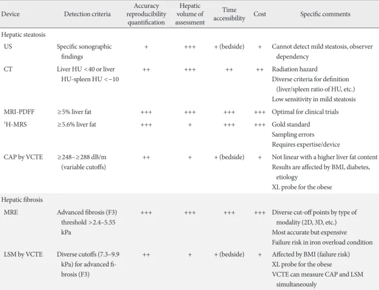
Table 2. Summary of the currently used imaging devices for the quantification of hepatic steatosis and fibrosis
Device Detection criteria Accuracy
reproducibility quantification
Hepatic volume of assessment
Time
accessibility Cost Specific comments Hepatic steatosis
US Specific sonographic findings
+ +++ + (bedside) + Cannot detect mild steatosis, observer dependency
CT Liver HU <40 or liver HU-spleen HU <−10
++ +++ ++ ++ Radiation hazard
Diverse criteria for definition (liver/spleen ratio of HU, etc.) Low sensitivity in mild steatosis
MRI-PDFF ≥5% liver fat +++ +++ +++ +++ Optimal for clinical trials
1H-MRS ≥5.6% liver fat +++ + +++ +++ Gold standard
Sampling errors Requires expertise/device CAP by VCTE ≥248–≥288 dB/m
(variable cutoffs)
++ + + (bedside) + Not linear with a higher liver fat content Results are affected by BMI, diabetes,
etiology
XL probe for the obese Hepatic fibrosis
MRE Advanced fibrosis (F3) threshold >2.4–5.55 kPa
+++ +++ +++ +++ Diverse cut-off points by type of modality (2D, 3D, etc.) Most accurate but expensive Failure risk in iron overload condition LSM by VCTE Diverse cutoffs (7.3–9.9
kPa) for advanced fi- brosis (F3)
++ + + (bedside) + Affected by BMI (failure risk) XL probe for the obese
VCTE can measure CAP and LSM simultaneously
US, ultrasonography; CT, computed tomography; MRI-PDFF, magnetic resonance imaging-proton density fat fraction; 1H-MRS, proton mag- netic resonance spectroscopy; CAP, controlled attenuation parameter; VCTE, vibration-controlled transient elastography; BMI, body mass in- dex; MRE, magnetic resonance elastography; LSM, liver stiffness measurement.
Currently, US, computed tomography (CT), MRI, 1H-MRS, and VCTE are available tools to measure NAFLD depending on the center or clinic (Table 2, Fig. 1).
Ultrasonography and computed tomography
US is the recommended first line screening method for pa- tients with T2DM by the European NAFLD guidelines [34].
US has interobserver variability and limited sensitivity to de- tect mild (<20%) steatosis [38], while optimum sensitivity for liver US was reported to be achieved at a liver fat content of
≥12.5% (sensitivity of approximately 80% to 85%) [39,40].
Similar to US, CT has limited sensitivity to detect mild ste- atosis (<30% liver fat). Radiation exposure is an additional drawback. Thus, CT scans cannot be recommended for the di- agnosis of hepatic steatosis [2].
Vibration-controlled transient elastography
VCTE measures the speed of a mechanically induced shear wave across the liver using pulse-echo ultrasonic acquisitions to obtain a liver stiffness measurement (LSM), as a marker of hepatic fibrosis, and ultrasonic attenuation through the liver to derive the CAP, as a marker of hepatic steatosis. VCTE is an easy-to-perform tool to obtain both LSM and CAP values us- ing an M (3.5 MHz, at 2.5 to 6.5 cm-depth) or XL (2.5 MHz, at 3.5 to 7.5 cm-depth) ultrasound probe. Although LSM and CAP values are relatively reliable and well-validated, these pa- rameters are affected by various patient factors, especially body mass index which may lead to an overestimation [41,42]. With the availability of the XL probe, which has been proven for use in patients with morbid obesity, the failure rate of VCTE for obtaining the LSM and CAP values was reported to be less
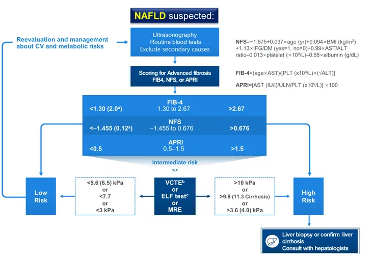
Exclude secondary causes
Fig. 1. Algorithm for non-alcoholic fatty liver disease (NAFLD) evaluation. CV, cardiovascular; NFS, NAFLD fibrosis score; BMI, body mass index; IFG, impaired fasting glucose; DM, diabetes mellitus; AST, aspartate aminotransferase; ALT, alanine aminotrans- ferase; FIB-4, fibrosis-4; PLT, platelet; APRI, AST to platelet ratio index; ULN, upper limit of normal; VCTE, vibration-controlled transient elastography; ELF, enhanced liver fibrosis; MRE, magnetic resonance elastography. aHigher cutoffs for patients aged >65 years, bVariable cutoffs have been suggested. Measured values are affected by body factors, cFurther validation is required.
than 5%, while the significant disagreement between the first and second readings for LSM and CAP when obtained back to back was 18% and 11%, respectively [43].
Magnetic resonance imaging-based techniques
MRI-PDFF can provide fast, quantitative, accurate, and gener- alized hepatic fat measurements for the entire liver, thereby overcoming the heterogeneity of fat deposition [44,45], while magnetic resonance elastography (MRE) is a useful diagnostic tool for the differentiation of histologically determined ad- vanced liver fibrosis from non-advanced fibrosis [46,47]. Addi- tionally, LSM measured by MRE may reflect whole liver pa- thology rather than that of a very small area. MRI-PDFF is be- coming the gold standard for hepatic steatosis quantification [48]. MRI-PDFF or 1H-MRS can measure liver fat more pre- cisely than biopsy [49,50]. In a secondary analysis of a clinical trial involving patients with biopsy-proven NASH, it was shown that histologic responders (≥2-point reduction in NAFLD activity score [NAS], without any worsening of fibrosis on liver biopsy) had a proportional reduction in the MRI- PDFF of 29.3% from baseline which was statistically significant when compared with histologic nonresponders [51]. However, at the present time, magnetic resonance techniques cannot be used for the assessment of NASH resolution or the exact assess- ment of fibrosis progression or improvement. Thus, these is- sues need to be addressed when designing future clinical trials.
Liver biopsy
Liver biopsy provides an accurate diagnosis in approximately 90% of patients with unexplained abnormalities revealed by liver function tests [52]. Noninvasive tools for fibrosis staging are not reliable for therapeutic or other decisions but may be helpful for excluding the probability of significant fibrosis and for predicting advanced fibrosis, thus guiding the decision to perform liver biopsy or not in a patient with NAFLD [53].
Further studies are required to determine whether an active NASH screening strategy in patients with T2DM and active therapy on the basis of the currently available evidence are cost-effective and beneficial for long-term CV and liver-related outcomes. Health care providers taking care of patients with diabetes are strongly encouraged to be vigilant for any signs and symptoms of chronic liver disease and, if indicated, further assessment of the stage of hepatic fibrosis should be conducted [54,55].
Further assessment after the diagnosis of hepatic steatosis Keynotes
- Staged approaches are recommended to determine the extent of liver fi- brosis in patients with NAFLD.
- To estimate hepatic fibrosis using nonimaging modalities, noninvasive biomarker models such as fibrosis-4 (FIB-4) or the NAFLD fibrosis score (NFS) are well-validated and widely used for screening high-risk patients.
- To assess liver fibrosis, VCTE is a point-of-care imaging device with moderate accuracy and high accessibility, while MRE has better accuracy performance, but it is not widely available.
The severity of hepatic fibrosis is the most powerful determi- nant of long-term outcomes, including mortality [31]; thus, the assessment of fibrosis is essential to manage patients with NAFLD. Although quantitative noninvasive imaging assess- ment of steatosis is feasible in the clinical setting, none of the current imaging technologies can reliably differentiate simple steatosis from NASH or detect its progression to early stage fi- brosis [56]. Several prediction scores have been developed and validated to identify or exclude advanced fibrosis (≥F3). First- line testing should use either FIB-4 or the NFS. As an alterna- tive, the AST to platelet ratio index [APRI] may also be used.
Those patients with indeterminate FIB-4 (1.3 to 2.67) or NFS scores (−1.455 to 0.676) require a second-line test in a context dependent manner: VCTE, serum enhanced liver fibrosis (ELF) score, or MRE. The ELF score is a surrogate index based on extracellular matrix panel consisting of plasma concentra- tions of hyaluronic acid, tissue inhibitor of metalloproteinase 1, and procollagen type III amino-terminal peptide [57] Howev- er, the ELF test seems to need further validation, even though European guidelines recommend the test [34]. Patients with high FIB-4 (>2.67) or NFS (>0.676) values should be consid- ered for referral to a specialist clinic irrespective of second-line tests (Fig. 1) [33]. Thus, the use of these scores in combination with imaging studies seems to be more reasonable. The cyto- keratin 18 fragment test did not outperform AST measure- ment in discriminating NASH from simple steatosis [58].
By using VCTE, the threshold values of CAP (≥263 to 288 dB/m) for steatosis and the LSM (≥8.6 to 9.6 kPa) for ad- vanced fibrosis have been variably reported [39,48]. The very low values of the LSM (e.g., <5.6 or <6.5 kPa combined with a result from other noninvasive tests) suggest an exclusion of moderate fibrosis (Fig. 1) [59]. Although the threshold levels for the changes of MRI-PDFF to define improvement of ste- atosis seem to be 25% to 30%, the cutoff value for percent change in MRE to define the improvement of fibrosis needs
further studies; it seems that more than a 20% difference is re- quired to be confident [60].
Genetic variant study Keynotes
- Genetic tests for the assessment of NAFLD are not officially recommend- ed, but identifying the carriers of high-risk genetic variants may be help- ful in some specific conditions.
Genetic factors are also important factors that determine sus- ceptibility to the development and progression of NAFLD, con- sidering that heritable factors account for approximately 50% of the interindividual differences in the prevalence of NASH with cirrhosis in a twin study [61]. Among several genetic risk fac- tors, single-nucleotide polymorphisms (SNPs) in patatin-like phospholipase domain–containing 3 (PNPLA3) (rs738409 c.444 C>G, p.I148M), transmembrane 6 superfamily, member 2 (TM6SF2) (rs58542926 c.449 C>T, p.E167K), and membrane bound O-acyltransferase domain containing 7-transmembrane channel-like 4 (MBOAT7) (rs641738 C>T) have been relatively well validated to promote the development of NAFLD and its progression (i.e., cirrhosis, HCC, or both) [62-66].
PNPLA3 encodes adiponutrin, a triglyceride (TG) lipase that regulates both TG and retinoid metabolism. The PNPLA3 I148M variant is resistant to proteasomal degradation by evad- ing ubiquitylation and accumulates on lipid droplets, which in- terferes with lipolysis and causes a change in phospholipid re- modeling [67]. The PNPLA3 SNP rs738409 is strongly associat- ed with hepatic steatosis, steatohepatitis, fibrosis, and HCC [66].
TM6SF2 is involved in very low-density lipoprotein (VLDL) se- cretion from hepatocytes. The SNP rs58542926 C>T in TM6SF2 results in a loss-of-function, inducing a higher liver TG content and lower circulating lipoproteins. As with PNPLA3, the TM6SF2 minor (T) allele is associated with greater hepatic steatosis, more severe NASH and greater hepatic fibrosis/cirrhosis, but intrigu- ingly, the more common major (C) allele is associated with the promotion of VLDL excretion, conferring an increased risk of dyslipidemia and cardiovascular disease (CAD) [65,68]. In line with this, in a large exome-wide association study of plasma lipids in more than 300,000 individuals, the PNPLA3 I148M and TM6SF2 E167K variants were strongly associated with he- patic steatosis and progression to NASH, cirrhosis, and HCC, but also with increased risk of diabetes, lower blood TG, lower low-density lipoprotein cholesterol (LDL-C) concentrations, and protection from CAD [66].
The MBOAT7 rs641738 T allele is associated with reduced MBOAT7 protein expression and has been shown to be associ- ated with an increase in the risk of steatosis and histologic liver damage in NAFLD (i.e., higher severity of necro-inflammation and fibrosis) independent of obesity [69]. The variant may also predispose patients to HCC in patients without cirrhosis [65,70]. The MBOAT7 gene encodes lysophosphatidylinositol (LPI) acyltransferase 1, known as LPIAT1 or MBOAT7, which selectively uses LPI and arachidonoyl-CoA to form 2-arachi- donoyl phosphatidylinositol (PI) [71,72]. Consistent with this function, lipidome changes in the plasma and liver of patients with NAFLD have been reported: decreases in plasma levels of PI (36:4), PI (38:3), and PI (38:5) and decreases in hepatic con- centrations of PI (36:4) and PI (38:3) in proportion to the num- ber of MBOAT7 variant alleles [69,73]. LPIAT1 contributes to the regulation of free arachidonic acid in the cell through the remodeling of phospholipids [74]. MBOAT7 deficiency is thus predicted to increase free polyunsaturated fatty acids and their pro-inflammatory eicosanoid lipids [70,75].
Interestingly, in an Italian cohort study that evaluated the re- lationship between HCC risk and the total number of risk al- leles including PNPLA3 I148M, TM6SF2 E167K, and MBOAT7 rs641738 T, there was a significant association between the number of risk alleles and HCC [70].
In addition, several protective variants have also been re- ported [76]. In particular, SNPs (rs72613567, rs62305723, and rs6834314) in HSD17B13, a gene that encodes the hepatic lip- id droplet protein 17β-hydroxysteroid dehydrogenase type 13, were reported to be associated with decreased inflammation, ballooning, and liver enzyme levels in patients with NAFLD [77,78]. Recently, HSD17B13 was identified as a lipid droplet enzyme retinol dehydrogenase, highlighting the importance of retinoid homeostasis in NAFLD and its progression [78].
The splice variant rs72613567 produces a truncated, nonfunc- tional protein that was associated with lower odds of various liver diseases and HCC [77]. It was also found that the isoform encoded by the protective allele is catalytically defective against estradiol [77].
Nutritional factors are very important even when consider- ing genetic factors, as adiposity has been shown to amplify the effect of the NAFLD risk alleles in PNPLA3, TM6SF2, and glu- cokinase regulatory protein (GCKR) [79,80]. Currently, genetic tests are not officially recommended, but identifying carriers of high risk genetic variants may be helpful in some specific con- ditions.
TREATMENT OF NAFLD
Keynotes
- Management for patients with T2DM and NAFLD should be aimed at reducing the risk factors associated with their high CV risk as well as de- creasing hepatic fat accumulation and delaying the progression of inflam- mation and fibrosis.
- Lifestyle modification is the first step in the management for all patients with T2DM and NAFLD to improve NAFLD as well as hyperglycemia, dyslipidemia, and blood pressure levels.
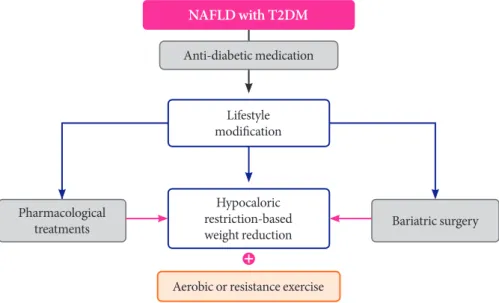
Epidemiological evidence suggests a strong bidirectional rela- tionship between T2DM and NAFLD. Furthermore, NAFLD resolution attenuates T2DM risk, but when obesity, insulin re- sistance, or hyperglycemia is combined with NAFLD, the T2DM risk is significantly increased [4]. In a long-term follow- up study of patients with NAFLD, diabetes and smoking, along with fibrosis, age, and absence of statin use, contributed to mortality, underscoring the need for a comprehensive ap- proach to patient management [32]. Additionally, spontaneous regression of NASH and even NASH-related fibrosis has been observed in clinical trials and may be related to lifestyle modi- fications and behavioral changes [81]. However, pharmacolog- ical treatment is more powerful than lifestyle modification for glucose control. Moreover, lifestyle modification plus anti-dia- betic drugs are likely to have a synergistic effect on reducing the risk factors associated with CV risk and decreasing hepatic fat accumulation in these patients, thereby delaying the pro- gression of inflammation and fibrosis. Therefore, all patients
with T2DM and NAFLD should be strongly encouraged to adopt both lifestyle changes and anti-diabetic medication use (Fig. 2) [3,82].
Weight reduction Keynotes
- Weight loss induced by either lifestyle modification or bariatric surgery improves glucometabolic profiles and reduces liver fat in subjects with T2DM.
- Regarding lifestyle modification, specific diets rather than calorie restric- tion or specific types of exercise protocols (aerobic or resistance) were not clearly defined for their efficacy at the present time.
It is well established that weight loss induced by either lifestyle modification or bariatric surgery in overweight or obese indi- viduals with T2DM results in significant improvement or reso- lution of T2DM and its comorbidities such as hypertension, and hyperlipidemia [3,83,84]. In addition, weight reduction has been associated with alleviated intrahepatic fat contents and aminotransferase levels [81]. Even relatively small amounts of weight loss can reduce liver fat and improve hepatic insulin resistance independent of any changes in insulin-stimulated peripheral glucose metabolism [85]. In adult populations with NAFLD, a systematic review that included a total of 23 studies evaluating the effect of lifestyle interventions, such as diet, physical activity, and/or exercise, on the hepatic indicators of steatosis, inflammation and fibrosis as well as glucose control/
insulin sensitivity showed that the reductions in liver fat and/
Fig. 2. Suggested algorithm for the management of patients with non-alcoholic fatty liver disease (NAFLD) and type 2 diabetes mellitus (T2DM).
NAFLD with T2DM Anti-diabetic medication
Bariatric surgery Pharmacological
treatments
Aerobic or resistance exercise Lifestyle
modification
Hypocaloric restriction-based weight reduction
or liver aminotransferase concentrations were strongly corre- lated with weight loss [86]. However, it should be noted that sustainable maintenance of weight loss with lifestyle interven- tions for long-term periods was achievable in only 3% to 6% of the subjects [9,15].
The magnitude of weight reduction Keynotes
- The improvement in histology is proportional to the magnitude of the weight loss, but this proportional trend might not be applicable to gluco- metabolic profiles.
- A magnitude of weight loss in the range of approximately 5% to 7% may clearly decrease steatosis but approximately 8% to 10% weight reduction is needed to reverse steatohepatitis.
In adult populations with NAFLD, a greater degree of weight loss, induced by either lifestyle modifications or bariatric sur- gical procedures, is associated with a greater improvement in histologic features [82]. Summarizing all current available re- ports, it appears that a magnitude of weight loss in the range of approximately 5% to 7% may clearly decrease steatosis and the associated metabolic parameters, but 8% to 10% weight reduc- tion is needed to reverse steatohepatitis [3,83,85-88]. Although a ≥7% weight loss also improved NAS, fibrosis was unchanged.
Furthermore, the highest rates of NAS reduction, steatohepati- tis resolution, and fibrosis regression occurred in patients with
≥10% weight loss [89]. The threshold of 7% weight loss was achieved by <50% of patients, even with intensive multidisci- plinary lifestyle intervention.
Lifestyle modification Keynotes
- The weight reduction achievable by dietary program remains the corner- stone of lifestyle modification for patients with T2DM and NAFLD.
- Caloric restriction, rather than dietary macronutrient composition, has a greater role in preventing the development and progression of T2DM, in reducing hepatic steatosis and in delaying inflammation and fibrosis.
- Exercise per se can reduce hepatic fat even in the absence of weight loss.
Both aerobic and resistance exercise leading to a similar weight reduction are similarly effective in hepatic fat reduction in patients with T2DM.
- Individually tailored approaches combining a mainly hypocaloric diet in conjunction with a progressive increase in aerobic exercise/resistance training are associated with the magnitude of the weight reduction.
Of the ways to achieve weight reduction, lifestyle modification that includes a programmed diet and exercise is an effective and sound treatment for all patients with NAFLD and NASH [34,82,90]. In addition, lifestyle modification improves hyper-
glycemia, atherogenic dyslipidemia, and blood pressure levels [91-93]. Of the lifestyle programs, however, weight reduction achievable by dietary programs or interventions remains the cornerstone of lifestyle modification for all patients with T2DM and NAFLD [34,82,90]. It seems that caloric restric- tion, rather than dietary macronutrient composition, has a greater role in reducing hepatic fat and delaying the progres- sion of inflammation and fibrosis in patients with NAFLD [81,83,85,86,94-96]. Regarding the macronutrient composi- tion, comparable effects have been observed with equally hy- pocaloric low-carbohydrate versus high-carbohydrate diets [97] and low-fat versus low-carbohydrate diets [98]. Exercise trials have reported an inconsistent amount of weight loss or changes in surrogate biomarkers achieved after exercise inter- ventions, but the heterogeneities of the baseline characteristics of the study subjects and the exercise protocols must also be considered [3,99,100]. Even with these findings, physical activ- ity, either aerobic or resistance type, should be strongly pro- moted for the management of fatty liver in patients with T2DM because the benefits are not exclusively contingent upon weight loss [101]. It seems that, despite the unclear bene- ficial roles of exercise on weight loss and hepatic fat reduction, exercise itself has been shown to improve liver enzymes and insulin resistance in all patients with diabetes with NAFLD and NASH [102,103]. A comprehensive approach with com- bined dietary and exercise interventions has shown significant benefit on NAFLD, especially for long-term intervention peri- ods. However, whether a combined lifestyle intervention has synergistic effects compared to the separate approaches of diet and exercise is unclear. A meta-analysis by Keating et al. [102]
showed that a combined dietary and exercise intervention had no significant pooled effects size (ES) when compared to diet alone (ES: –0.05; 95% confidence interval, –0.38 to 0.27; P=
0.76). However, another systematic review that included eigh- teen studies to investigate whether a diet-plus-exercise inter- vention for a minimum of 6 months was more effective for weight loss than a diet-only intervention among obese or over- weight adults showed that a combined diet-plus-exercise inter- vention provided greater long-term weight loss than a diet-on- ly intervention [104]. Although the effects of lifestyle modifica- tion in the absence of weight loss on NAFLD and NASH might not be clear, it is clear that lifestyle changes with either or both a hypocaloric diet and exercise reduce the risk of CV disease and the onset and progression of T2DM [34,82,105].
Pharmacologic treatment
For patients with T2DM and NAFLD, however, pharmacologi- cal treatment is more powerful than lifestyle modification for glucose control. Moreover, lifestyle modification plus anti-dia- betic drugs are likely to offer synergistic benefits for reducing the risk factors associated with their high CV risk [3,91] as well as decreasing hepatic fat accumulation and delaying the pro- gression of inflammation and fibrosis. Therefore, especially in patients with T2DM, all patients should adopt both lifestyle changes and anti-diabetic medication use [82].
Anti-diabetic agents Keynotes
- Patients with T2DM and NAFLD should adopt both lifestyle changes and anti-diabetic medication use.
- There are no definite pharmacologic treatments for NAFLD in patients with T2DM.
- Metformin is not recommended for treating NAFLD in patients with T2DM.
- Currently, pioglitazone could be a 1st-line option to improve NASH in patients with T2DM with some improvement in fibrosis.
- Evidence that sodium-glucose cotransporter 2 (SGLT2) inhibitors im- prove NAFLD/NASH with fibrosis in patients with T2DM is emerging.
Although not generally recommended, these agents may be considered as an initial treatment option in specific conditions for the treatment of NAFLD in patients with T2DM.
- Glucagon-like peptide-1 receptor agonists (GLP-1 RAs) have some evi- dence of improving NAFLD/NASH with fibrosis, but these agents are not generally recommended for the treatment of NAFLD in patients with T2DM.
- Dipeptidyl peptidase-4 (DPP-4) inhibitors are not recommended for treating NAFLD in patients with T2DM.
1) Metformin
Studies on metformin use in NAFLD failed to show any im- provement in liver histology in patients with NAFLD and NASH [106,107]. Thus, metformin is not recommended for treating NAFLD in patients with T2DM. However, T2DM is now considered an independent risk factor for HCC and met- formin may have a protective role in HCC occurrence [108].
2) Thiazolidinediones
Pioglitazone, one of the thiazolidinediones, is a peroxisome proliferator-activated receptor (PPAR)-γ agonist with insulin- sensitizing effects. An randomized controlled trial (RCT) eval- uating the efficacy and safety of long-term pioglitazone (45 mg/day) treatment in patients with biopsy-proven NASH and prediabetes or T2DM showed that compared with the placebo group, only patients with T2DM showed a significant resolu- tion of NASH (60% vs. 16%) and reduction in fibrosis from
baseline [109]. Currently available data from previous studies suggest that, although it may reduce fibrosis, the clinical effect of pioglitazone may come from the suppression of rapid fibro- sis progression rather than from fibrosis regression by turning off the metabolic drivers of liver fibrosis in T2DM [109,110].
Pioglitazone could be a first-line therapy for patients with NASH and T2DM. However, pioglitazone should not be rou- tinely used to treat NAFLD in patients with T2DM because of limited data and the common side effects of weight gain, fluid retention, and bone mass loss.
Lobeglitazone is currently being prescribed for T2DM in Korea. In a multicenter, prospective clinical trial, patients with T2DM and NAFLD treated with lobeglitazone (0.5 mg daily) for 24 weeks had improved liver enzymes and ameliorated he- patic fat contents assessed by VCTE-CAP [111].
3) Sodium-glucose cotransporter 2 inhibitors
SGLT2 inhibitors, including dapagliflozin, canagliflozin, empa- gliflozin, ipragliflozin, and luseogliflozin are being used in- creasingly in the treatment of T2DM; they promote weight loss which is an attractive property for the treatment of patients with NAFLD. Although SGLT2 inhibitors are not yet generally recommended for the treatment of NAFLD in patients with T2DM, emerging data suggest that SGLT2 inhibitors reduce the risk of progression of NAFLD [112], as the following results have been reported: a decrease in hepatic fat content [113,114];
a decrease in AST and ALT [114,115]; a decrease in the mea- sures of fibrosis, such as the FIB-4 index [115,116], the fibrosis index calculated from hyaluronic acid and type IV collagen 7S [116], and VCTE-measured LSM [117]; an improvement in he- patic insulin sensitivity [114]; and histology [118]. Well-de- signed RCTs are needed to elucidate whether SGLT2 inhibitors should be used as the first-line drug choice in patients with NAFLD/NASH with T2DM. In the meantime, these agents may be considered as an initial option to treat NAFLD in pa- tients with T2DM and specific cardio-renal conditions [119].
4) Glucagon-like peptide-1 receptor agonists
GLP-1 RAs, including exenatide, lixisenatide and liraglutide, are promising candidates for the treatment of NAFLD and NASH because they can reduce weight and enhance insulin action. However, GLP-1 RAs are not generally recommended for the treatment of NAFLD in patients with T2DM because of the limited data to date [112]. In a well-performed clinical trial in patients with biopsy-proven NASH, liraglutide (1.8 mg dai-
ly) treatment for 48 weeks was associated with greater resolu- tion of steatohepatitis and less progression of fibrosis [120].
5) Dipeptidyl peptidase-4 inhibitors
DPP-4 inhibitors including sitagliptin, vildagliptin, linagliptin, saxagliptin, gemigliptin, alogliptin, teneligliptin, and anagliptin are commonly used in patients with T2DM, but there are few studies about the efficacy of DPP-4 inhibitors in patients with T2DM and NAFLD; they are not yet believed to have a benefi- cial effect on NAFLD. Only vildagliptin showed a clinically sig- nificant decrease in hepatic TG levels measured by MRI-PDFF (27% with vildagliptin, from 7.3%±1.0% [baseline] to 5.3%±
0.9% [endpoint]), unrelated to changes in body weight, during 6 months of therapy in patients with T2DM and NAFLD [121].
Mixed results have been reported for sitagliptin. A pilot clinical study demonstrated that sitagliptin could improve hepatocyte ballooning and liver enzymes in patients with T2DM and NASH after 1 year of treatment [122]. However, many other studies have failed to show an effect of sitagliptin treatment on liver fat content or liver stiffness [123,124].
Non-antidiabetic agents Keynotes
- Vitamin E is not recommended to treat NAFLD in patients with T2DM.
- Lipid-lowering agents such as statins, ezetimibe, fibrates, niacin, omega-3 polyunsaturated fatty acids, and colesevelam did not improve hepatic ste- atosis in patients with NAFLD.
- Ursodeoxycholic acid (UDCA) and pentoxifylline are not recommended for the treatment of NAFLD or NASH.
- Obeticholic acid may improve NASH without progression of fibrosis in patients with T2DM and NASH. Further studies are required to prove its efficacy and long-term effects as well as its adverse effects.
1) Vitamin E
Vitamin E is an antioxidant that prevents liver injury by block- ing intrinsic apoptotic pathways and protecting against oxida- tive stress. Vitamin E improves liver histology in nondiabetic adults with biopsy-proven NASH [125], but is not recom- mended in patients with diabetes because of a lack of evidence [5,82]. In addition, there are some concerns that long-term use of vitamin E may be associated with increased all-cause mor- tality, an increased incidence of hemorrhagic stroke, and an in- creased risk of prostate cancer.
2) Lipid-lowering agents
Although statins are beneficial in the management of dyslipid- emia and high CV risk as well as in reducing the risk of HCC
and mortality, especially in patients with diabetes, lipid-lower- ing agents such as statins, ezetimibe, fibrates, niacin, omega-3 polyunsaturated fatty acids, and colesevelam did not improve hepatic steatosis in patients with NAFLD [5,82,126]. There was also no evidence for the use of one of these agents for the treat- ment of NAFLD in patients with T2DM.
3) Ursodeoxycholic acid
UDCA, a naturally occurring bile acid, reduced oxidative stress and had antiapoptotic properties. Although several stud- ies reported that UDCA improved liver enzymes and hepatic steatosis in patients with NAFLD [127,128], both conventional (13 to 15 mg/kg daily) and high (23 to 28 mg/kg daily) doses of UDCA failed to induce histologic improvement in patients with NASH [129,130].
4) Pentoxifylline
Pentoxifylline inhibits a number of proinflammatory cytokines including tumor necrosis factor-α. Although pentoxifylline (400 mg three times a day) improved the histological features of NASH in 55 adults with biopsy-confirmed NASH, only 9.1% of participants had T2DM [131]; another study reported no histological improvement in 30 patients, so larger studies are needed to establish the role of pentoxifylline in the man- agement of patients with NASH and T2DM [132].
5) Obeticholic acid
Obeticholic acid is a natural agonist of the farnesoid X receptor and decreases insulin resistance and hepatic steatosis in animal models. Obeticholic acid (25 mg daily) treatment for 72 weeks, where 52.7% of patients had T2DM, improved liver histology in patients with NASH but increased LDL-C levels and caused pruritus [133]. Further studies are needed to clarify the long- term benefit and safety of obeticholic acid in patients with T2DM and NAFLD.
6) Carnitine
Carnitine is a modulator of mitochondrial free fatty acid trans- port and oxidation and has antioxidative activity in hepato- cytes [98]. In patients with NASH, L-carnitine treatment (1 g daily) for 24 weeks improved liver enzymes and histological manifestations [134]. Treatment with the carnitine-orotate complex (824 mg, three times daily) for 12 weeks in patients with T2DM and NAFLD improved serum ALT and improved hepatic steatosis as assessed by CT in 78 patients [135].
7) Elafibranor
Elafibranor is a dual agonist of PPAR-α and PPAR-δ and im- proves insulin resistance in liver and peripheral tissue. In a phase 2b trial, elafibranor treatment (120 mg daily) for 52 weeks tended to induce resolution of NASH without fibrosis progression despite some methodological limitations. The pre- defined endpoint was not met in the intention to treat popula- tion and 39.1% of study participants had T2DM [136].
8) Cenicriviroc
Cenicriviroc, a dual antagonist of the C-C chemokine receptor types 2 and 5, has potent anti-inflammatory and antifibrotic activity in animal models. In a phase 2b study, in which half of subjects had T2DM and two-thirds had metabolic syndrome, cenicriviroc treatment (150 mg daily) for 1 year achieved im- provement in fibrosis and no worsening of steatohepatitis compared with placebo [137].
9) Anti-obesity drugs
Because weight reduction is a key component for the treatment of NAFLD, anti-obesity drugs can be potential candidates.
However, few studies have investigated the efficacy of anti- obesity drugs for the treatment of NAFLD. Orlistat (120 mg three times daily for 6 months), an inhibitor of fat absorption, showed improved steatosis assessed by US and confirmed by liver biopsy in patients with NAFLD [138]. Another study evaluated the effect of orlistat on NAFLD in patients who re- ceived a 1,400 kcal/day diet plus vitamin E (800 IU) daily. Orli- stat (120 mg three times a day) did not enhance weight loss or improve liver enzymes and histopathology in fifty overweight subjects (10% of subjects had T2DM) [139].
Bariatric surgery Keynotes
- In addition to its effects on body weight and metabolic profiles, bariatric surgery can decrease long-term mortality related to diabetes, CV diseas- es, and cancers.
- Bariatric surgery can be considered in obese patients with NAFLD and T2DM to resolve NASH and improve fibrosis.
Bariatric surgery has been accepted as a treatment option in obese patients with T2DM who do not achieve durable weight loss and improvement in comorbidities (including hyperglyce- mia) with nonsurgical methods [119]. A meta-analysis showed that the majority of patients undergoing bariatric surgery ap-
pear to improve or completely resolve the histopathological features of steatosis, inflammation, and ballooning. Fibrosis also improved by a weighted mean decrease of 11.9% in the in- cidence of fibrosis [140]. Another recent meta-analysis includ- ing 3,093 biopsies also reported the resolution of steatosis in 66% of patients, inflammation in 50%, ballooning degenera- tion in 76%, and fibrosis in 40% [141]. With regard to proce- dure selection, there is still a lack of data on which method, i.e., sleeve gastrectomy or Roux-en Y gastric bypass, is more effec- tive. However, considering the safety of the surgery itself, sleeve gastrectomy is recommended in patients with NASH cirrhosis [82,142,143].
CONFLICTS OF INTEREST
No potential conflict of interest relevant to this article was re- ported.
ORCID
Byung-Wan Lee https://orcid.org/0000-0002-9899-4992 Bong-Soo Cha https://orcid.org/0000-0003-0542-2854 Dae Ho Lee https://orcid.org/0000-0002-8832-3052
ACKNOWLEDGMENTS
None
REFERENCES
1. Loomba R, Abraham M, Unalp A, Wilson L, Lavine J, Doo E, Bass NM; Nonalcoholic Steatohepatitis Clinical Research Net- work. Association between diabetes, family history of diabe- tes, and risk of nonalcoholic steatohepatitis and fibrosis. Hepa- tology 2012;56:943-51.
2. Lee YH, Cho Y, Lee BW, Park CY, Lee DH, Cha BS, Rhee EJ.
Nonalcoholic fatty liver disease in diabetes. Part I: epidemiol- ogy and diagnosis. Diabetes Metab J 2019;43:31-45.
3. Bril F, Cusi K. Management of nonalcoholic fatty liver disease in patients with type 2 diabetes: a call to action. Diabetes Care 2017;40:419-30.
4. Han E, Lee YH. Non-alcoholic fatty liver disease: the emerg- ing burden in cardiometabolic and renal diseases. Diabetes Metab J 2017;41:430-7.
5. Chalasani N, Younossi Z, Lavine JE, Charlton M, Cusi K, Ri-
nella M, Harrison SA, Brunt EM, Sanyal AJ. The diagnosis and management of nonalcoholic fatty liver disease: practice guid- ance from the American Association for the Study of Liver Diseases. Hepatology 2018;67:328-57.
6. Browning JD, Szczepaniak LS, Dobbins R, Nuremberg P, Hor- ton JD, Cohen JC, Grundy SM, Hobbs HH. Prevalence of he- patic steatosis in an urban population in the United States: im- pact of ethnicity. Hepatology 2004;40:1387-95.
7. Vernon G, Baranova A, Younossi ZM. Systematic review: the epidemiology and natural history of non-alcoholic fatty liver disease and non-alcoholic steatohepatitis in adults. Aliment Pharmacol Ther 2011;34:274-85.
8. Younossi ZM, Koenig AB, Abdelatif D, Fazel Y, Henry L, Wymer M. Global epidemiology of nonalcoholic fatty liver disease-Meta-analytic assessment of prevalence, incidence, and outcomes. Hepatology 2016;64:73-84.
9. Park SH, Jeon WK, Kim SH, Kim HJ, Park DI, Cho YK, Sung IK, Sohn CI, Keum DK, Kim BI. Prevalence and risk factors of non-alcoholic fatty liver disease among Korean adults. J Gas- troenterol Hepatol 2006;21(1 Pt 1):138-43.
10. Jeong EH, Jun DW, Cho YK, Choe YG, Ryu S, Lee SM, Jang EC. Regional prevalence of non-alcoholic fatty liver disease in Seoul and Gyeonggi-do, Korea. Clin Mol Hepatol 2013;19:266- 72.
11. Portillo-Sanchez P, Bril F, Maximos M, Lomonaco R, Bier- nacki D, Orsak B, Subbarayan S, Webb A, Hecht J, Cusi K.
High prevalence of nonalcoholic fatty liver disease in patients with type 2 diabetes mellitus and normal plasma aminotrans- ferase levels. J Clin Endocrinol Metab 2015;100:2231-8.
12. Hyysalo J, Mannisto VT, Zhou Y, Arola J, Karja V, Leivonen M, Juuti A, Jaser N, Lallukka S, Kakela P, Venesmaa S, Simonen M, Saltevo J, Moilanen L, Korpi-Hyovalti E, Keinanen-Kiu- kaanniemi S, Oksa H, Orho-Melander M, Valenti L, Fargion S, Pihlajamaki J, Peltonen M, Yki-Jarvinen H. A population- based study on the prevalence of NASH using scores validated against liver histology. J Hepatol 2014;60:839-46.
13. Williams CD, Stengel J, Asike MI, Torres DM, Shaw J, Contre- ras M, Landt CL, Harrison SA. Prevalence of nonalcoholic fat- ty liver disease and nonalcoholic steatohepatitis among a largely middle-aged population utilizing ultrasound and liver biopsy: a prospective study. Gastroenterology 2011;140:124- 31.
14. Younossi ZM, Marchesini G, Pinto-Cortez H, Petta S. Epide- miology of nonalcoholic fatty liver disease and nonalcoholic steatohepatitis: implications for liver transplantation. Trans-
plantation 2019;103:22-7.
15. Kim BY, Jung CH, Mok JO, Kang SK, Kim CH. Prevalences of diabetic retinopathy and nephropathy are lower in Korean type 2 diabetic patients with non-alcoholic fatty liver disease. J Diabetes Investig 2014;5:170-5.
16. Mohan V, Farooq S, Deepa M, Ravikumar R, Pitchumoni CS.
Prevalence of non-alcoholic fatty liver disease in urban south Indians in relation to different grades of glucose intolerance and metabolic syndrome. Diabetes Res Clin Pract 2009;84:84- 91.
17. Kim SK, Choi YJ, Huh BW, Park SW, Lee EJ, Cho YW, Huh KB.
Nonalcoholic fatty liver disease is associated with increased ca- rotid intima-media thickness only in type 2 diabetic subjects with insulin resistance. J Clin Endocrinol Metab 2014;99:1879- 84.
18. Targher G, Bertolini L, Rodella S, Tessari R, Zenari L, Lippi G, Arcaro G. Nonalcoholic fatty liver disease is independently as- sociated with an increased incidence of cardiovascular events in type 2 diabetic patients. Diabetes Care 2007;30:2119-21.
19. Kim CH, Park JY, Lee KU, Kim JH, Kim HK. Fatty liver is an independent risk factor for the development of type 2 diabetes in Korean adults. Diabet Med 2008;25:476-81.
20. Sung KC, Kim SH. Interrelationship between fatty liver and insulin resistance in the development of type 2 diabetes. J Clin Endocrinol Metab 2011;96:1093-7.
21. Sung KC, Wild SH, Byrne CD. Resolution of fatty liver and risk of incident diabetes. J Clin Endocrinol Metab 2013;98:3637-43.
22. Yamazaki H, Tsuboya T, Tsuji K, Dohke M, Maguchi H. Inde- pendent association between improvement of nonalcoholic fatty liver disease and reduced incidence of type 2 diabetes.
Diabetes Care 2015;38:1673-9.
23. Bhatia LS, Curzen NP, Calder PC, Byrne CD. Non-alcoholic fatty liver disease: a new and important cardiovascular risk factor? Eur Heart J 2012;33:1190-200.
24. Singh S, Allen AM, Wang Z, Prokop LJ, Murad MH, Loomba R. Fibrosis progression in nonalcoholic fatty liver vs nonalco- holic steatohepatitis: a systematic review and meta-analysis of paired-biopsy studies. Clin Gastroenterol Hepatol 2015;13:643- 54.
25. McPherson S, Hardy T, Henderson E, Burt AD, Day CP, Anst- ee QM. Evidence of NAFLD progression from steatosis to fi- brosing-steatohepatitis using paired biopsies: implications for prognosis and clinical management. J Hepatol 2015;62:1148- 55.
26. Angulo P. Nonalcoholic fatty liver disease. N Engl J Med 2002;
346:1221-31.
27. Mittal S, Sada YH, El-Serag HB, Kanwal F, Duan Z, Temple S, May SB, Kramer JR, Richardson PA, Davila JA. Temporal trends of nonalcoholic fatty liver disease-related hepatocellu- lar carcinoma in the veteran affairs population. Clin Gastro- enterol Hepatol 2015;13:594-601.
28. Pocha C, Kolly P, Dufour JF. Nonalcoholic fatty liver disease- related hepatocellular carcinoma: a problem of growing mag- nitude. Semin Liver Dis 2015;35:304-17.
29. Lee SS, Jeong SH, Byoun YS, Chung SM, Seong MH, Sohn HR, Min BY, Jang ES, Kim JW, Park GJ, Lee YJ, Lee KH, Ahn S. Clinical features and outcome of cryptogenic hepatocellular carcinoma compared to those of viral and alcoholic hepatocel- lular carcinoma. BMC Cancer 2013;13:335.
30. Allen AM, Therneau TM, Larson JJ, Coward A, Somers VK, Kamath PS. Nonalcoholic fatty liver disease incidence and im- pact on metabolic burden and death: a 20 year-community study. Hepatology 2018;67:1726-36.
31. Ekstedt M, Hagstrom H, Nasr P, Fredrikson M, Stal P, Kecha- gias S, Hultcrantz R. Fibrosis stage is the strongest predictor for disease-specific mortality in NAFLD after up to 33 years of follow-up. Hepatology 2015;61:1547-54.
32. Angulo P, Kleiner DE, Dam-Larsen S, Adams LA, Bjornsson ES, Charatcharoenwitthaya P, Mills PR, Keach JC, Lafferty HD, Stahler A, Haflidadottir S, Bendtsen F. Liver fibrosis, but no other histologic features, is associated with long-term out- comes of patients with nonalcoholic fatty liver disease. Gas- troenterology 2015;149:389-97.
33. Newsome PN, Cramb R, Davison SM, Dillon JF, Foulerton M, Godfrey EM, Hall R, Harrower U, Hudson M, Langford A, Mackie A, Mitchell-Thain R, Sennett K, Sheron NC, Verne J, Walmsley M, Yeoman A. Guidelines on the management of abnormal liver blood tests. Gut 2018;67:6-19.
34. European Association for the Study of the Liver (EASL); Euro- pean Association for the Study of Diabetes (EASD); European Association for the Study of Obesity (EASO). EASL-EASD- EASO clinical practice guidelines for the management of non- alcoholic fatty liver disease. Diabetologia 2016;59:1121-40.
35. Prati D, Colli A, Conte D, Colombo M. Spectrum of NAFLD and diagnostic implications of the proposed new normal range for serum ALT in obese women. Hepatology 2005;42:1460-1.
36. Kwo PY, Cohen SM, Lim JK. ACG clinical guideline: evaluation of abnormal liver chemistries. Am J Gastroenterol 2017;112:18- 35.
37. Terrault NA, Lok ASF, McMahon BJ, Chang KM, Hwang JP,
Jonas MM, Brown RS Jr, Bzowej NH, Wong JB. Update on prevention, diagnosis, and treatment of chronic hepatitis B:
AASLD 2018 hepatitis B guidance. Hepatology 2018;67:1560- 99.
38. Hernaez R, Lazo M, Bonekamp S, Kamel I, Brancati FL, Gual- lar E, Clark JM. Diagnostic accuracy and reliability of ultraso- nography for the detection of fatty liver: a meta-analysis. Hep- atology 2011;54:1082-90.
39. Stefan N, Haring HU, Cusi K. Non-alcoholic fatty liver disease:
causes, diagnosis, cardiometabolic consequences, and treat- ment strategies. Lancet Diabetes Endocrinol 2019;7:313-24.
40. Bril F, Ortiz-Lopez C, Lomonaco R, Orsak B, Freckleton M, Chintapalli K, Hardies J, Lai S, Solano F, Tio F, Cusi K. Clinical value of liver ultrasound for the diagnosis of nonalcoholic fat- ty liver disease in overweight and obese patients. Liver Int 2015;35:2139-46.
41. Petta S, Ciminnisi S, Di Marco V, Cabibi D, Camma C, Licata A, Marchesini G, Craxi A. Sarcopenia is associated with severe liver fibrosis in patients with non-alcoholic fatty liver disease.
Aliment Pharmacol Ther 2017;45:510-8.
42. Shen F, Zheng RD, Shi JP, Mi YQ, Chen GF, Hu X, Liu YG, Wang XY, Pan Q, Chen GY, Chen JN, Xu L, Zhang RN, Xu LM, Fan JG. Impact of skin capsular distance on the perfor- mance of controlled attenuation parameter in patients with chronic liver disease. Liver Int 2015;35:2392-400.
43. Vuppalanchi R, Siddiqui MS, Van Natta ML, Hallinan E, Brandman D, Kowdley K, Neuschwander-Tetri BA, Loomba R, Dasarathy S, Abdelmalek M, Doo E, Tonascia JA, Kleiner DE, Sanyal AJ, Chalasani N; NASH Clinical Research Net- work. Performance characteristics of vibration-controlled transient elastography for evaluation of nonalcoholic fatty liv- er disease. Hepatology 2018;67:134-44.
44. Kim KY, Song JS, Kannengiesser S, Han YM. Hepatic fat quantification using the proton density fat fraction (PDFF):
utility of free-drawn-PDFF with a large coverage area. Radiol Med 2015;120:1083-93.
45. Capitan V, Petit JM, Aho S, Lefevre PH, Favelier S, Loffroy R, Hillon P, Krause D, Cercueil JP, Guiu B. Macroscopic hetero- geneity of liver fat: an MR-based study in type-2 diabetic pa- tients. Eur Radiol 2012;22:2161-8.
46. Loomba R, Wolfson T, Ang B, Hooker J, Behling C, Peterson M, Valasek M, Lin G, Brenner D, Gamst A, Ehman R, Sirlin C.
Magnetic resonance elastography predicts advanced fibrosis in patients with nonalcoholic fatty liver disease: a prospective study. Version 2. Hepatology 2014;60:1920-8.
47. Kim D, Kim WR, Talwalkar JA, Kim HJ, Ehman RL. Ad- vanced fibrosis in nonalcoholic fatty liver disease: noninvasive assessment with MR elastography. Radiology 2013;268:411-9.
48. Caussy C, Alquiraish MH, Nguyen P, Hernandez C, Cepin S, Fortney LE, Ajmera V, Bettencourt R, Collier S, Hooker J, Sy E, Rizo E, Richards L, Sirlin CB, Loomba R. Optimal thresh- old of controlled attenuation parameter with MRI-PDFF as the gold standard for the detection of hepatic steatosis. Hepa- tology 2018;67:1348-59.
49. Nasr P, Forsgren MF, Ignatova S, Dahlstrom N, Cedersund G, Leinhard OD, Noren B, Ekstedt M, Lundberg P, Kechagias S.
Using a 3% proton density fat fraction as a cut-off value increas- es sensitivity of detection of hepatic steatosis, based on results from histopathology analysis. Gastroenterology 2017;153:53-5.
50. Bannas P, Kramer H, Hernando D, Agni R, Cunningham AM, Mandal R, Motosugi U, Sharma SD, Munoz del Rio A, Fer- nandez L, Reeder SB. Quantitative magnetic resonance imag- ing of hepatic steatosis: validation in ex vivo human livers.
Hepatology 2015;62:1444-55.
51. Patel J, Bettencourt R, Cui J, Salotti J, Hooker J, Bhatt A, Her- nandez C, Nguyen P, Aryafar H, Valasek M, Haufe W, Hooker C, Richards L, Sirlin CB, Loomba R. Association of noninva- sive quantitative decline in liver fat content on MRI with his- tologic response in nonalcoholic steatohepatitis. Therap Adv Gastroenterol 2016;9:692-701.
52. Bravo AA, Sheth SG, Chopra S. Liver biopsy. N Engl J Med 2001;344:495-500.
53. Gunn NT, Shiffman ML. The use of liver biopsy in nonalco- holic fatty liver disease: when to biopsy and in whom. Clin Liver Dis 2018;22:109-19.
54. Wong VW, Chalasani N. Not routine screening, but vigilance for chronic liver disease in patients with type 2 diabetes. J Hepatol 2016;64:1211-3.
55. Corey KE, Klebanoff MJ, Tramontano AC, Chung RT, Hur C.
Screening for nonalcoholic steatohepatitis in individuals with type 2 diabetes: a cost-effectiveness analysis. Dig Dis Sci 2016;
61:2108-17.
56. Bedossa P, Patel K. Biopsy and noninvasive methods to assess progression of nonalcoholic fatty liver disease. Gastroenterol- ogy 2016;150:1811-22.
57. Lichtinghagen R, Pietsch D, Bantel H, Manns MP, Brand K, Bahr MJ. The Enhanced Liver Fibrosis (ELF) score: normal values, influence factors and proposed cut-off values. J Hepa- tol 2013;59:236-42.
58. Tada T, Kumada T, Toyoda H, Saibara T, Ono M, Kage M.
New scoring system combining the FIB-4 index and cytokera- tin-18 fragments for predicting steatohepatitis and liver fibro- sis in patients with nonalcoholic fatty liver disease. Biomarkers 2018;23:328-34.
59. Siddiqui MS, Vuppalanchi R, Van Natta ML, Hallinan E, Kowdley KV, Abdelmalek M, Neuschwander-Tetri BA, Loom- ba R, Dasarathy S, Brandman D, Doo E, Tonascia JA, Kleiner DE, Chalasani N, Sanyal AJ; NASH Clinical Research Net- work. Vibration-controlled transient elastography to assess fi- brosis and steatosis in patients with nonalcoholic fatty liver disease. Clin Gastroenterol Hepatol 2019;17:156-63.
60. Jayakumar S, Middleton MS, Lawitz EJ, Mantry PS, Caldwell SH, Arnold H, Mae Diehl A, Ghalib R, Elkhashab M, Abdel- malek MF, Kowdley KV, Stephen Djedjos C, Xu R, Han L, Mani Subramanian G, Myers RP, Goodman ZD, Afdhal NH, Charlton MR, Sirlin CB, Loomba R. Longitudinal correlations between MRE, MRI-PDFF, and liver histology in patients with non-alcoholic steatohepatitis: analysis of data from a phase II trial of selonsertib. J Hepatol 2019;70:133-41.
61. Loomba R, Schork N, Chen CH, Bettencourt R, Bhatt A, Ang B, Nguyen P, Hernandez C, Richards L, Salotti J, Lin S, Seki E, Nelson KE, Sirlin CB, Brenner D; Genetics of NAFLD in Twins Consortium. Heritability of hepatic fibrosis and steato- sis based on a prospective twin study. Gastroenterology 2015;
149:1784-93.
62. Krawczyk M, Rau M, Schattenberg JM, Bantel H, Pathil A, Demir M, Kluwe J, Boettler T, Lammert F, Geier A; NAFLD Clinical Study Group. Combined effects of the PNPLA3 rs738409, TM6SF2 rs58542926, and MBOAT7 rs641738 vari- ants on NAFLD severity: a multicenter biopsy-based study. J Lipid Res 2017;58:247-55.
63. Koo BK, Joo SK, Kim D, Bae JM, Park JH, Kim JH, Kim W.
Additive effects of PNPLA3 and TM6SF2 on the histological severity of non-alcoholic fatty liver disease. J Gastroenterol Hepatol 2018;33:1277-85.
64. Romeo S, Kozlitina J, Xing C, Pertsemlidis A, Cox D, Pennac- chio LA, Boerwinkle E, Cohen JC, Hobbs HH. Genetic varia- tion in PNPLA3 confers susceptibility to nonalcoholic fatty liver disease. Nat Genet 2008;40:1461-5.
65. Del Campo JA, Gallego-Duran R, Gallego P, Grande L. Ge- netic and epigenetic regulation in nonalcoholic fatty liver dis- ease (NAFLD). Int J Mol Sci 2018;19:911.
66. Liu DJ, Peloso GM, Yu H, Butterworth AS, Wang X, Mahajan A, Saleheen D, Emdin C, Alam D, Alves AC, Amouyel P, Di Angelantonio E, Arveiler D, Assimes TL, Auer PL, Baber U,