The Volume of Subscapularis Muscle Remains Unaffected by Supraspinatus Tendon Tears: Three-dimensionally Reconstructed Magnetic Resonance Imaging Analysis
Yong Cheol Jun, Young Lae Moon , Havinder Dev Bhardwaj1, Jae Hwan Lim, Dong Hyuk Cha
Department of Orthopedic Surgery, Chosun University School of Medicine, Gwangju, Korea, 1Department of Orthopedic Surgery, Punjab Institute of Medical Sciences, Jalandhar, Punjab, India
Background: This study aimed to compare the subscapularis muscle volume between the intact groups (group I) and supraspinatus ten- don tear groups (group T) based on the sex and three different age groups.
Methods: Subjects with a group I and subjects with group T without any other lesions were retrospectively evaluated from among pa- tients who received a magnetic resonance imaging (MRI) scan between January 2011 and December 2013. The MRI scans were studied by a consultant radiologist. The subscapularis muscle volume was compared according to the age and sex; the age groups were catego- rized as patients in their 40s, 50s, and 60s. The volume of subscapularis muscle was measured by three-dimensional reconstructed im- ages acquired through the axial section of 1.5T MRI.
Results: No statistically significant differences were observed between subscapularis muscle volume of the group I and group T, except for male patients in their 50s (group I: 100,650 mm3 vs. group T: 106,488 mm3) and 60s (group I: 76,347 mm3 vs. group T: 99,549 mm3) (p<0.05). Males had a larger mean volume of subscapularis muscle than females, and the subscapularis muscle volume decreased in a linear manner with increasing age.
Conclusions: Decrease in subscapularis muscle volume was observed with increasing age, and the impact of supraspinatus tear on sub- scapularis muscle volume is age and sex dependent.
(Clin Shoulder Elbow 2019;22(1):3-8)
Key Words: Rotator cuff tear; Muscle atrophy; Quantitative assessment
Copyright © 2019 Korean Shoulder and Elbow Society. All Rights Reserved.
Clinics in Shoulder and Elbow Vol. 22, No. 1, March, 2019 https://doi.org/10.5397/cise.2019.22.1.3
Received May 15, 2018. Revised October 21, 2018. Accepted November 5, 2018.
Correspondence to: Young Lae Moon
Department of Orthopedic Surgery, Chosun University Hospital, 365 Pilmun-daero, Dong-gu, Gwangju 61453, Korea
Tel: +82-62-220-3147, Fax: +82-62-226-3379, E-mail: [email protected], ORCID: https://orcid.org/0000-0002-4487-9884 IRB approval: Chosun University Hospital (No. CHOSUN 2015-11-003-002).
Financial support: None. Conflict of interests: None.
Introduction
Due to the inherent interaction with each other, the four rotator cuff muscles serve as the main dynamic stabilizers of the humeral head.1-3) The antagonist subscapularis and infraspinatus/
teres minor muscles build a force couple that centers the hu- meral head in an anteroposterior direction.4,5) Any asymmetric tonus of the agonists results in altered glenohumeral shear force leading to humeral head displacement from its center of rotation in the glenoid cavity, thereby resulting in the other antagonist muscle weakness and atrophy.5,6) Also, changes in muscle size
and joint strength are known to occur throughout the adult lifes- pan. Muscle volume, especially of the shoulder, decreases with age. The linear relationship, however, between muscle volume and age remains uncertain.7-9)
The subscapularis muscle is the largest and most powerful muscle among the rotator cuff muscles; it is also named the
“forgotten tendon” as it rarely tears. This muscle plays a key role as an anterior structure of the transverse force couple along with the infraspinatus and teres minor muscles, which are posterior to the shoulder joint.10,11) Among the posterior structures of the transverse force couple, the importance of the infraspinatus and
teres minor muscles are widely recognized; however, signifi- cance of the supraspinatus muscle as the posterior structure of the shoulder joint remains unclear.
Recently, magnetic resonance imaging (MRI) has been widely applied to evaluate the shoulder joint, and has been highly use- ful in understanding anatomical structures.12-15) Several methods have been introduced to measure the volume of the rotator cuff muscles. Tingart et al.16) separated each muscle of the rotator cuff in cadaver experiments and then measured the amount of overflowing water when each muscle was soaked in water. Juul- Kristensen et al.17) evaluated the muscle volume in 20 patients using MRI. According to the data collected by Juul-Kristensen et al.17) and Tingart et al.,16) no differences were observed between the volume of muscle obtained by soaking in water in the ca- daver experiments and the volume of muscle obtained by three- dimensional (3D) reconstruction with MRI. Currently, rotator cuff atrophy is assessed using a multi-layered approach involving these methods.
As a posterior structure of the shoulder, the supraspinatus tear affects the posterior shoulder force couple, and could also affect the anterior shoulder muscle volume (subscapularis). This retro- spective study was undertaken to evaluate if the supraspinatus tendon tear affects the subscapularis muscle volume. We com- pared the subscapularis muscle volume between groups, with and without supraspinatus tendon tear, by considering sex and age.
Methods
Subject Enrollment
Between January 2011 to December 2013, 1,592 patients underwent MRI due to shoulder pain. We analyzed MRI read- ings of 1,218 patients in their 40s to 60s; 374 patients were ex- cluded since they did not meet the age requirement. The results
were studied by a musculoskeletal radiologist having an experi- ence of more than 30 years. Among the 1,218 patients enrolled, 990 patients were excluded due to abnormal MRI findings, including supraspinatus tendon tear. Of the 228 patients finally enrolled for evaluation, 96 patients had only supraspinatus ten- don tear (group T) and 132 patients showed no abnormality (in- tact) in the MRI (group I). Considering the age, 60 subjects were randomly selected from both groups, where 20 each belonged to the 40s, 50s, and 60s age groups (Fig. 1). Subjects were ran- domly selected from each subgroup using random sampling in Excel (Microsoft, Redmond, WA, USA).
Demographics
A total of 120 patients were evaluated, including 60 shoul- der joints in female patients and 60 shoulder joints in male patients, with an overall mean age of 54.3 ± 9.29 years (range, 40–69 years). Totally, 60 participants had a normal supraspinatus tendon (group I: age range, 40–69 years; mean, 55.1 ± 9.47 years), and 60 patients had a supraspinatus tendon tear (group T:
age range, 40–69 years; mean, 53.6 ± 10.32 years). The three categorized age groups consisted of 20 patients each.
Three-dimensional Reconstruction and Volume Measurement
We conducted a quantitative assessment of the subscapularis muscle volume using 3D reconstruction of MRIs in patients in- cluded in the group T and group I. The MRIs used in this study were obtained through an Avanto 1.5-T MRI (Siemens, Erlangen, Germany). The slice size of the images was 3.0 mm.
Briefly, muscle contours were assigned to every transverse slide of the MRI by using the ‘‘Livewire’’ option on the MIMIC 10.01 (for Intel x86 Platform III+; Materialise NV, Leuven, Bel- gium) medical imaging software. Volume masks were calculated from the assigned contours on the transverse slides. The sub-
Eligibility: 1,592
Assessment 1 radiologist
Remaining group T: 96 only supraspinatus tear
Comparison group I: 132 subjects showing normal MRI finding
Exclusion: 1,364 Age out of their 40s to 60s: 374 Abnormal MRI finding (40s to 60s): 990
Other rotator cuff tear: 341 (including supraspinatus tear) Calcific tendinitis: 304 Adhesive capsulitis: 248 Paralabral cyst: 61 Labral lesion: 36
Randomized
selection: 60 Randomized
selection: 60
Fig. 1. Flow chart of subject selection. Group I showed no abnormality (intact) and group T had only supraspinatus tendon tear in the magnetic resonance imaging.
MRI: magnetic resonance imaging.
scapularis muscle was outlined on each scan, proceeding from the medial border of the scapular to the lesser tuberosity, which belongs to the subscapularis tendon. As the outline was hand- drawn in the MIMIC program, accuracy was low. To increase the accuracy, 3 orthopedic surgeons participated in taking measure- ments (Fig. 2, 3). Interobserver reliability was evaluated using in- traclass correlation coefficients (ICCs) introduced by Shrout and Fleiss18) Interobserver reliability was relatively high, with an ICCs of 0.836.
Statistical Analysis
All data were statistically analyzed using the IBM SPSS ver.
21.0 software (IBM Corp., Armonk, NY, USA). Continuous vari- ables between the two groups was compared by the t-test (Table 1), and linear regression was used to analyze the correlation
between age and subscapularis muscle volume according to sex (Table 2-4). Statistical significance was accepted at p<0.05.
Results
No significant differences were detected between the groups with respect group I and group T among participants in their 40s (p>0.05). However, a significant difference was observed between both groups when considering male in the 50s and 60s (p<0.05). Subscapularis muscle volume loss tended to increase with age in the group I, as indicated by linear regression analysis (Fig. 4, 5). However, this linear decrease was not observed in the group T (Table 2, 3).
2.09 70.29
-19.00 49.21
-4.94
-19.00
63.26
49.21 A
R
P
L
A
R
P
L
A
R
P
L
A
R
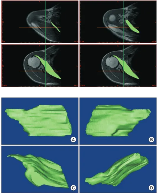
P
L
Fig. 2. Green label: Schematic surface of the subscapularis muscle border in a transverse section; Green colored schematic surface of the subscapularis muscle on transverse sec- tion can be seen here; Drawing the outline of the subscapularis muscle in a transverse section.
A B
C D
Fig. 3. Three-dimensional (3D) reconstruc- tion image of subscapularis muscle. (A) 3D reconstruction of the image of the subscapu- laris muscle is presented. Subscapularis 3D reconstruction image; (A) is the anterior aspect of the 3D reconstruction image of subscapularis muscle, (B) is the posterior aspect, (C) is the anteroinferior aspect, (D) is the anterosuperior aspect.
Discussion
The primary goal of this study was to explore the relationship between the subscapularis muscle volume and supraspinatus tendon tear. Results of this study indicate no significant relation- ship between supraspinatus tendon tear and volume of sub- scapularis muscle in females. However, a higher subscapularis muscle volume was observed in group T than group I of males in their 50s and 60s.
We hypothesized that the posterior section of the supra- spinatus interacts with the subscapularis muscle as a posterior
structure which contributes to some degree of anterior-posterior stability. Furthermore, the supraspinatus tendon tear affects the volume of the subscapularis muscle loss, thereby leading to a tear.19)
According to Bergin et al.,19) subscapularis tendon abnormality is related to chronicity of supraspinatus tendon tears. In patients with supraspinatus tear, the dynamic anterior instability increases due to the elevated posterior shoulder force couple weakness, resulting in sub-coracoid impingement and consequently a sub- scapularis tendon tear.19) MRI scans reveal the existence of bone marrow edema of lesser tuberosity; however, the study of bone marrow edema was not included in the current study.
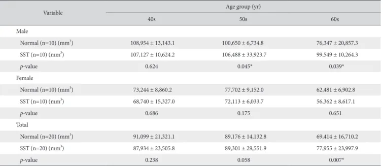
Contrary to our hypothesis, our study revealed different out- comes. We initially hypothesized that supraspinatus tendon tear affects the reduction of the subscapularis muscle volume. How- ever, we did not account for the degree of supraspinatus tendon tear size, tear thickness, torn tendon retraction degree, occupa- tion, activities, and body skeletal mass.20) Hence, our data re- vealed no significant relationship between supraspinatus tendon tear and volume of subscapularis muscle in the group T of males Table 1. Age Specific Subscapularis Muscle Volume and t-test of Supraspinatus Tendon Tear Patients and Normal Participants
Variable Age group (yr)
40s 50s 60s
Male
Normal (n=10) (mm3) 108,954 ± 13,143.1 100,650 ± 6,734.8 76,347 ± 20,857.3
SST (n=10) (mm3) 107,127 ± 10,624.2 106,488 ± 33,923.7 99,549 ± 10,264.3
p-value 0.624 0.045* 0.039*
Female
Normal (n=10) (mm3) 73,244 ± 8,860.2 77,702 ± 9,152.0 62,481 ± 6,902.8
SST (n=10) (mm3) 68,740 ± 15,327.0 72,113 ± 6,033.7 56,362 ± 8,617.1
p-value 0.686 0.175 0.651
Total
Normal (n=20) (mm3) 91,099 ± 21,321.1 89,176 ± 14,132.8 69,414 ± 16,710.2
SST (n=20) (mm3) 87,934 ± 23,505.8 89,301 ± 29,551.9 77,955 ± 23,997.9
p-value 0.238 0.058 0.007*
Values are presented as mean ± standard deviation.
SST: supraspinatus tendon tear.
*Significant decrease in muscle volume with increasing age; linear regression analysis (p<0.05).
Table 2. Linear Regression Analysis of the Volume of Subscapularis Muscle in Normal Participants (group I)
Sex Number Mean (mm3) R2 p-value
Male 30 95,317.37 0.414 0.000*
Female 30 71,142.83 0.226 0.008*
Group I showed no abnormality (intact) in the magnetic resonance imaging.
*p<0.05.
Table 3. Linear Regression Analysis of the Volume of Subscapularis Muscle in Patients with SST (group T)
Sex Number Mean (mm3) R2 p-value
Male 30 104,388.37 0.024 0.414
Female 30 65,738.93 0.146 0.037*
Group T had only SST.
SST: supraspinatus tendon tear.
*p<0.05.
Table 4. Linear Regression Analysis of the Total Volume of Subscapularis Muscle (group I and group T)
Sex Total number Mean (mm3) R2 p-value
Male 60 99,852.87 0.142 0.003*
Female 60 11,672.06 0.181 0.001*
Group I showed no abnormality (intact) and group T had only supraspinatus tendon tear in the magnetic resonance imaging.
*p<0.05.
in their 40s. Since the supraspinatus tendon tear might be small in the 40s with a shorter duration of tear, its effect on the volume of the subscapularis muscle may be negligible. Furthermore, in the group comprising males in their 50s and 60s, group T had higher subscapularis muscle volume than group I. We also con- sidered that males included in the group T had a higher level of labor intensity or sports activities, and these factors might affect in increasing the volume of subscapularis muscle before onset of supraspinatus tear. However, we also considered that this result could be due to the small number of samples, thereby resulting in selection bias which plays a role in enhancing the subscapu- laris muscle volume in group T than in group I. Furthermore, our data showed that the subscapularis muscle volume in group T of female was lower than in group I, but was statistically not signifi- cant. We believe this could be that most female are housewives or working with lower labor intensity jobs. When considering sex of the female, the subscapularis muscle volume seemed more affected by the supraspinatus tendon tear than by life activities.
In this study, we found that although the subscapularis muscle volume reduces with aging, it was not related to cuff tear. This finding is supported by previous muscle studies that demon- strate that the aging process results in a loss of muscle mass, with subsequent replacement by fat and connective tissue.21,22) Regardless of the sex, reduced muscle volume and aging were correlated for subjects in their 40s, 50s, and 60s. With the on- set of advancing age, muscle tissue is gradually lost, resulting in diminished mass and strength; this condition is referred to as sarcopenia.21,23) One study presented that patients with cuff tears have sarcopenia, and patients with large to massive tears have a significantly inferior sarcopenic index than those with small to medium tears.24) They measured the whole-body muscle mass as per the Janssen25) method of bioelectrical impedance analysis equation, which is different from the analysis used in our current
paper, in that we only calculated the subscapularis muscle.24) Another paper explains that rotator cuff atrophy is related to increasing age and not to tear severity. For patients without ro- tator cuff tears, the prevalence of fatty infiltration and atrophy increased with aging.26) Further evaluation is required to deter- mine the pathophysiology and progression of muscle atrophy in rotator cuff muscles.
Although cadaver studies provide a detailed understanding of the effects of supraspinatus tendon tear on the subscapularis muscle atrophy or tendon tear, it is necessary to achieve meaning- ful results. In this study, we measured the muscle volume of the rotator cuff (such as subscapularis muscle) through MRI. Since only the muscle part is measured (except for the surrounding fatty tissue), the accuracy is relatively high.27) Considering various other factors such as tear size, jobs, sports activities, height, and weight that influence the muscle mass will yield more meaning- ful results.
This study has some limitations. Most notably, being retro- spective in design, we are unable to make any causal claims explaining the relationship between supraspinatus tear and sub- scapularis muscle volume. Second, this study has a small sample size and does not exclude factors that might affect muscle volume, such as height, weight, occupation, and sports activi- ties. Third, we did not account duration of the disease and size (i.e., full-thickness tear or partial thickness) of the supraspinatus tendon tear. Fourth, muscle volume measurements using the MIMIC program did not consider fatty infiltration in the muscles, hence the measured volume value does not reflect the intrinsic atrophy of the muscles accurately.
Conclusion
Analysis applying 3D reconstruction by MRI reveals decreas-
40 45 50 55 60 65 70
Subscapularismusclevolume(mm)3
Age (yr) 160,000
140,000 120,000 100,000 80,000 60,000 40,000 20,000 0 180,000
R linear regression=0.1422
Fig. 4. Linear regression analysis of the age and subscapularis muscle volume in males. The average volume of subscapularis muscle is decreased in males, with aging.
40 45 50 55 60 65 70
Subscapularismusclevolume(mm)3
Age (yr) 100,000
80,000
60,000
40,000 120,000
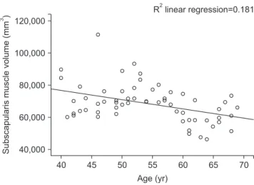
R linear regression=0.1812
Fig. 5. Linear regression analysis of age and subscapularis muscle volume in females. The average volume of subscapularis muscle is decreased in females, with aging.
ing subscapularis muscle volume with aging, regardless of the sex. The supraspinatus tear was seen to affect the subscapularis muscle volume in a certain group of age and sex.
References
1. Favre P, Senteler M, Hipp J, Scherrer S, Gerber C, Snedeker JG.
An integrated model of active glenohumeral stability. J Biomech.
2012;45(13):2248-55. doi: 10.1016/j.jbiomech.2012.06.010.
2. Lippitt S, Matsen F. Mechanisms of glenohumeral joint stabil- ity. Clin Orthop Relat Res. 1993;(291):20-8. doi: 10.1097/
00003086-199306000-00004.
3. McMahon PJ, Lee TQ. Muscles may contribute to shoulder dislocation and stability. Clin Orthop Relat Res. 2002;(403 Suppl):S18-25. doi: 10.1097/00003086-200210001-00003.
4. Ackland DC, Pandy MG. Lines of action and stabilizing poten- tial of the shoulder musculature. J Anat. 2009;215(2):184-97.
doi: 10.1111/j.1469-7580.2009.01090.x.
5. Thompson WO, Debski RE, Boardman ND 3rd, et al. A biomechanical analysis of rotator cuff deficiency in a ca- daveric model. Am J Sports Med. 1996;24(3):286-92. doi:
10.1177/036354659602400307.
6. Hansen ML, Otis JC, Johnson JS, Cordasco FA, Craig EV, War- ren RF. Biomechanics of massive rotator cuff tears: implications for treatment. J Bone Joint Surg Am. 2008;90(2):316-25. doi:
10.2106/JBJS.F.00880.
7. Holzbaur KR, Murray WM, Gold GE, Delp SL. Upper limb muscle volumes in adult subjects. J Biomech. 2007;40(4):742- 9. doi: 10.1016/j.jbiomech.2006.11.011.
8. Saul KR, Vidt ME, Gold GE, Murray WM. Upper limb strength and muscle volume in healthy middle-aged adults. J Appl Bio- mech. 2015;31(6):484-91. doi: 10.1123/jab.2014-0177.
9. Vidt ME, Daly M, Miller ME, Davis CC, Marsh AP, Saul KR.
Characterizing upper limb muscle volume and strength in old- er adults: a comparison with young adults. J Biomech. 2012;
45(2):334-41. doi: 10.1016/j.jbiomech.2011.10.007.
10. Burkhart SS. Arthroscopic treatment of massive rotator cuff tears. Clinical results and biomechanical rationale. Clin Orthop Relat Res. 1991;(267):45-56.
11. Lo IK, Burkhart SS. The comma sign: an arthroscopic guide to the torn subscapularis tendon. Arthroscopy. 2003;19(3):334-7.
doi: 10.1053/jars.2003.50080.
12. Morag Y, Jamadar DA, Miller B, Dong Q, Jacobson JA. The subscapularis: anatomy, injury, and imaging. Skeletal Radiol.
2011;40(3):255-69. doi: 10.1007/s00256-009-0845-0.
13. Pfirrmann CW, Zanetti M, Weishaupt D, Gerber C, Hodler J. Subscapularis tendon tears: detection and grading at MR arthrography. Radiology. 1999;213(3):709-14. doi: 10.1148/
radiology.213.3.r99dc03709.
14. Tuoheti Y, Itoi E, Minagawa H, et al. Quantitative assessment of thinning of the subscapularis tendon in recurrent anterior
dislocation of the shoulder by use of magnetic resonance im- aging. J Shoulder Elbow Surg. 2005;14(1):11-5. doi: 10.1016/
j.jse.2004.04.009.
15. Warner JJ, Higgins L, Parsons IM 4th, Dowdy P. Diagnosis and treatment of anterosuperior rotator cuff tears. J Shoulder Elbow Surg. 2001;10(1):37-46. doi: 10.1067/mse.2001.112022.
16. Tingart MJ, Apreleva M, Lehtinen JT, Capell B, Palmer WE, Warner JJ. Magnetic resonance imaging in quantitative analysis of rotator cuff muscle volume. Clin Orthop Relat Res. 2003;
(415):104-10. doi: 10.1097/01.blo.0000092969.12414.e1.
17. Juul-Kristensen B, Bojsen-Moller F, Finsen L, et al. Muscle sizes and moment arms of rotator cuff muscles determined by mag- netic resonance imaging. Cells Tissues Organs. 2000;167(2- 3):214-22. doi: 10.1159/000016784.
18. Shrout PE, Fleiss JL. Intraclass correlations: uses in assess- ing rater reliability. Psychol Bull. 1979;86(2):420-8. doi:
10.1037/0033-2909.86.2.420.
19. Bergin D, Parker L, Zoga A, Morrison W. Abnormalities on MRI of the subscapularis tendon in the presence of a full-thick- ness supraspinatus tendon tear. AJR Am J Roentgenol. 2006;
186(2):454-9. doi: 10.2214/AJR.04.1723.
20. Melis B, Nemoz C, Walch G. Muscle fatty infiltration in rotator cuff tears: descriptive analysis of 1688 cases. Orthop Traumatol Surg Res. 2009;95(5):319-24. doi: 10.1016/j.otsr.2009.05.001.
21. Lexell J, Taylor CC, Sjöström M. What is the cause of the age- ing atrophy? Total number, size and proportion of different fiber types studied in whole vastus lateralis muscle from 15- to 83-year-old men. J Neurol Sci. 1988;84(2-3):275-94. doi:
10.1016/0022-510X(88)90132-3.
22. Reimers CD, Harder T, Saxe H. Age-related muscle atro- phy does not affect all muscles and can partly be compen- sated by physical activity: an ultrasound study. J Neurol Sci.
1998;159(1):60-6. doi: 10.1016/S0022-510X(98)00134-8.
23. Rosenberg IH. Sarcopenia: origins and clinical relevan ce. J Nutr. 1997;127(5 Suppl):990s-1s. doi: 10.1093/jn/127.5.990S.
24. Chung SW, Yoon JP, Oh KS, et al. Rotator cuff tear and sarco- penia: are these related? J Shoulder Elbow Surg. 2016;25(9):
e249-55. doi: 10.1016/j.jse.2016.02.008.
25. Janssen I. Influence of sarcopenia on the development of physical disability: the Cardiovascular Health Study. J Am Geriatr Soc. 2006;54(1):56-62. doi: 10.1111/j.1532- 5415.2005.00540.x.
26. Barry JJ, Lansdown DA, Cheung S, Feeley BT, Ma CB. The relationship between tear severity, fatty infiltration, and muscle atrophy in the supraspinatus. J Shoulder Elbow Surg.
2013;22(1):18-25. doi: 10.1016/j.jse.2011.12.014.
27. Matsumura N, Oguro S, Okuda S, et al. Quantitative assess- ment of fatty infiltration and muscle volume of the rotator cuff muscles using 3-dimensional 2-point Dixon magnetic reso- nance imaging. J Shoulder Elbow Surg. 2017;26(10):e309-18.
doi: 10.1016/j.jse.2017.03.019.