ABSTRACT
Sublingual immunotherapy (SLIT) is an effective treatment for allergic diseases. However, the mechanism by which this therapy exhibits its efficacy has not been fully delineated. To elucidate the mechanisms of SLIT in the treatment of cedar pollinosis (CP), we performed a multivariate analysis of microarray data on mRNA expression in CD4+ T cells and basophils.
Although 2-year treatment with SLIT using cedar extracts was effective in > 70% of patients with CP, the remaining patients did not respond to this therapy. The mRNA expression levels in peripheral CD4+ T cells and basophils from both high- and non-responder patients before and after undergoing SLIT were comparatively studied using microarray analysis. By processing the data using serial multivariate analysis, an apoptosis pathway was extracted in both CD4+ T cells and basophils. Conclusively, the strong treatment effectiveness of SLIT in patients with CP may be caused by the induction of apoptosis in CD4+ T cells and basophils in these patients (Trial registry at University Hospital Medical Information Network Clinical Trials Registry Database, UMIN000016532).
Keywords: Allergic rhinitis; cytokines; immunoglobulin E
INTRODUCTION
Cedar pollinosis (CP) is a specific seasonal allergic disease, especially in Japan, that afflicts up to 30% of people during February to April.1,2 Besides symptom-relieving medications, allergen-specific immunotherapy (AIT) is one of the most effective treatments for CP. Many years after administration of subcutaneous immunotherapy (SCIT) using standardized cedar pollen extract since the 1960s,3 the first sublingual immunotherapy (SLIT) was approved for use in 2014.4 There is much clinical and scientific evidence regarding the effectiveness and safety of SLIT for patients with CP;5-7 additionally, this therapy is easier to administer and safer to use than SCIT. In cases treated with SCIT, systemic injections were required and severe side effects including fatal anaphylaxis were feared. However, the mechanisms by which SLIT and SCIT exert their efficacies have not been fully elucidated.
Despite the usefulness of SLIT, it has been reported that up to 30% of treated patients do not respond to this therapy.8,9 We have also confirmed this fact in our recent clinical
Brief Communication
Received: Feb 19, 2018 Revised: Apr 22, 2018 Accepted: May 14, 2018 Correspondence to Minoru Gotoh, MD, PhD
Department of Otorhinolaryngology, Nippon Medical School, 1-1-5 Sendagi, Bunkyo-ku, Tokyo 113-8603, Japan.
Tel: +81-3-3822-2131 Fax: +81-3-5814-6207 E-mail: [email protected]
Copyright © 2018 The Korean Academy of Asthma, Allergy and Clinical Immunology • The Korean Academy of Pediatric Allergy and Respiratory Disease
This is an Open Access article distributed under the terms of the Creative Commons Attribution Non-Commercial License (https://
creativecommons.org/licenses/by-nc/4.0/) which permits unrestricted non-commercial use, distribution, and reproduction in any medium, provided the original work is properly cited.
ORCID iDs Minoru Gotoh
https://orcid.org/0000-0002-1040-5103 Osamu Kaminuma
https://orcid.org/0000-0003-1211-0410 Takachika Hiroi
https://orcid.org/0000-0002-8814-2657 Kimihiro Okubo
https://orcid.org/0000-0002-9900-4284 Disclosure
There are no financial or other issues that might lead to conflict of interest.
Minoru Gotoh ,1,2* Osamu Kaminuma ,2,3 Takachika Hiroi ,2 Kimihiro Okubo 1,2
1Department of Otorhinolaryngology, Nippon Medical School, Tokyo, Japan
2Allergy and Immunology Project, The Tokyo Metropolitan Institute of Medical Science, Tokyo, Japan
3Center for Life Science Research, University of Yamanashi, Yamanashi, Japan
Microarray-Based Multivariate Analysis of the Effectiveness of
Sublingual Immunotherapy for Cedar
Pollinosis
study;10,11 nevertheless, the existence of patients who are high-responders (HRs) or non- responders (NRs) to SLIT may be helpful in understanding the mechanisms of SLIT. Thus, by detailed comparison between HRs and NRs, we have delineated an essential biological signaling pathway responsible for the efficacy of SLIT. Although the difference between the HR and NR groups were not obvious even in the transcriptomic analysis,11 using a multivariate analysis of microarray data on mRNA expression in CD4+ T cells and basophils, we have suggested the involvement of apoptosis-related pathways in the efficacy of SLIT in patients with CP.
MATERIALS AND METHODS
Clinical study of SLIT for the treatment of CP
The study design, recruitment of patients, administration of cedar pollen extracts and evaluation of clinical efficacies have been described elsewhere.10 Briefly, 202 patients with Japanese CP, who lived in Tokyo, Japan, were over 20 years of age, and were being treated at the Nippon Medical School Hospital, the Tokyo Metropolitan Hospitals and clinics in Tokyo were included in this study. All patients showed symptoms of allergic rhinitis such as sneezing, rhinorrhea and nasal congestion during the pollen season from February to April for at least 3 consecutive years. They were positive on skin-tests as well as for immunoglobulin E (IgE) against the cedar pollen allergen. The study was registered in the University Hospital Medical Information Network Clinical Trials Registry Database (UMIN000016532) and was conducted in accordance with the Declaration of Helsinki and Good Clinical Practice guidelines. All experimental procedures were approved by the ethical committee of Tokyo Metropolitan Institute of Medical Science (Approval No.17-10), Nippon Medical School Hospital (No.18-3), Tokyo Metropolitan Komagome Hospital (No.18-473), Tokyo Metropolitan Hiroo Hospital (No.18-14), Tokyo Metropolitan Fuchu Hospital (No.18- 2), Tokyo Metropolitan Otsuka Hospital (No.2006-1), and Tokyo Metropolitan Health and Medical Treatment Corporation Ebara Hospital (No.18-1). Endo ENT Clinic and Hirooka Clinic were also participated in this study under the supervision of Nippon Medical School Hospital. All patients provided informed written consent prior to participation.
All subjects were individually administered an allergen from cedar pollen.10 Briefly, patients held the allergen under their tongues for 2 minutes and then spat it out. Starting with 1 drop consisting of 2 Japanese allergy units (JAU) per mL, the amount of allergen administered to the patients was gradually increased up to 20 drops of 2,000 JAU per mL as the maintenance dose at 5 weeks. The administration of the allergen started from July 2006 and further continued for 2 years.
Nasal symptoms of individual patients observed between February 1 and April 30 in 4 pollen seasons from 2006 to 2009 were investigated using the Japanese guideline for allergic rhinitis.1,12 Briefly, the numbers of sneezes and nose-blowing, and the extents of nasal congestion and eye itchiness were daily recorded by each patient, using an allergy diary according to the classification developed by Okuda.13 The quality of life (QOL) of the patients were determined 3 times, at the end of February, the middle of March, and the middle of April, respectively, for each year using a Japan Rhinoconjunctivitis Quality of Life Questionnaire No.1 (JRQLQ No1). At the end of each pollen season, the clinical efficacy of SLIT was evaluated based on the descriptions in the allergy diaries and the JRQLQ No1 as previously described.6
Cell preparation and microarray analysis
The preparation of CD4+ T cells and basophils, and the subsequent microarray analysis of mRNA expression have been described elsewhere.10,11 Briefly, blood samples were obtained twice, namely, in June 2006 and 2008, respectively, just prior to the start of and following the end of allergen administration. CD4+ T cells and basophils were purified from peripheral blood mononuclear cells as CD4+ and CD123+CD11clow cell populations, respectively, using a FACSAria cell sorting system (BD Biosciences, San Diego, CA, USA).
Total RNA was extracted from the stored cells. Microarray analyses for the extracted RNA from HRs and NRs, before and after undergoing SLIT, were comparably performed using GeneChip Human Gene 1.0 ST Array (Agilent Technologies, Santa Clara, CA, USA) with GeneChip Scanner 3000 7G (Affymetrix Inc., Santa Clara, CA, USA) and Affymetrix GeneChip Command Console Software. Just before the microarray analysis, the quality of RNA was examined by Qubit RNA IQ Assay Kit (ThermoFisher Scientific, Waltham, MA, USA).
Multivariate pathway analysis
The microarray data from CD4+ T cells were processed using the orthogonal partial least squares (OPLS) analysis with SIMCA-P™ software (Umetrics, Umeå, Sweden). Highly reliable markers that may distinguish between the 2 groups were extracted by OPLS-discriminant analysis (OPLS- DA). The target group pairs were 1) HRs before and after undergoing SLIT, 2) NRs before and after undergoing SLIT, and 3) HRs and NRs before undergoing SLIT. The resulting candidate markers were processed for pathway analysis using the MetaCore™ software (version 6.24, build 67895; Thomson Reuters, Philadelphia, PA, USA). This analysis was repeated using the microarray data from basophils to compare HRs and NRs after undergoing SLIT.
RESULTS
The detailed results of this clinical study have been described elsewhere.10 Consistent with the results of previous investigations,14,15 in our study approximately 30% of patients did not show any improvements in their symptoms even after undergoing the 2-year treatment with SLIT.
The first 33 HR patients whose conditions were extremely improved and the last 34 NR patients whose conditions were either unchanged or exacerbated were selected. After the exclusion of the samples from cypress pollen-specific IgE-positive patients and those with damaged RNA or DNA, 25 samples each from HRs and NRs were processed for microarray analyses.
Using OPLS-DA with a confidence interval of 0.4 for the microarray data from CD4+ T cells, 1,145, 1,475 and 652 genes were extracted in the comparison between HRs before and after undergoing SLIT (Supplementary Fig. S1A), NRs before and after SLIT (Supplementary Fig. S1B), and HRs and NRs before undergoing SLIT (Supplementary Fig. S1C), respectively. The HRs and NRs after undergoing SLIT were not distinguishable by OPLS-DA.
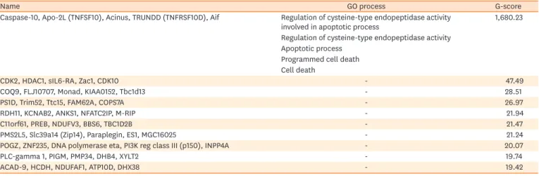
These extracted genes were analyzed by pathway analysis. In the comparison between HRs before and after undergoing SLIT, an apoptosis pathway involving Caspase-10, Apo-2L (TNFSF10), Acinus, tumor necrosis factor (TNF)-related apoptosis-inducing ligand (TRAIL) receptor with a truncated death domain (TRUNDD, TNF receptor superfamily member 10D [TNFRSF10D]) and Aif was identified with a G-score of 1,680.23 (Table 1 and Figure A). However, no significant pathways with high G-scores were determined in the comparison between NRs before and after undergoing SLIT or between HRs and NRs before undergoing SLIT (data not shown).
Using OPLS-DA with a confidence interval of 0.4 for the microarray data from basophils, 780 genes were extracted in the comparison between HRs and NRs after undergoing SLIT (Supplementary Fig. S2). Then, as in the case of microarray data from CD4+ T cells, an apoptosis induction pathway involving TNF-receptor (R)1, tBid, receptor for advanced glycation end products (RAGE), Bid and Caspase-3 was identified with a G-score of 133.08 (Table 2 and Figure B).
DISCUSSION
To delineate the mechanisms by which SLIT is effective for the treatment of CP, biomarker sets that are different between HRs and NRs both before and after undergoing therapy were investigated by multivariate analysis of microarray data on mRNA expression in CD4+ T cells and basophils.
CD4+ T cells play crucial roles in antigen recognition; therefore, they are implicated in the effects of AIT.16-20 By comparing data on HRs before and after undergoing SLIT, an apoptosis Table 1. Top 10 list of GO process networking molecules differentially expressed in CD4+ T cells of HR patients between before and after SLIT
Name GO process G-score
Caspase-10, Apo-2L (TNFSF10), Acinus, TRUNDD (TNFRSF10D), Aif Regulation of cysteine-type endopeptidase activity
involved in apoptotic process 1,680.23
Regulation of cysteine-type endopeptidase activity Apoptotic process
Programmed cell death Cell death
CDK2, HDAC1, sIL6-RA, Zac1, CDK10 - 47.49
COQ9, FLJ10707, Monad, KIAA0152, Tbc1d13 - 28.51
PS1D, Trim52, Ttc15, FAM62A, COPS7A - 26.97
RDH11, KCNAB2, ANKS1, NFATC2IP, M-RIP - 21.94
C11orf61, PREB, NDUFV3, BBS6, TBC1D2B - 21.47
PMS2L5, Slc39a14 (Zip14), Paraplegin, ES1, MGC16025 - 21.24
POGZ, ZNF235, DNA polymerase eta, PI3K reg class III (p150), INPP4A - 20.07
PLC-gamma 1, PIGM, PMP34, DHB4, XYLT2 - 19.74
ACAD-9, HCDH, NDUFAF1, ATP10D, DHX38 - 19.42
The predicted GO processes were only shown for the highest G-score networking molecules (MetaCore™ version 6.24, build 67895).
GO, gene ontology; HR, high-responders; SLIT, sublingual immunotherapy; TRUNDD, tumor necrosis factor-related apoptosis-inducing ligand receptor with a truncated death domain; TNFRSF10D, tumor necrosis factor receptor superfamily member 10D.
Table 2. Top 10 list of GO process networking molecules differentially expressed in basophils after SLIT between HR and NR patients
Name GO process G-score
TNF-R1, tBid, RAGE, Bid, Caspase-3 Apoptotic process 133.08
Programmed cell death Regulation of apoptotic process Regulation of programmed cell death Cell death
StARD5, MTND2, Lunatic fringe, FAM78A, ZNF174 - 42.19
VAMP1, SZT2, PIKE (CENTG1), TFIIIC110, ZNF526 - 37.28
Lunatic fringe, Fringe, Radical fringe, Manic fringe glycosyltransferase - 37.02
PNUTS, DATF1, FIC1, Diacylglycerol kinase, zeta, CREB1 - 32.62
TNF-R1, EGLN2, E4F1, RFC1, GPAM10 - 31.59
IL-17 receptor, ITGA5, CCDC142, Bystin, USP19 - 31.27
RFP, GSTM2, ZFP276, HSPA1B, E4F1 - 26.30
Zibra, Bid, MKP-X, Dcp1b, USP11 - 26.30
ITGA5, ETS2, ETS, PUR-alpha, HSP70 - 26.03
The predicted GO processes were only shown for the highest G-score networking molecules (MetaCore™ version 6.24, build 67895).
GO, gene ontology; SLIT, sublingual immunotherapy; HR, high-responders; NR, non-responders.
pathway was identified. Consistent with this finding, the AIT-induced apoptosis in the T cells of atopic patients has previously been reported.21-23 However, several other groups have demonstrated that Fas-induced apoptosis is unrelated to the effects of AIT.24,25 We recently demonstrated that some taste receptors expressed on CD4+ T cells were related to the efficacy of SLIT. Although further functional analysis is required to confirm those possibilities including the contribution of taste receptors, our present findings support the hypothesis that the effect of SLIT is achieved, at least in part, by the induction of apoptosis in CD4+ T cells.
The involvement of basophils in the pathogenesis of allergic diseases has recently been discussed.26,27 Like in CD4+ T cells, an apoptosis-inducing pathway has been identified in basophils too; this is an intriguing observation. The apoptosis of T cells is a well-known
Extracellular Membrane Cytoplasm
Nucleus Extracellular Membrane
Cytoplasm
Nucleus
B A
Figure. The pathway in CD4+ T cells and basophils that is expected to be related to the efficacy of SLIT. The candidate markers extracted from CD4+ T cells (A) as shown in Supplementary Fig. S1 that may distinguish HRs before and after undergoing SLIT and those from basophils (B) as shown in Supplementary Fig. S2 that may distinguish HRs and NRs after undergoing SLIT were processed for pathway analysis using the MetaCore™ software. The most reliable pathway, wherein the extracted markers are connected by a bold line, is shown.
SLIT, sublingual immunotherapy; HR, high-responders; NR, non-responders.
biological response, but the same has not been commonly recognized in basophils. However, Matsumoto et al.28 and Förster et al.29 recently reported the presence of a Fas-mediated apoptosis pathway in basophils. Since basophils play important roles in the pathogenesis of allergic diseases,30 the induction of apoptosis in basophils may be an additional mechanism for the efficacy of treatment with SLIT.
In conclusion, our present findings suggest the possibility that the effectiveness of SLIT in the treatment of CP is mainly caused by the induction of apoptosis in CD4+ T cells and basophils. Further investigations are needed to prove this hypothesis and to study whether apoptosis is induced in T cells and basophils by the same mechanisms.
ACKNOWLEDGMENTS
This work was supported by a Grant-in-Aid for JSPS KAKENHI (No. 15K08626) to Minoru Gotoh, and funding from the Towa Foundation for Food Science & Research, the Hoyu Science Foundation, the Tojuro Iijima Foundation for Food Science and Technology, and the Shimabara Science Foundation to Osamu Kaminuma.
SUPPLEMENTARY MATERIALS
Supplementary Fig. S1
Multivariate pathway analysis of microarray data from CD4+ T cells.
Click here to view
Supplementary Fig. S2
Multivariate pathway analysis of microarray data from basophils.
Click here to view
REFERENCES
1. Fujieda S, Kurono Y, Okubo K, Ichimura K, Enomoto T, Kawauchi H, et al. Examination, diagnosis and classification for Japanese allergic rhinitis: Japanese guideline. Auris Nasus Larynx 2012;39:553-6.
PUBMED | CROSSREF
2. Yamada T, Saito H, Fujieda S. Present state of Japanese cedar pollinosis: The national affliction. J Allergy Clin Immunol 2014;133:632-639.e5.
PUBMED | CROSSREF
3. Sohn MH. Efficacy and safety of subcutaneous allergen immunotherapy for allergic rhinitis. Allergy Asthma Immunol Res 2018;10:1-3.
PUBMED | CROSSREF
4. Mitobe Y, Yokomoto Y, Ohashi-Doi K. Safety evaluation of standardized allergen extract of Japanese cedar pollen for sublingual immunotherapy. Regul Toxicol Pharmacol 2015;71:529-40.
PUBMED | CROSSREF
5. Horiguchi S, Okamoto Y, Yonekura S, Okawa T, Yamamoto H, Kunii N, et al. A randomized
controlled trial of sublingual immunotherapy for Japanese cedar pollinosis. Int Arch Allergy Immunol 2008;146:76-84.
PUBMED | CROSSREF
6. Okubo K, Gotoh M, Fujieda S, Okano M, Yoshida H, Morikawa H, et al. A randomized double-blind comparative study of sublingual immunotherapy for cedar pollinosis. Allergol Int 2008;57:265-75.
PUBMED | CROSSREF
7. Fujimura T, Yonekura S, Horiguchi S, Taniguchi Y, Saito A, Yasueda H, et al. Increase of regulatory T cells and the ratio of specific IgE to total IgE are candidates for response monitoring or prognostic biomarkers in 2-year sublingual immunotherapy (SLIT) for Japanese cedar pollinosis. Clin Immunol 2011;139:65-74.
PUBMED | CROSSREF
8. Kim ST. Outcome of sublingual immunotherapy in patients with allergic rhinitis sensitive to house dust mites. Allergy Asthma Immunol Res 2015;7:99-100.
PUBMED | CROSSREF
9. Okamoto Y, Okubo K, Yonekura S, Hashiguchi K, Goto M, Otsuka T, et al. Efficacy and safety of sublingual immunotherapy for two seasons in patients with Japanese cedar pollinosis. Int Arch Allergy Immunol 2015;166:177-88.
PUBMED | CROSSREF
10. Gotoh M, Kaminuma O, Nakaya A, Katayama K, Motoi Y, Watanabe N, et al. Identification of biomarker sets for predicting the efficacy of sublingual immunotherapy against pollen-induced allergic rhinitis. Int Immunol 2017;29:291-300.
PUBMED | CROSSREF
11. Gotoh M, Kaminuma O, Nakaya A, Katayama K, Watanabe N, Saeki M, et al. Involvement of taste receptors in the effectiveness of sublingual immunotherapy. Allergol Int. Forthcoming 2018.
PUBMED | CROSSREF
12. Okubo K, Kurono Y, Fujieda S, Ogino S, Uchio E, Odajima H, et al. Japanese guideline for allergic rhinitis.
Allergol Int 2011;60:171-89.
PUBMED | CROSSREF
13. Okuda M. Epidemiology of Japanese cedar pollinosis throughout Japan. Ann Allergy Asthma Immunol 2003;91:288-96.
PUBMED | CROSSREF
14. Di Lorenzo G, Mansueto P, Pacor ML, Rizzo M, Castello F, Martinelli N, et al. Evaluation of serum s-IgE/
total IgE ratio in predicting clinical response to allergen-specific immunotherapy. J Allergy Clin Immunol 2009;123:1103-10, 1110.e1-e4.
PUBMED | CROSSREF
15. Manzotti G, Lombardi C. Allergen immunotherapy as a drug: the new deal of grass allergen tablets from clinical trials to current practice. Eur Ann Allergy Clin Immunol 2013;45:34-42.
PUBMED
16. Moingeon P, Mascarell L. Induction of tolerance via the sublingual route: mechanisms and applications.
Clin Dev Immunol 2012;2012:623474.
PUBMED | CROSSREF
17. Cappella A, Durham SR. Allergen immunotherapy for allergic respiratory diseases. Hum Vaccin Immunother 2012;8:1499-512.
PUBMED | CROSSREF
18. Jutel M, Van de Veen W, Agache I, Azkur KA, Akdis M, Akdis CA. Mechanisms of allergen-specific immunotherapy and novel ways for vaccine development. Allergol Int 2013;62:425-33.
PUBMED | CROSSREF
19. Allam JP, Bieber T, Novak N. Dendritic cells as potential targets for mucosal immunotherapy. Curr Opin Allergy Clin Immunol 2009;9:554-7.
PUBMED | CROSSREF
20. Jutel M, Kosowska A, Smolinska S. Allergen immunotherapy: past, present, and future. Allergy Asthma Immunol Res 2016;8:191-7.
PUBMED | CROSSREF
21. Guerra F, Carracedo J, Madueño JA, Sanchez-Guijo P, Ramírez R. Allergens induce apoptosis in lymphocytes from atopic patients. Hum Immunol 1999;60:840-7.
PUBMED | CROSSREF
22. Guerra F, Carracedo J, Solana-Lara R, Sánchez-Guijo P, Ramírez R. TH2 lymphocytes from atopic patients treated with immunotherapy undergo rapid apoptosis after culture with specific allergens. J Allergy Clin Immunol 2001;107:647-53.
PUBMED | CROSSREF
23. Gardner LM, O'Hehir RE, Rolland JM. High dose allergen stimulation of T cells from house dust mite- allergic subjects induces expansion of IFN-gamma+ T Cells, apoptosis of CD4+IL-4+ T cells and T cell anergy. Int Arch Allergy Immunol 2004;133:1-13.
PUBMED | CROSSREF
24. Kinikli G, Ateş A, Turgay M, Aydoğan N, Tokgöz G. Serum-soluble Fas antigen level in patients with allergic rhinitis: its relation to specific immunotherapy. Allergy Asthma Proc 2006;27:145-7.
PUBMED
25. Ciepiela O, Zawadzka-Krajewska A, Kotula I, Wasik M, Demkow U. Sublingual immunotherapy in asthma does not influence lymphocyte sensitivity to Fas stimulation. Eur J Med Res 2010;15 Suppl 2:17-20.
PUBMED
26. Falcone FH, Zillikens D, Gibbs BF. The 21st century renaissance of the basophil? Current insights into its role in allergic responses and innate immunity. Exp Dermatol 2006;15:855-64.
PUBMED | CROSSREF
27. Jahnsen FL, Farkas L, Lund-Johansen F, Brandtzaeg P. Involvement of plasmacytoid dendritic cells in human diseases. Hum Immunol 2002;63:1201-5.
PUBMED | CROSSREF
28. Matsumoto K, Maeda A, Bochner BS, Wakiguchi H, Saito H. Induction of apoptosis in human basophils by anti-Fas antibody treatment in vitro. Int Arch Allergy Immunol 2008;146 Suppl 1:40-6.
PUBMED | CROSSREF
29. Förster A, Falcone FH, Gibbs BF, Preussner LM, Fiebig BS, Altunok H, et al. Anti-Fas/CD95 and tumor necrosis factor-related apoptosis-inducing ligand (TRAIL) differentially regulate apoptosis in normal and neoplastic human basophils. Leuk Lymphoma 2013;54:835-42.
PUBMED | CROSSREF
30. Karasuyama H, Yamanishi Y. Basophils have emerged as a key player in immunity. Curr Opin Immunol 2014;31:1-7.
PUBMED | CROSSREF