유방의 유두상 병변은 비교적 드문 질환으로 조직검사를 시 행하는 양성종양의 10%, 악성종양의 1-2%를 차지하며(1-4) 경피적 핵생검으로는 정확한 병리학적 진단이 어려워(5) 대부 분은 외과적 절제 생검을 시행해 왔다(6). 그러나 최근에 유두 상 병변을 진단하는데 있어 병리 결과와 영상 소견이 일치한다 면 수술적 절제 없이 경피적 핵생검으로도 충분하다는 보고들 이 있다(7, 8). 입체정위 유도 하에서는 11 게이지 진공흡인생 검법이 14 게이지 자동총생검법보다 조직학적 저평가율이나 재 조직검사율이 낮아 더 효과적인 것으로 알려져 있다(9-13).
그러나 초음파 유도 하 14 게이지 자동총생검법에 대한 11 게 이지 진공흡인생검법의 이점에 대해서는 아직 밝혀진 바가 없 다(14). 초음파 유도 하에서 11 게이지 진공흡인생검법이 14
게이지 자동총생검법보다 조직학적 저평가, 재조직검사율, 위 음성률에서 더 유용하다는 보고가 있으나 통계학적으로는 차 이가 없었다(15). 더욱이 유두상 병변을 진단하고 치료하는 데 있어 초음파 유도 하 14 게이지 자동총생검법에 대한 11 게이 지 진공흡인생검법의 이점에 대해서는 아직 밝혀진 바가 없다.
이 연구에서 저자들은 유두상 병변을 진단하는 데 있어 초음파 유도 하에 14 게이지 자동총생검법을 시행한 군과 11 게이지 진공흡인생검법을 시행한 군의 성적을 비교하고자 하였다.
대상과 방법
2003년 1월부터 2005년 4월까지 행해진 1,723개의 병변에 대한 초음파 유도 하 경피적 생검 중 Tavassoli의 기준에 따 라 유두종, 유두종증(papillomatosis), 비정형성 유두종, 비침윤
유방의 유두상 병변: 초음파 유도 하 14 게이지 자동총생검법과 11 게이지 진공흡인생검법의 비교1
고은숙・조나리야・양상규・김도연・문우경
목적: 유방의 유두상 병변에 대한 초음파 유도 하 14 게이지 자동총생검법과 11 게이지 진공 흡인생검법의 성적을 비교하고자 한다.
대상과 방법: 2003년 1월부터 2005년 4월까지 초음파 유도 하 핵생검법을 시행한 1,723개의
병변 중 98개(5.7%)가 병리학적으로 유두상 병변으로 보고되었다. 이 중에서 65개는 14 게이 지 자동총생검법으로 33개는 11 게이지 진공흡인생검법으로 진단되었다. 14 게이지 자동총생 검법을 시행 받은 군의 54%(35/65)와 11 게이지 진공흡인생검법을 시행 받은 군의 15%(5/33) 가 절제생검을 받았다. 두 군의 병리 소견과 함께 수술 소견, 그리고 추적 검사 소견을 분석하 고 두 군간의 조직학적 저평가율(histologic underestimation rate)과 재조직검사율(repeat biopsy rate)을 비교하였다. 재조직검사율은 재조직검사수를 전체 조직검사수로 나누어서 구했 다. 비정형 관상피 증식증 저평가(ADH underestimation)는 핵생검에서 비정형성 유두종이 나 왔으나 수술에서 암이 나온 경우로 관상피내암 저평가(DCIS underestimation)는 핵생검에서 관상피내암이 나왔으나 수술에서 침윤성 암이 나온 경우로 정의했다.
결과: 재조직검사율은 14 게이지 자동총생검법의 경우 42%(27/65)였으며 11 게이지 진공흡 인생검법의 경우 9.1%(3/33)였다. 비정형 관상피 증식증 저평가는 14 게이지 자동총생검법에 서 50%(7/14)였으며 11 게이지 진공흡인생검법에서는 0%(0/4)였다. 관상피내암 저평가는 14 게이지 자동총생검법의 경우 14%(1/7)였으며 11 게이지 진공흡인생검법의 경우 0%(0/2) 였다. 위음성율은 두 군 모두 0%였다.
결론: 유방의 유두상 병변에 대한 초음파 유도 하 11 게이지 진공흡인생검법이 14 게이지 자 동총생검법보다 조직학적 저평가율과 재조직검사율 측면에서 성적이 더 우수하다.
1서울대학교 의과대학 방사선과학교실, 방사선의학연구소
이 논문은 2005년 11월 1일 접수하여 2006년 3월 22일에 채택되었음.
성 유두상암, 침윤성 유두상암을 포함한 모든 유두상 병변을 대상으로 하였다(6). 미세유두상 관상피내암(micropapillary DCIS)과 침윤성 미세유두상암 (invasive micropapillary carcinoma)은 연구대상에 포함되지 않았다.
이 기간 동안 행해진 1,723 건의 조직검사 중 유두상 병변 으로 진단된 경우는 98개(5.7%)였다. 이들 중 65개는 14 게 이지 자동총 (Pro-Mag 2.2, Manan Medical Products, Northbrook, IL, U.S.A.)을 이용했으며 33개는 11 게이지 진 공흡인생검법 (Mammotome; Ethicon-Endosurgery, Cincinnati, OH, U.S.A.)을 이용해 진단되었다. 조직검사 방법 은 임상의사와 환자의 선호도 반영되었지만 조직검사를 시행 하는 영상의학과 의사의 판단에 의해 주로 결정되었다. 진공흡 인생검법은 진공흡인생검법이 더 유리하다고 알려진 석회화를 포함한 병변, 유관 내 병변, 1 cm이하의 작은 병변에 주로 사 용했으며 자동총생검법은 병변이 여러 개이거나 병변의 위치 가 유륜하 또는 액와부일 때 주로 사용했다(16-18). 모든 조 직검사는 환자가 바로 눕거나 비스듬히 누운 상태에서 10- 또 는 12-MHz의 고해상도 선형 탐촉자(Kretz-Medison, Seoul;
HDI 5000, Advanced Technology Labaratories, Bothell, WA, U.S.A.)를 이용했다. 평균 조직 수는 14 게이지 자동총생검법 의 경우 5.48(범위, 3-10)개였고 11 게이지 진공흡인생검법 의 경우 9.8(범위, 4-20)개였다. 11 게이지 진공흡인생검법 의 경우 가능한 많은 양의 조직을 얻는 것을 원칙으로 했으며 전체 33개의 병변 중 15개는 완전 제거를 했다. 진공흡인생검 법 후 병변이 있던 자리에 클립은 넣지 않았다. 대상 병변의 평균 크기는 14 게이지 자동총생검법의 경우 12 mm(범위, 5-38 mm)였고 11 게이지 진공흡인생검법의 경우 11 mm(범 위, 5-25 mm)였다. 이들 간의 통계학적으로 유의한 차이는 없었다(p= 0.202). 대상 병변의 Breast Imaging Reporting and Data System(BI-RADS) 분류는 14 게이지 자동총생검 법의 경우 category 3가 6개(9%), category 4가 59개(91%) 였다. 11 게이지 진공흡인생검법의 경우 category 3가 6개 (18%), category 4가 26개(79%), category 5가 1개(3%)였 다.
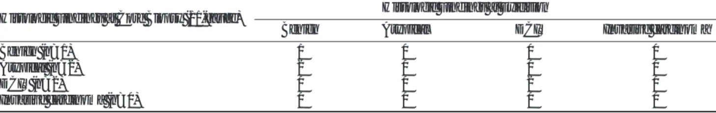
65개의 14 게이지 자동총생검법 결과 42개는 양성(유두종 이 41개, 유두종증이 1개), 15개는 비정형성 병변(비정형성 유 두종 또는 양성 병변이면서 비정형성을 보이는 병변)이었고 8 개가 악성(유두상 관상피내암이 7개, 침윤성 유두상암이 1개) 이었다 (Table 1).
33개의 11 게이지 진공흡인생검법 결과 27개가 양성(유두
종이 25개, 유두종증이 2개), 4개가 비정형성 병변(비정형성 유두종 또는 양성 병변이면서 비정형성을 보이는 병변)이었으 며 2개가 악성(모두 유두상 관상피내암)이었다(Table 1).
조직검사 결과 비정형성이나, 관내 또는 침윤성 암이 포함된 경우는 모든 예에서 외과적 절제를 권고하였다. 양성 병변이라 도 병리의사나 임상의사 또는 환자가 원하는 경우 외과적으로 절제되었다. 양성인 병리결과가 영상소견과 부합한다면 6, 12, 24개월의 추적검사를 권고했다. 전체 55개의 양성 병변 중 30 개(55%)에서 유방촬영술 또는 초음파 추적검사가 이뤄졌으며 각각 14 게이지 자동총생검법의 경우 29개 중 14개(48%)에 서, 11 게이지 진공흡인생검법의 경우 26개 중 16개(62%)에 서였다. 14 게이지 자동총생검법을 시행한 한 개의 비정형 병 변은 수술도 추적검사도 이루어지지 않았다. 평균 추적 기간은 14 게이지 자동총생검법의 경우 11.3개월, 11 게이지 진공흡 인생검법의 경우 12.6개월이었다.
“조직학적 저평가”란 경피적 생검 결과는 비정형 관상피 증 식증(atypical hyperplasia, ADH)이었으나 수술에서 암이 나 온 경우(비정형 관상피 증식증 저평가, ADH underesti- mation) 또는 경피적 생검 결과는 관상피내암(관상피내암 저 평가, ductal carcinoma in situ, DCIS)이 나왔으나 수술에서 침윤성 암으로 나온 경우(DCIS underestimation)로 정의한다 (12, 19, 20). 저평가율은 수술을 한 각각의 고위험 병변 중 수술에서 암이 나온 수를 수술을 한 전체 고위험 병변의 수로 나누어서 구했다. 재조직검사가 이뤄졌으면 처음 조직검사와의 시간 간격과 그 이유를 조사하였다. 즉각적 재조직검사 (immediate rebiopsy)란 첫 번째 영상 추적검사가 이뤄지기 전 재조직검사가 시행되는 경우를 말하고 지연 재조직검사 (delayed rebiopsy)란 첫 번째 영상 추적검사 후 재조직검사 가 이뤄지는 경우를 의미한다. 재조직검사가 이뤄지는 이유로 는 고위험 병변, 불충분한 검체, 영상-조직소견 간 불일치 (discordance), 추적 영상에서 병변의 진행 등이 있을 수 있다.
재조직검사율은 재조직검사수를 전체 조직검사수로 나누어서 구했다(13). 14 게이지 자동총생검법군과 11 게이지 진공흡 인 생검법군 간의 조직학적 저평가와 재조직검사율을 비교하 였다.
결 과
14 게이지 자동총생검법의 경우 65개 중 35개에서 수술을 했으며(53.8%, 양성 13/42, 비정형성 14/15, 악성 8/8) 11 게 이지 진공흡인생검법의 경우 33개 중 5개에서 수술을 했다 (15.2%, 양성 1/27, 비정형성 2/4, 악성 2/2). 전체 양성 병변 69개 중 25개(36.2%, 14 게이지 자동 총에서 15개, 11 게이 지 진공흡인 생검법에서 10개)는 추적검사도 수술도 이뤄지지 않았다. 14 게이지 자동총생검법 결과 양성이었던 13개의 병 변 중 수술 후 악성으로 바뀐 경우는 없었으나 양성 유두종이 었던 2개에서 수술 후 비정형성 유두종으로 바뀌었다. 14개의 비정형성 병변은 수술 결과 비정형성 유두종 6개, 비정형성이 없는 양성 병변 1개, 유두상 관상피내암 5개, 침윤성 암 2개였 Table 1. Histology at Core Biopsy
Gun* VA
Benign 42 (65%) 27 (82%)
Atypical 15 (23%) 04 (12%)
Malignant 08 (12%) 2 (6%)
Total 65 33
Gun*; 14-gauge automated gun biopsy VA ; 11-gauge vacuum-assisted biopsy
다. 8개의 악성 병변은 수술 결과 유두상 관상피내암 5개, 비 정형성 유두종 1개, 침윤성 암 2개였다.
11 게이지 진공흡인생검법에서 양성이면서 수술한 1개의 유 두종증의 경우 수술 후에도 양성이었다. 3개의 비정형성 병변 은 수술 결과 양성 유두종 2개, 섬유선종 1개였다. 11 게이지 진공흡인생검법에서 비정형성으로 나온 나머지 두 개의 병변 은 영상 추적검사에서 병변의 진행을 보이지 않고 안정적이었 다. 2개의 유두상 관상피내암은 수술 후에도 유두상 관상피내 암이었다. 두 군의 핵생검과 수술 후 병리 소견을 요약하면 각 각 Table 2, 3과 같다.
14 게이지 자동총생검법의 경우 양성인 병변 13개와 비정 형성 병변 12개에서 재조직검사가 시행되었으며 모든 예에서 즉각적 재조직검사였다. 양성 병변의 재조직검사의 이유는 만 져지는 종괴나 유두 분비 등의 이유로 환자나 임상의사가 원 해서였다. 11 게이지 진공흡인생검법의 경우 양성 병변 1개와 비정형성 병변 2개에서 재조직검사가 이뤄졌고 추적검사에서 병변의 진행을 보였던 한 개를 제외하고는 즉각적 재조직검사 였다. 재조직검사율은 14 게이지 자동총생검법의 경우 42% (27/65)였고 11 게이지 진공흡인생검법의 경우 9.1%(3/33)였다. 비정형 관상피 증식증 저평가율은 14 게이 지 자동총생검법의 경우 50%(7/14)였고 11 게이지 진공흡인 생검법의 경우 0%(0/4)였다. 관상피내암 저평가율은 14 게이 지 자동총생검법의 경우 14%(1/7)였고 11 게이지 진공흡인 생검법의 경우 0%(0/2)였다(Table 4).
고 찰
유두상 병변은 섬유혈관줄기를 가지며 유관 내로 돌출하는 유두 모양의 상피세포의 증식이다(6, 21). Haagensen 등(22) 은 유두상 종양을 양성 관내 유두종과 악성 유두상암의 두 가 지 주된 형태로 나눴다. Tavassoli(6)는 유두상 병변을 더 넓 은 스펙트럼으로 나눴다. 여기에는 유두종, 유두종증, 경화성 유두종(sclerosing papilloma), 그리고 비정형 유두종이 포함
된다. 또한, 유두종에서 발생한 암(carcinoma arising in a papilloma), 유두상 관상피내암, 침윤성 유두상암도 포함된다.
이들 병변간은 형태학적으로 유사한 양상을 보이기 때문에 이 들을 병리조직학적 소견으로 양성과 악성 병변을 감별하는 것 은 어려우며 판단할 조직의 양이 적을 경우 더욱 그러하다.
Tavassoli(6)는 유두상 병변에서 양성 병변을 악성 병변과 구 별하는 가장 중요한 요소는 병변의 강 내쪽을 따라 존재하는 한 층의 근상피세포(myoepithelial cell)의 존재라고 하였다.
가장 흔한 양성 유두상 종양은 유두종으로 엽상의 섬유혈관 기질과 증식성의 유관 상피세포로 구성된다(1). 유두종은 유 륜하의 큰 유관에서 또는 유방 말초부위의 작은 유관에서 발 견될 수 있다(6, 21). 단일성 유두종은 대부분 중심부에 위치 하며 유방촬영에서 잘 보이지 않으며 종종 유두분비와 관련이 있다. 말초 유두종은 더 흔하게 말초부위에 위치하며 증상이 없는 경우가 많다(23). 다발성 말초 유두종이 있는 여성은 유 방암의 발생 위험이 커진다는 주장도 제기되고 있다(6). 유두 상암은 전체 유방암의 2% 이내로 주로 유두상 관상피내암으 로 나타난다 (6, 24).
본 연구에서는 영상 유도 하 조직검사의 5.7%가 유두상 병 변으로 3%였던 Liberman 등(7)이나 4.9%였던 Mercado 등 (16)의 보고보다 약간 높게 나타났다. 또한, 조직 검사에서 양 성 유두상 병변이 나온 경우 수술적 절제 없이 임상적 또는 영 상 추적검사로 안전하게 관리할 수 있다는 이전에 나온 보고 들과도 일치한다(7, 16). 14 게이지 자동총생검법과 11 게이 지 진공흡인생검법 모두에서 양성으로 나온 병변이 수술 후 악
Table 2. Surgical Findings of 14-gauge Automated Gun Biopsy
Histologic Findings at Core Biopsy (14-gauge) Histologic Findings at Exicision
Benign Atypical DCIS Invasive carcinoma
Benign (n=13) 11 2 0 0
Atypical (n=14) 1 6 5 2
DCIS (n=7) 0 1 5 1
Invasive carcinoma (n=1) 0 0 0 1
Table 3. Surgical Findings of 11-gauge Vacuum-Assisted Biopsy
Histologic Findings at Core Biopsy (11-gauge) Histologic Findings at Exicision
Benign Atypical DCIS Invasive carcinoma
Benign (n=1) 1 0 0 0
Atypical (n=2) 2 0 0 0
DCIS (n=2) 0 0 2 0
Invasive carcinoma (n=0) 0 0 0 0
Table 4. Histologic Underestimation & Repeat Biopsy Rate
Gun* VA
ADH underestimation 50% (7/14)0 0% (0/4) DCIS underestimation 14% (1/7)00 0% (0/2) Repeat biopsy rate 42% (27/65) 9.1% (3/33).
Gun*; 14-gauge automated gun biopsy VA ; 11-gauge vacuum-assisted biopsy
성으로 밝혀지는 경우는 하나도 없었다. 따라서 이전의 보고들 과 종합해 볼 때 경피적 핵생검에서 비정형성을 포함하지 않 는 양성 유두상 병변으로 진단된 경우 즉각적인 외과적 절제 의 필요 없이 영상 추적검사로도 안전하게 관리할 수 있다. 그 러나 비정형성 병변은 두 군 합쳐서 7개(39%, 7/18)의 병변 이 수술 후 관상피내암이나 침윤성 암으로 밝혀졌으며 7개 모 두 14 게이지 자동총생검법을 시행한 경우였다. 따라서 조직 검사 결과 비정형 유두종이나 비정형성을 가진 유두상 병변이 라면 특히 14 게이지 자동총생검법의 경우 반드시 수술적 절 제가 있어야 한다.
조직검사에서의 저평가는 검사 특성상 피하기 어려운 제한 점으로 잘 알려져 있다(25, 26). 14 게이지 자동총생검법으로 비정형성으로 나와 수술을 시행한 14개 중 7개가 수술 후 유 두상 관상피내암이나 침윤성 암으로 바뀌었다. 그러나 11 게 이지 진공흡인생검법의 경우 수술 후 암으로 나온 경우가 없 었다. 유사하게 14 게이지 자동총생검법에서 유두상 관상피내 암으로 나온 7개 중 한 개가 수술 후 침윤성 암으로 밝혀졌으 나 11 게이지 진공흡인생검법의 경우 2개 모두 수술 후에도 관상피내암이었다. 이런 조직학적 저평가는 획득하는 조직의 양을 늘리거나, 더 굵은 게이지의 바늘을 이용하거나, 진공흡 인생검법을 사용하면 줄어들 것으로 생각된다(12). 따라서 조 직학적 저평가를 줄이기 위해서는 11 게이지 진공흡인생검법 의 사용이 요구된다.
본 연구의 제한점은 다음과 같다. 첫째: 본 연구에 포함된 대상의 수가 11 게이지 진공흡인생검법이 작았으며 특히 비정 형성과 악성 병변이 각각 4개, 2개로 14 게이지 자동총생검법 에 비해 작았다. 따라서 저평가율이 다소 과장되었을 수 있다.
둘째: 진공흡인생검법이 작은 병변과 미세석회화 검출에 주로 사용되었고 큰 종괴는 자동총을 많이 이용하여 기법 선택에 치 우침이 있었을 수 있다는 것이다. 그러나 본 연구에서는 두 군 간의 병변의 크기에 유의한 차이가 없었으므로 큰 영향을 미 치지는 않았을 것으로 생각된다.
결론적으로 조직검사 결과가 양성이면서 영상소견과 부합한 다면 수술적 절제 없이 영상추적만으로도 안전하게 관리될 수 있다. 그러나 비정형성 병변이라면 조직학적 저평가의 위험 때 문에 수술적 절제가 요구된다. 또한, 14 게이지 자동총생검법 에 비해 11 게이지 진공흡인생검법이 조직학적 저평가와 재조 직검사 측면에서 성적이 우수하였다.
참 고 문 헌
1. Rosen PP. Benign papillary tumors. In Rosen PP. Rosen’s breast pathology. Philadelphia: Lippincott-Raven, 1997:67-104
2. Rosen PP. Papillary carcinoma. In Rosen PP. Rosen’s breast patholo- gy. Philadelphia: Lippincott-Raven, 1997:335-354
3. Gadd MA. Papillary lesions. In Harris JR, Lippman ME, Morrow M, Hellman S. Diseases of the breast. Philadelphia: Lippincott- Raven, 1996:42-45
4. Haagensen CD. Solitary intraductal papilloma. In Haagensen CD.
Diseases of the breast. Philadelphia: Saunders, 1986:136-175 5. Ellis IO, Elston CW, Pinder SE. Papillary lesions. In Elston CW,
Ellis IO. Systemic Pathology: the breast, 3rd ed. Edinburgh:
Churchill Livingstone, 1998;133-146
6. Tavassoli FA. Papillary lesions. In Tavassoli FA. Pathology of the breast. New York: Elsevier, Norwalk; Conn: 1992;193-227 7. Liberman L, Bracero N, Vuolo MA, Dershaw DD, Morris EA,
Abramson AF, et al. Percutaneous large-core biopsy of papillary breast lesions. AJR Am J Roentgenol 1999; 172:331-337
8. Philpotts LE, Shaheen NA, Jain KS, Carter D, Lee CH. Uncommon high-risk lesions of the breast diagnosed at stereotactic core-needle biopsy: clinical importance. Radiology 2000; 216:831-837
9. Philpotts LE, Lee CH, Horvath LJ, Lange RC, Carter D, Tocino I.
Underestimation of breast cancer with 11-gauge vacuum suction biopsy. AJR Am J Roentgenol 2000;175:1047-1050
10. Jackman RJ, Burbank F, Parker SH, Evans WP 3rd, Lechner MC, Richardson TR, et al. Atypical ductal hyperplasia diagnosed at stereotactic breast biopsy: improved reliability with 14-gauge, di- rectional, vacuum-assisted biopsy. Radiology 1997;204:485-488 11. Philpotts LE, Shaheen NA, Carter D, Lange RC, Lee CH.
Comparison of rebiopsy rates after stereotactic core needle biopsy of the breast with 11-gauge vacuum suction probe versus 14-gauge needle and automatic gun. AJR Am J Roentgenol 1999;172:683-687 12. Burbank F. Stereotactic breast biopsy of atypical ductal hyperpla- sia and ductal carcinoma in situ lesions: improved accuracy with directional, vacuum-assisted biopsy. Radiology 1997;202:843-847 13. Jackman RJ, Nowels KW, Rodriguez-Soto J, Marzoni FA Jr,
Finkelstein SI, Shepard MJ. Stereotactic, automated, large-core needle biopsy of nonpalpable breast lesions: false-negative and his- tologic underestimation rates after long-term follow-up. Radiology 1999;210:799-805
14. Philpotts LE, Hooley RJ, Lee CH. Comparison of automated versus vacuum-assisted biopsy methods for sonographically guided core biopsy of the breast. AJR Am J Roentgenol 2003;180:347-351 15. Cho N, Moon WK, Cha JH, Kim SM, Kim SJ, Lee SH, et al.
Sonographically guided core biopsy of the breast: comparison of 14-gauge automated gun and 11-gauge directional vacuum-assisted biopsy methods. Korean J Radiol 2005;6:102-109
16. Mercado CL, Hamele-Bena D, Singer C, Koenigsberg T, Pile- Spellman E, Higgins H, et al. Papillary lesions of the breast: evalua- tion with stereotactic directional vacuum-assisted biopsy.
Radiology 2001;221:650-655
17. Liberman L, Smolkin JH, Dershaw DD, Morris EA, Abramson AF, Rosen PP. Calcification retrieval at stereotactic, 11-gauge, direc- tional, vacuum-assisted breast biopsy. Radiology 1998;208:251-260 18. Parker SH, Klaus AJ, McWey PJ, Schilling KJ, Cupples TE,
Duchesne N, et al. Sonographically guided directional vacuum-as- sisted breast biopsy using a handheld device. AJR Am J Roentgenol 2001;177:405-408
19. Liberman L, Dershaw DD, Glassman JR, Abramson AF, Morris EA, La Trenta LR, et al. Analysis of cancers not diagnosed at stereotactic core breast biopsy. Radiology 1997;203:151-157 20. Burbank F, Parker SH. Methods for evaluating the quality of an
image-guided breast biopsy program. Semin Breast Dis 1998;1:71- 83
21. Page DL. Papilloma and related lesions. In Page DL, Anderson TJ.
Diagnostic histopathology of the breast. Edinburgh, Scotland;
Churchill Livingstone, 1987;104-119
22. Haagensen CD, Stout AP, Phillips JS. The papillary neoplasms of the breast. I. Benign intraductal papilloma. Ann Surg 1951;133:18- 36
23. Cardenosa G, Eklund GW. Benign papillary neoplasms of the breast: mammographic findings. Radiology 1991;181:751-755 24. Schneider JA. Invasive papillary breast carcinoma: mammograph-
ic and sonographic appearance. Radiology 1989;171:377-379 25. Lee CH, Carter D, Philpotts LE, Couce ME, Horvath LJ, Lange RC,
et al. Ductal carcinoma in situ diagnosed with stereotactic core needle biopsy: can invasion be predicted? Radiology 2000;217:466- 470
26. Jackman RJ, Burbank F, Parker SH, Evans WP 3rd, Lechner MC, Richardson TR, et al. Stereotactic breast biopsy of nonpalpable le- sions: determinants of ductal carcinoma in situ underestimation rates. Radiology 2001;218:497-502
J Korean Radiol Soc 2006;54:537-541
Address reprint requests to : Woo Kyung Moon, M.D., Department of Radiology, College of Medicine Seoul National University and The Institute of Radiation Medicine, Seoul National University Medical Research Center
28, Yeongeon-dong, Jongno-gu, Seoul 110-744, Korea.
Tel. 82-2-2072-2114 Fax. 82-2-743-6385 E-mail: [email protected]
Papillary Lesions of the Breast: Comparison of the US-guided 14-Gauge Automated Gun Method and the 11-Gauge Directional
Vacuum-Assisted Biopsy Method
1Eun Sook Ko, M.D., Nariya Cho, M.D., Sang-Kyu Yang, M.D., Do Youn Kim, M.D., Woo Kyung Moon, M.D.
1Department of Radiology, College of Medicine Seoul National University and The Institute of Radiation Medicine, Seoul National University Medical Research Center
Purpose: To compare the outcomes of US-guided 14-gauge automated biopsy and 11-gauge vacuum-assisted biopsy for the papillary lesions of the breast.
Materials and Methods: We retrospectively reviewed the US-guided core biopsies of 1,723 consecutive breast lesions that were treated from January 2003 to April 2005. Ninety-eight lesions (5.7%) were pathologically re- ported as papillary lesions. The biopsies were performed with using a 14-gauge automated gun on 65 lesions or with using an 11-gauge vacuum-assisted device on 33 lesions. Thirty-five lesions (54%, 35/65) of 14-gauge auto- mated gun biopsies and 5 lesions (15%, 5/33) of 11-gauge vacuum-assisted biopsies underwent surgery. The histologic findings were compared with the surgical, imaging and follow-up findings. The histologic underesti- mation rate, the repeat biopsy rate and the false negative rate were compared between the two groups. The re- peat biopsy rate was determined by dividing the total number of core biopsies into the number of repeat biop- sies. “ADH underestimation”was defined as a lesion yielding atypical ductal hyperplasia on percutaneous biopsy and carcinoma at surgery, and “DCIS underestimation”was defined as a lesion yielding ductal carcino- ma in situ on percutaneous biopsy and invasive carcinoma at surgery.
Results: The repeat biopsy rate was 42% (27/65) for the 14-gauge automated gun biopsies and 9.1% (3/33) for the 11-gauge vacuum-assisted biopsies. The ADH underestimation rate was 50% (7/14) for the 14-gauge auto- mated gun biopsies and 0% (0/4) for the 11-gauge vacuum-assisted biopsies. The DCIS underestimation was 14% (1/7) for the 14-gauge automated gun biopsies and 0% (0/2) for the 11-gauge vacuum-assisted biopsies.
The false negative rate was 0% for these two groups.
Conclusion: For the papillary lesions of the breast, the outcomes of the US-guided core biopsies performed with the 11-gauge vacuum-assisted device were better than those of the biopsies performed with the 14-gauge automated gun, in terms of underestimation and repeat biopsy.
Index words :Breast, biopsy Breast, US Breast, neoplasms