Ultrasonographically-Guided Biopsy after Digital Mammographically Guided Two-Dimensional
Localization of Breast Microcalcifications
1Jung Hee Shin, M.D.1, 2, Hye-Young Choi, M.D.2, Hyun-Ah Kim, M.D.3, Boo-Kyung Han, M.D.1, Woo Kyung Moon, M.D.4
1Department of Radiology, Samsung Medical Center, Sunkyunkwan University, School of Medicine
2Department of Radiology and Medical Research Institute, Ewha Womans University, College of Medicine, Ewha University Mokdong Hospital
3Department of General Surgery and Medical Research Institute, Ewha Womans University, College of Medicine, Ewha University Mokdong Hospital
4Department of Radiology and Clinical Research Institute, Seoul National University Hospital and the Institute of Radiation Medicine, Seoul National University Medical Research Center, Seoul, Korea
Received January 8, 2007 ; Accepted July 23, 2007
Address reprint requests to : Hye-Young Choi, M.D., Department of Radiology and Medical Research Institute, Ewha Womans University, College of Medicine, Ewha University Mokdong Hospital
911-1, Mok-dong, Yangcheon-gu, Seoul 158-710, Republic of Korea.
Tel. 82-2-2650-5038 Fax. 82-2-2650-5302 E-mail: [email protected]
Purpose: To investigate the efficacy of ultrasound (US)-guided core biopsies after digi- tal mammography-guided two-dimensional localization (DM-2DL) of breast microcal- cifications.
Materials and Methods: Twenty-two patients with 23 suspicious microcalcifications underwent US-guided core biopsies after DM-2DL, to mark the sites on the skin where microcalcifications had been found (craniocaudal and mediolateral (or latero- medial) views). Of the 23 lesions, 4 were sampled using a 14-gauge automated gun and the other 19 were sampled using an 8-gauge vacuum-assisted device. The lesions were categorized into two groups: those with and those without microcalcifications ob- served on US. The success rate for correctly sampling microcalcifications on the speci- men radiograph in the two groups was assessed and their pathologic outcomes were investigated.
Results: Of the 23 lesions, 16 were invisible and 7 were visible to ultrasonographic mi- crocalcifications. The sampling success rate for the specimen radiographs was 100%
for ultrasonographic visible microcalcifications and 88% (14/16) for lesions invisible to ultrasonography after DM-2DL (p = 1.000). The cancer rate of individuals with micro- calcifications observed on US (57%, 4/7) was greater than in individuals without visi- ble microcalcifications (13%, 2/16) (p=0.045).
Conclusion: Although some microcalcifications are invisible on US, a US-guided biop- sy after DM-2DL is a useful method for the successful sampling of the microcalcifica- tions.
Index words :Calcifications Mammography Biopsy
Breast microcalcifications are difficult to detect by ul- trasonography (US) and the diagnosis of suspicious mammographic microcalcifications usually requires a stereotactic biopsy or surgery with needle localization.
To our knowledge, the stereotactic biopsy is generally the first diagnostic choice for suspicious microcalcifica- tions not seen on US; however, this method has limita- tions in small lesions and women with insufficient breast thickness (1). Above all, because most hospitals in Korea do not have a stereotactic device, we devised an- other diagnostic way to obtain a biopsy of mammo- graphic microcalcifications. A US-guided biopsy has sev- eral advantages, which include speed, comfort, no radia- tion exposure, and real-time needle visualization. The detection of the potential areas of suspicious microcalci- fications, which are not identified by US require the fastest and most available methods in order to render the histological confirmation of a breast lesion. With the current digital technology, a full-field digital mammog- raphy can rapidly be displayed, and the images can be manipulated. In addition, this method decreases the time needed for needle localization (2, 3). If a localiza- tion technique for invisible ultrasonographic microcalci- fications can be optimized, a US-guided biopsy might be preferred and become a more popular method, even with microcalcification cases which do not permit for a stereotactic biopsy.
This study investigated the effectiveness of the US- guided core biopsy after a digital mammography-guided two-dimensional localization (DM-2DL) of breast micro- calcifications.
Materials and Methods
Patients and lesions
From May 2005 to February 2006, 306 patients under- went a US-guided breast biopsy for lesions with suspi- cious imaging, clinical findings, or probably benign le- sions (in patients who were extremely anxious) at our in- stitution. Among them, 22 consecutive women with 23 suspicious microcalcifications prospectively underwent US-guided core biopsies after a DM-2DL. The institu- tional review board approved this study, and each pa- tient provided informed consent before undergoing a biopsy. At the time of diagnosis, each patient was noti- fied of the suspicious lesion, which required a biopsy, and consented to undergo an ultrasonographic evalua- tion of the suspicious area to determine if the lesion could be biopsied under ultrasonographic guidance. Of
the 23 lesions, 16 were invisible and 7 were visible to ul- trasonographic microcalcifications. Of the 16 invisible microcalcifications, one lesion was subsequently detect- ed by a DM-2DL.
The patients selected for this study ranged in age from 33 to 58 years (mean age: 42 years). The mammographic microcalcifications were categorized according to the le- sion size, distribution, number, morphology and the as- sociated findings using BI-RADS descriptors (4). The suspicious microcalcifications requiring a biopsy were classified as BIRADS category 4 or 5, and subsequently interpreted by two breast imaging radiologists indepen- dently.
Localization and Biopsy Procedure
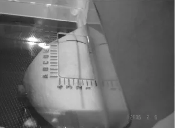
Overall, the two breast imaging radiologists initially evaluated the 23 lesions with bilateral whole breast ul- trasonography. A digital mammography-guided two- di- mensional localization was performed to mark the skin where the microcalcifications were located (craniocau- dal and mediolateral (or lateromedial) views), using a fenestrated compression paddle (Senographe DS, General Electric Medical Systems, Buc, France). The pa- tient remained stationary with their breast under com- pression while drawing the skin marker. A radiologist marked the expected location of the calcifications with an indelible pen drawn on an orthogonal line defined by an optical localizer for two-dimensional biopsy for each view (Fig. 1). Once the skin was marked, all US exami- nations marked as a suspicious area were analyzed us- ing a 5-12 MHz linear array transducer (HDI 5000;
Fig. 1. Skin mark for the digital mammography guided two-di- mensional localization. The black line over the skin was drawn orthogonally in the craniocaudal view, as defined by an optical localizer over the location of the microcalcifications.
Philips Medical Systems, Bothell, WA) with full knowl- edge of the mammographic location. The presence of any US visible microcalcifications was documented un- der the skin marking. The US-guided biopsy was per- formed using a 14-gauge automated gun (Pro-Mag 2.2, Manan Medical Products, Northbrook, IL, USA) in 4 of the lesions, or an 8-gauge vacuum-assisted device (Mammotome, Biopsys/Ethicon endosurgery In., Cincinnati, OH, USA) in the 19 remaining lesions, which were targeted on the fiducial marker. The criteria for using a 14-gauge automatic gun were microcalcifica- tions with a wide extent (> 3cm) or microcalcifications associated with a mass. For each microcalcification, at least five core specimens were taken when using an 8- gauge vacuum-assisted device, whereas seven core spec- imens were taken when using a 14-gauge automated gun. Overall, a mean of 8.5 specimens (range, 5 to 25 specimens) per lesion were obtained. The depth of each lesion was divided into the anterior, middle and posteri- or portion through a mammography. A radiologist deter- mined that the lesion depth was used to guide the path of the needle, and was aligned such that the skin marker indicating the microcalcifications were in the path of the needle as it was fired. For the vacuum-assisted tech- nique, the probe was placed beneath the skin marker, and was positioned such that the calcification area was located superficially to the tissue-cutting notch of the probe. Once the sampling was complete, specimen radi- ographies were performed in order to confirm the pres- ence of calcifications, and if they were observed on a mammography. If the lesion in question included calci- fications, the samples were sorted and placed into sepa- rate cassettes labeled “with” or “without” calcifications
to further assist the pathologist. Each biopsy specimens was placed into a jar containing formalin. The biopsy was considered a success if the radiologist felt that the calcification from the original image was definitely pre- sent on the specimen image. Conversely, the biopsy was considered a failure if no convincing calcification was present. Moreover, a failed biopsy indicated that no mi- crocalcifications were present in any specimens despite performing the biopsy trial three times. All but one pa- tient underwent surgery after being diagnosed with a malignancy, on the core biopsy or when the initial and repeat core biopsy was nondiagnostic. The average du- ration of a US-guided biopsy after DM-2DL microcalci- fications was 20 minutes. However, this was dependant on the correctness of the target. Twenty-three lesions, including bilateral lesions in one of the patients, were biopsied using a 14-gauge automated gun in 4 lesions, whereas an 8-gauge vacuum-assisted device was used in 19 lesions. After the biopsies, each lesion was catego- rized into two groups: those with and those without mi- crocalcifications seen on US. The success rate in captur- ing microcalcifications on a specimen radiograph was assessed, for the two groups, using the Fisher’s exact test. Their pathologic outcomes were investigated. The patients with a benign diagnosis were recommended to undergo a mammographic follow-up every 6 months for 2 years, upon which, patients are identified as being sta- ble. An excision with needle localization was recom- mended if the biopsy result showed a radiologic-patho- logic discordance.
Table 1. Mammographic Findings of Microcalcifications
Mammographic Findings (n = 23) US Invisible Microcalcifications (n=16) US Visible Microcalcifications (n=7) Extent of microcalcifications (mm)
Mean ± SD 10.75 ± 1.02 16.71 ± 1.38
(range, 3-36) (range, 5-43)
Number of microcalcifications
05-15 06 0
16-50 08 4
>50 02 3
Morphology of microcalcifications
Pleomorphic 03 5
Amorphous 12 2
Punctate 01 0
Distribution of microcalcifications
Clustered 13 3
Segmental 03 4
Associated findings
Mass 00 2
Results
Imaging Findings
Suspicious microcalcifications viewed by a mammog- raphy were interpretered as being clustered amorphous (10), segmental amorphous (4), clustered pleomorphic (5), segmental pleomorphic (3), and clustered punctate (1). The latter was considered to be a suspicious finding because of a new lesion found at the ipsilateral breast conservation operation bed, and was confirmed patho- logically as being a fibrosis with calcifications. The mammographic findings are listed in Table 1. The le- sions ranged in size between 3 to 43 mm (mean, 13 mm). Twenty lesions were assigned a BI-RADS category 4 (suspicious), whereas three lesions were assigned BI- RADS category 5 (highly suggestive of a malignancy).
The 20 category 4 lesions included 4A in 13 and 4B in 8.
The echogenic dots, representing calcificiations on US, could not be found in 16 patients with US invisible mi- crocalcifications. Moreover, of the 7 patients with US visible microcalcifications, 5 demonstrated multiple echogenic dots on US at the site corresponding to the mammographic calcifications, with the remaining two patients showing a hypoechoic mass and calcifications on US.
Results of US-Guided Biopsy after DM-2DL
Of the 23 lesions, 16 ultrasonographic microcalcifica- tions were invisible, whereas 7 were visible. The suc- cess rate for the identification of microcalcifications on the specimen radiograph was 100% in ultrasonographic visible microcalcifications and 88% (14/16) in those in- visible by biopsies after DM-2DL (Fig. 2). The difference between the visible and invisible ultrasonographic mi- crocalcifications was not statistically significant (p = 1.000). In the two failed cases, one was followed up and there were no changes for 24 months. The remaining patient underwent a surgical excision for lesions in both breasts because of a contralateral malignancy on a biop- sy result, and was confirmed as a contralateral ductal carcinoma in situ and an ipsilateral fibrocystic change.
The cancer rate (57%, 4/7) of the microcalcifications seen on US was a higher than that (13%, 2/16) of those without microcalcifications (p=0.054). There were no statistically significant differences in the success rate of biopsies between a 14-gauge automated gun (100%, 4/4) and an 8-gauge vacuum-assisted device (89%, 17/19) (p
= 1.000).
Histopathological Findings
As a result of the biopsies, the histological findings in 16 patients with US invisible microcalcifications are de- scribed as follows: 2 ductal carcinoma in situ (DCIS), 2 atypical ductal hyperplasias, 4 fibrosis, 5 fibrocystic dis-
A
B Fig. 2. A 46-year-old woman with a fibrocystic disease.
A. A magnified view of the right breast shows fewer than 15 clustered amorphous microcalcifications, which were invisible on sonography (not shown).
B. As US-guided biopsy using an 8-gauge vacuum-assisted device follow- ing a digital mammography-guided two-dimensional localization success- fully biopsied microcalcifications (circle) in one of five specimens.
eases, 1 fibroadenoma, 1 adenosis, and 1 intraductal hy- perplasia. The histological findings of the 7 patients with US visible microcalcifications were described as fol- lows: 3 invasive ductal carcinomas, 1 DCIS, 2 fibrocys- tic changes, and 1 adenosis. The microcalcifications, which could be observed on the specimen radiograph, were present on the pathological section in all cases.
Two cases showed radiologic-pathologic discordance, upon which was confirmed by the fibrocystic disease.
The other case underwent was followed up by 6 exami- nations at every 6 month interval and has been stable for 2 years. All benign lesions matchedwith radiologic- pathologic concordance have been followed up for a mean of 13 months (range, 6-25 months).
No false-negative results were noted. There were no underestimations of the disease in the two atypical duc- tal hyperplasia and three noninvasive carcinoma cases.
Discussion
If the stereotactic biopsy of the suspicious microcalci- fications is not performed for various technical reasons or because of a lack of equipment, an alternative method will be needed to make a histological confirma- tion of microcalcifications prior to surgery.
Using state-of the-art US equipment and a higher fre- quency transducer, radiologists are now identifying mi- crocalcifications more frequently by ultrasonography (5- 7). Moreover, these microcalcifications are usually demonstrated within the hypoechoic masses that facili- tate the detection of echogenic microcalcifications (7).
The hybrid digital mammography/US guided biopsy ap- pears to offer a simple means of ensuring the accurate sampling of breast microcalcifications without the need for a surgical excision or stereotactic biopsy. Our suc- cess rate (88%) for retrieving ultrasonographically invisi- ble microcalcifications during US-guided biopsies, after DM-2DL, is comparable to that of retreiving ultrasono- graphically visible microcalcifications.
A US-guided biopsy for an invisible lesion without as- sociated findings is limited, although a radiologist did recognize the mammographic location of microcalcifica- tions. However, targeting and retrieving microcalcifica- tions by a US-guided biopsy, after DM-2DL, increases the confidence that the correct area is being sampled.
In this study, if microcalcifications were not docu- mented on the initial specimen radiograph, the proce- dure was considered incomplete, and additional cores were required. Inadequate radiological localization may
lead to the removal of an excessively large amount of tis- sue, which is not the optimal result for a diagnostic pro- cedure, for microcalcifications that might turn out to be benign, whereas the increase in the number of cores can contribute to the need to retrieve microcalcifications within the biopsy specimens. However, there were no significant complications encountered in this study.
Some reports showed that ultrasonography can identi- fy clustered microcalcifications in breast cancers (5,8- 10). Similar to previous reports, this study demonstrated that the cancer rate observed by US (57%) was higher than in cases without microcalcifications (13%).
In this study, we were able to use the vacuum-assisted device for US-guided biopsies of microcalcifications in- visible to ultrasonography. The vacuum-assisted device enables the radiologist to obtain samples more quickly, and required less precision in the placement of the nee- dle for retrieval of microcalcifications due to its ability to suction tissue from the adjacent areas. In addition, this device can also reduce the potential for sampling errors, as well as the underestimation of the disease introduced by the multi-pass techniques (11-13). In the case of a stereotactic biopsy on a prone table, failure to retrieve microcalcifications was least common with 11-gauge vacuum-assisted devices when compared to 14-gauge core and 14-gauge vacuum (14). Although the compara- tive analysis of 11-gauge and 8-gauge vacuum-assisted devices for biopsy of microcalcifications under US or stereotactic guidance hasnot yet been seen, we have of- ten used 8-gauge vacuum-assisted devices in order to ob- tain enough tissue to include microcalcifications not de- tected on US. A more expansive study is necessary to determine whether the 8-gauge vacuum-assisted devices could actually reduce false negatives or the underesti- mation of disease compared with 11-gauge vacuum-as- sisted devices.
Further limitations of this study included the relative- ly small study sample size and the limited application of the multiple potentially significant covariates in the dif- ferent patients, lesion types, radiologists and technolo- gists performing the procedures. The several obstacles experienced in successfully performing a DM-2DL in- cludes microcalcifications near the nipple or at the far periphery, which are difficult to localize with DM-2DL.
The skin marking of these lesions should be performed in only one view, if possible. When DM-2DL is per- formed, the breast should be naturally positioned and compressed. If the breast is pulled too much or is fo- cused toward the lesion at the time of the mammogra-
phy, the skin marker will not correspond to the possible mammographic area, which in turn, may lead to an in- correct ultrasonographic biopsy track. After failing to biopsy following a DM2DL, additional excisions, in- crease in cost, as well as an increase in the patient’s anx- iety. Of the two failed biopsy cases in this study, one was followed up with no change for 24 months because the microcalcifications were faint, very small, amor- phous, and had a number of 5, which was considered to be of a low concern for a malignancy. The remaining pa- tients underwent a surgical excision and were further confirmed to have a contralateral ductal carcinoma and an ipsilateral fibrocystic change.
Despite no false negatives or underestimations in this study, the long- term follow up of the benign microcalci- fications, which were diagnosed by a needle biopsy is necessary, especially if a biopsy is performed during the operator`s learning period (15).
In conclusion, although microcalcifications are invisi- ble on US, a US-guided biopsy after DM-2DL is a useful method for the successful sampling of microcalcifica- tions.
References
1. Liberman L, Fahs MC, Dershaw DD, Bonaccio E, Abramson AF, Cohen MA, et al. Impact of stereotaxic core breast biopsy on cost of diagnosis. Radiology 1995;195:633-637
2. Dershaw DD, Fleischman RC, Liberman L, Deutch B, Abramson AF, Hann L. Use of digital mammography in needle localization procedures. AJR Am J Roentgenol 1993;161:559-562
3. Yang WT, Whitman GJ, Johnson MM, Bolanos-Clark M, Kushwaha AC, Hunt KK, et al. Needle localization for excisional biopsy of breast lesions: comparison of effect of use of full-field digital versus screen-film mammographic guidance on procedure
time. Radiology 2004;231:277-281
4. American College of Radiology. Breast Imaging Reporting and Data System (BI-RADS) Atlas. Reston, VA: American College of Radiology, 2003
5. Moon WK, Im JG, Koh YH, Noh DY, Park IA. US of mammo- graphically detected clustered microcalcifications. Radiology 2000;217:849-854
6. Soo MS, Baker JA, Rosen EL. Sonographic detection and sono- graphically guided biopsy of breast microcalcifications. AJR Am J Roentgenol 2003;180:941-948
7. Cheung YC, Wan YL, Chen SC, Lui KW, Ng SH, Yeow KM.
Sonographic evaluation of mammographically detected microcal- cifications without a mass prior to stereotactic core needle biopsy.
J Clin Ultrasound 2002;30:323-331
8. Yang WT, Suen M, Ahuja A, Metreweli C. In vivo demonstration of microcalcification in breast cancer using high resolution ultra- sound. Br J Radiol 1997;70:685-690
9. Ranieri E, D’Andrea MR, D’Alessio A, Bergomi S, Caprio G, Calabrese GB, et al. Ultrasound in the detection of breast cancer associated with isolated clustered microcalcifications, mammo- graphically identified. Anticancer Res 1997;17:2831-2835
10. Rickard MT. Ultrasound of malignant breast microcalcifications:
role in evaluation and guided procedures. Australas Radiol 1996;40:26-31
11. Meyer JE, Smith DN, DiPiro PJ, Denison CM, Frenna TH, Harvey SC, et al. Stereotactic breast biopsy of clustered microcalcifications with a directional, vacuum-assisted device. Radiology 1997;204:
575-576
12. Burbank F. Stereotactic breast biopsy of atypical ductal hyperpla- sia and ductal carcinoma in situ lesions: improved accuracy with directional, vacuum-assisted biopsy. Radiology 1997;202:843-847 13. Jackman RJ, Burbank F, Parker SH, Evans WP 3rd, Lechner MC,
Richardson TR, et al. Atypical ductal hyperplasia diagnosed at stereotactic breast biopsy: improved reliability with 14-gauge, di- rectional, vacuum-assisted biopsy. Radiology 1997;204:485-488 14. Jackman RJ, Rodriguez-Soto J. Breast microcalcifications: retrieval
failure at prone stereotactic core and vacuum breast biopsy—fre- quency, causes, and outcome. Radiology 2006;239:61-70
15. Han BK, Choe YH, Ko YH, Nam SJ, Kim JH, Yang JH. Stereotactic core-needle biopsy of non-mass calcifications: outcome and accu- racy at long-term follow-up. Korean J Radiol 2003;4:217-223
대한영상의학회지 2008;58:181-187
유방 미세석회화의 디지털 유방촬영기 유도하 2차원적 위치결정술후 초음파 유도하 생검
11성균관의대 의과대학 삼성서울병원 영상의학과
2이화여대 의과대학 이대목동병원 영상의학과
3이화여대 의과대학 이대목동병원 일반외과
4서울의대 의과대학 서울대병원 영상의학과
신정희1, 2・최혜영2・김현아3・한부경1・문우경4
목적: 유방 미세석회화의 대한 디지털 유방촬영술 유도 하 2차원적 위치결정(DM-2DL) 후 초음파 유도 하 생검 술의 유용성에 대해 알아보고자 하였다.
대상과 방법: 23군데의 의심되는 미세석회화를 가진 22명의 환자는 디지털 유방촬영술 유도 하 2차원적 위치결정 후 초음파 유도 하 생검술을 시행받았다. DM-2DL은 미세석회화가 상하위촬영과 내외위촬영(혹은 외내위촬영)에 서 보이는 부위의 피부에 표시하고자 사용되었다. 23개의 병변 중 4개는 14게이지 자동총을, 19개는 8게이지 진 공보조장치를 사용하여 조직검사를 시행했다. 병변들은 초음파에서 미세석회화가 보이는 군과 보이지 않는 군으로 나누었다. 두 군에서 표본촬영술시 얼마나 미세석회화가 포함되었는지 그 성공률을 알아보았다. 또한, 병리적 결과 를 알아보았다.
결과: 23개의 병변 중에 초음파에서 미세석회화가 보이지 않는 경우는 16이고 보이는 경우는 7이었다. DM-2DL 후 조직검사에서 표본촬영술시 미세석회화를 얻는 성공률은 초음파에서 보이는 경우는 100%이고 보이지 않는 경 우는 88%이었다(p=1.000). 초음파에서 보이는 미세석회화(57%, 4/7)는 보이지 않는 미세석회화(13%, 2/16)보 다 악성률이 더 높았다(p=0.045).
결론: 디지털 유방촬영술 유도하 2차원적 위치결정 후 초음파 유도 하 생검술은 미세석회화가 초음파에서 보이지 않더라도 미세석회화의 성공적인 채취를 위해 유용한 검사이다.