822
통신저자:이 상 훈
서울시 강동구 상일동 149번지
경희대학교 동서신의학병원 정형외과학교실, 척추센터 TEL: 02-440-8778ㆍFAX: 02-440-6296 E-mail: [email protected]
Address reprint requests to Sang-Hun Lee, M.D.
Department of Orthopaedic Surgery, Kyung Hee University Hospital, 149, Sangil-dong, Gangdong-gu, Seoul 134-090, Korea
Tel: +82-2.440-8778, Fax: +82.2-440-6296 E-mail: [email protected]
Spontaneous Vertebral Column Dislocation in Neurofibromatosis
- A Case Report -
Ki-Tack Kim, M.D., Sang-Hun Lee, M.D., Kyung-Soo Suk, M.D., Jung-Hee Lee, M.D., Eun-Min Seo, M.D., and Bi-O Jeong, M.D.
Department of Orthopaedic Surgery, School of Medicine, Kyung Hee University, Seoul, Korea
신경섬유종증에서 발생한 자발성 척주 탈구
- 증례 보고 -
김기택ㆍ이상훈ㆍ석경수ㆍ이정희ㆍ서은민ㆍ정비오 경희대학교 동서신의학병원 정형외과학교실, 척추센터
The dystrophic type of neurofibromatosis is a well-known bizarre deformity of the spine. There has been little literature about spontaneous vertebral column dislocation in the thoracic spine with progressive neurological symptoms. The authors present a case of thoracic spine dislocation vertically and transversely with intact posterior elements. A 35-year-old woman had stooping and back pain for five years. She developed motor weakness of both lower extremities three months ago. Plain X-ray and three-dimensional CT scans showed dislocation between T5 and T6 vertebrae, that were which was translated transversely and vertically with severe resorption of vertebral pedicles of T5 and T6. After skeletal traction, motor weakness was improved. And we performed two-staged anterior and posterior fusion to stabilize severe kyphoscoliosis. The patient was improved neurologically with free ambulation state at 24-month follow-up. Combined anterior and posterior fusion was an effective method for the treatment spontaneous vertebral column dislocation in the thoracic spine with progressive neurological symptoms.
Key Words: Neurofibromatosis, Spontaneous vertebral column dislocation, Dystrophic kyphoscoliosis
Scoliosis in neurofibromatosis is considered as the most common spinal manifestation, with the inci- dence ranging from 2% to 36%1). The dystrophic type is less common than the non-dystrophic type, difficult to manage. It is characterized by an an- gular curve of a short segment with bizarre de- formities of the spine. There have been only several cases of vertebral column dislocation in neurofibromatosis. We present a case of neuro- fibromatosis which had its vertebral column dis- located vertically and transversely with intact
posterior elements in the thoracic spine and pro- gressive neurological symptoms.
CASE REPORT
A 35-year-old woman with five-year history of progression of hump in her back had sudden and progressive paraparesis which began three months ago. The patient was unable to ambulate without support. She had been diagnosed as neurofibro- matosis five years ago. There was no antecedent trauma.
Fig. 1. The anteroposterior and lateral radiograph shows a tho- racic kyphoscoliosis and 95 degrees of segmental kyphosis involving T4 through T7.
Fig. 2. Three dimensional CT scan shows a complete dislo- cation of the fourth and fifth vertebrae on the sixth and se- venth anterolaterally, with the body of the T4 and 5 situated immediately anterior and right side to that of T6 and 7. All the posterior structures are preser- ved with smooth scoliotic curve.
On the physical examination, multiple cafe-au- lait spots were all over her skin which was diag- nosed as neurofibromatosis with a biopsy finally.
The muscle strength during extension of the hip and knee joints were grade 2 and 3, respectively,
and below the ankle joint were grade 4 for both sides. She had sensory loss of the trunk and the lower extremity. It was 20% of the normal between T6 and L2 dermatomes and 50% for below L3 dermatome. For lower-extremities, reflexes were
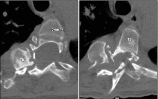
Fig. 3. Two consecutive axial CT images showing severe des- truction and resorption of ver- tebral pedicles of T5 and 7.
Fig. 4. Two consecutive coronal MR images show the continuity of spinal canal and sparing the spinal cord with dural ectasia around the deformity.
markedly hyperactive, and the Babinski reflexes were positive. There was sustained ankle clonus bilaterally.
The segmental kyphosis involving T4 through T7 was 95 degrees on plain radiographs (Fig. 1).
Three-dimensional CT scans revealed a complete
dislocation of the fourth and fifth vertebrae on the sixth and seventh anterolaterally, with the body of the T4 and T5 located immediately anterior and right side to that of T6 and T7 (Fig. 2). Axial im- ages of CT scans showed severe destruction and resorption of vertebral pedicles of T5 and T6. The
Fig. 5. The anteroposterior and lateral radiograph taken after the first procedure (posterior pedicle screw instrumentation with fusion from T2 to T12).
left pedicle of T4 and the right pedicle of T7 showed incomplete irregular resorption (Fig. 3). MRI showed dural ectasia around the lesion (Fig. 4). The pos- terior structures including spinous process, la- mina, and facet joints were all aligned along the smooth scoliotic curve without any subluxation or dislocation.
For the correction of kyphotic deformity, halo- pelvic traction was applied with gradually in- creased weight. Paraparesis improved at eight pounds and worsened at 15 pounds of traction. The weight was maintained at eight pounds until operation. After ten days of traction, we planned two-staged in situ posterior fusion followed by anterior fusion. The first procedure included pos- terior pedicle screw instrumentation and fusion from T2 to T12 vertebrae with a posterior ap- proach. The neurological status was not aggra- vated postoperatively.
The second procedure was performed three weeks later, using high thoracic periscapular ap- proach with the patient in the left lateral decubitus position. After removal of interposed fibro-osseous tissues around T4-T7 vertebral bodies, in situ fusion was performed without reduction of kypho- scoliosis. The resected fourth rib was grafted as a strut bone between T4 and T7 vertebral body and additional cancellous chip bone graft was done around the strut graft. The neurological findings remained normal after operation. The segmental kyphosis involving T4 through T7 was corrected to 48 degrees on lateral radiographs (Fig. 5).
The body jacket cast was applied postoperative for four months. The patient improved neuro- logically with free ambulation state at 24-months follow-up.
DISCUSSION
Neurofibromatosis is a disorder of neural crest cells involving not only neuroectoderm and meso-
derm but also endoderm, caused by the defect in the gene called neurofibromin on the long arm of chromosome 173). The characteristics of dystrophic changes including enlargement of foramen, scal- loping of vertebral bodies, rib deformities, and structural defect in pedicles are well known. One of the possible etiologies for this condition is regional dystrophy and dysplasia affecting both bone and meninges that have been referred to as mesodermal dysplasia2,8). Although subluxation or dislocation of spinal column in neurofibromatosis is very rare, progressive dystrophy of bony elements, dural ectasia, and laxity of ligamentous structure may cause structural instability of the vertebral column6,8,9).
In our review of literature, we found only several cases that had vertebral column dislocation8,9). Rockower, et al.6) reported two cases of spinal dis- location after minor trauma in children who had neurofibromatosis. Stone, et al. reported one case, a nine year-old boy who had dislocation of the first
on the second thoracic vertebra and no neurological deficit8). Winter9) reported a case that had dis- location of the ninth on the tenth thoracic ver- tebra.
The mechanism of the spontaneous dislocation in neurofibromatosis is uncertain. Simpson, et al7). reported that three cases with a traumatic bilateral pedicular fracture in the thoracic spine maintained the continuity of posterior elements and the cord whereas the body was displaced. In their cases, the spinal cord was not compressed and showed normal neurological findings that might be a possible explanation for the spinal column dislocation in neurofibromatosis. After severe osseous erosion and resorption of vertebral pedicles, separation of the vertebral body may occur without disruption of posterior elements and the spinal cord in neuro- fibromatosis.
For the treatment of a patient who has flexible kyphotic deformities with mild neurologic deficit, preoperative halo traction could be recom- mended4). In our case, vertebral bodies were seve- rely displaced and translated and, we attempted a closed reduction of the vertebral column by halo- pelvic traction, which improved the neurology of the patient at eight pounds. However, neurological deficits of the patient were deteriorated with weights more than 15 pounds. Thus, we performed in situ anterior and posterior fusion after minimal skeletal traction. Although posterior fusion alone may provide stability for the patients with non-dystrophic or dystrophic curves without ky- phosis, combined anterior and posterior fusion has been generally recommended to stabilize dystrophic curves in neurofibromatosis5). In a study of Parsini, et al5), the failure rate of posterior fusion was higher in the patients with kyphosis greater than 50 degrees, and the addition of planned anterior fusion decreased the failure rate more than 50%.
Winter, et al10). emphasized the importance of
combined anterior and posterior fusion at an early stage and explained the use of small amount of bone graft in limited area was a major reason for pseudarthrosis. In conclusion, the dystrophic ky- phoscoliosis with neurofibromatosis and loss of bilateral pedicles may cause the vertebral column dislocation with progressive neurological deficit.
Combined anterior and posterior fusion was an effective method for the treatment of dystrophic deformity.
REFERENCES
1. Akbarnia BA, Gabriel KR, Beckman E, Chalk D: Pre- valence of scoliosis in neurofibromatosis. Spine, 17(Suppl 8):
S244-S248, 1992.
2. Crawford AH, Al-Sayyad MJ: Miscellaneous conditions of the cervical spine. Neurofibromatosis, juvenile rheumatoid arthritis, and Rickets. In: Clark R ed. The cervical spine. 4th ed. Philadelphia, Lippincort Williams and Wilkins: 481-507, 2004.
3. Goldberg NS, Collins FS: The hunt for the neurofibro- matosis gene. Arch Dermatol, 127: 1705-1707, 1991.
4. Kim HW, Weinstein SL: Spine update. The management of scoliosis in neurofibromatosis. Spine, 22: 2770-2776, 1997.
5. Parisini P, Silvestre M, Greggi T, Paderni S, Cervellati S, Savini R: Surgical correction of dystrophic spinal curves in neurofibromatosis. Spine, 24: 2247-2253, 1999.
6. Rockower S, McKay D, Nason S: Dislocation of the spine in neurofibromatosis. A report of two cases. J Bone Joint Surg Am, 64: 1240-1242, 1982.
7. Simpson AH, Williamson DM, Golding SJ, Houghton GR: Thoracic spine translocation without cord injury. J Bone Joint Surg Br, 72: 80-83, 1990.
8. Stone JW, Bridwell KH, Shackelford GD, Abramson CL: Dural ectasia associated with spontaneous dislocation of the upper part of the thoracic spine in neurofibromatosis. A case report and review of the literature. J Bone Joint Surg Am, 69: 1079-1083, 1987.
9. Winter RB: Spontaneous dislocation of a vertebra in a patient who had neurofibromatosis. Report of a case with dural ectasia.
= 국문초록=
신경섬유종 척추측만증의 이영양성 만곡은 다양한 비전형적인 형태를 보이나 진행성 신경학적 증상을 보이는 자발성 척추체 탈구에 대해서는 보고된 바가 거의 없다. 저자들은 외상력 없이 척추의 후방구조물의 연속성은 유지된 상태에서 흉추체의 탈구를 보인 증례를 보고하는 바이다. 5년간 지속된 체간의 전방굴곡 및 요통을 호소하는 35세 여자로 최근 3개월 전부터 진행성 양측 하지 운동마비소견을 보였으며 방사선 소견 상 흉추 5, 6번 척추경의 심한 골흡수 소견 및 흉추 5, 6번 사이의 관상면 및 축상면 전위를 보이는 탈구 소견을 나타내었 다. 골견인 후 전, 후방 유합술을 시행하였으며 술 후 신경학적 증상은 호전을 보여 술 후 24개월에 독립 보행이 가능하였다.
색인 단어: 신경섬유종, 자발성 척추체 탈구, 이영양성 후측만증
J Bone Joint Surg Am, 73: 1402-1404, 1991.10. Winter RB, Lonstein JE, Anderson M: Neurofibromatosis
hyperkyphosis: a review of 33 patients with kyphosis of 80 degrees or greater. J Spinal Disord, 1: 39-49, 1988.