An acute aortic thrombosis in the absence of athero- sclerosis, aortic dissection, or aneurysm is an infrequent clinical entity and has been rarely reported in the litera- ture. Although the incidence of an acute aortic thrombo- sis is very low, it should be recognized carefully because of serious complications such as peripheral and visceral embolisms (1). We report two cases of an acute aortic thrombosis in lung cancer patients treated with chemotherapy and a review of the literature.
Case Report
Case 1
A 66-year-old man with a history of small cell lung cancer presented to the hospital for a third trial of chemotherapy. The patient had been treated by chemotherapy consisting of ifospamide, carboplatin and etoposide for two sessions. An follow-up enhanced chest
computed tomography (CT) scan demonstrated a marked decrease of the size of the mass. However, the CT scan (Fig. 1A, B) showed a newly developed intralu- minal-filling defect of a thrombus from the mid thoracic aorta to just above the celiac trunk. The aortic thrombus did not accompany an aneurysm, dissection or athero- sclerotic change. Two months ago, there was no aortic thrombus seen on a CT scan (Fig. 1C). As the patient had thrombocytopenia and did not show any complica- tions associated with the aortic thrombosis, the physi- cian decided to proceed with close follow-up and no an- ticoagulation therapy.
Case 2
A 65-year-old man with a history of non-small cell lung cancer visited the hospital for a follow-up en- hanced chest CT. The patient was treated with chemotherapy consisting of carboplatin and taxel for six sessions. A follow-up CT scan (Fig. 2A, B) showed no significant interval change in the lung as compared with the previous CT scan, but demonstrated a newly devel- oped intraluminal filling defect of a thrombus, about 6 cm in length at the level of the thoracoabdominal de-
J Korean Radiol Soc 2007;57:337-340
─ 337 ─
Acute Aortic Thromboses Occurring in Cancer Patients Treated with Chemotherapy
1Kyung-Ryeol Lee, M.D., Dong-Wook Sung, M.D.
1Department of Diagnostic Radiology, Kyung Hee University Hospital Received April 29, 2007 ; Accepted August 16, 2007
Address reprint requests to : Dong-Wook Sung, M.D., Department of Diagnostic Radiology, Kyung Hee University Hospital, 1 Hoeki-dong, Dongdamun-gu, Seoul 130-702, Korea.
Tel. 82-2-958-8616 Fax. 82-2-968-0787 E-mail: [email protected]
An acute aortic thrombosis in the absence of atherosclerosis, aortic dissection, or aneurysm is an infrequent clinical entity and has been rarely reported in the literature.
However, because of serious complications such as an embolism that can be fatal, one should always pay attention to the possibility of its occurrence. We report two cases of an acute aortic thrombosis of lung cancer patients treated with chemotherapy and a review of the literature.
Index words :Aorta Thrombosis
Chemotherapy, cancer Acute disease
scending aorta just above the celiac trunk. Forty-five days prior, a CT scan (Fig. 2C) showed no aortic throm- bus, aneurysm, dissection or atherosclerotic change. As the patient had thrombocytopenia and did not show any complications associated with the aortic thrombosis, the physician decided to proceed with close follow-up and no anticoagulation therapy.
Discussion
An acute aortic thrombosis in the absence of athero- sclerosis, aortic dissection, or aneurysm is an infrequent clinical entity and has been rarely reported in the litera- ture. Although the incidence of an acute aortic thrombo- sis is very low, it should be recognized carefully because of its serious complications such as a peripheral and vis- ceral embolism. Machleder et al. (1) reported an inci- dence of 0.45% of a non-aneurysmal aortic mural thrombus in 10,671 consecutive autopsies, with 17% of
these patients having an incidence of A distal emboliza- tion.
In the pathogenesis of an aortic thrombosis associated with atherosclerotic disease, atherosclerotic lesions have an important role in the formation of the thrombus as a nidus. However, the pathogenesis of a primary aortic thrombus has not yet been defined. The two cases did not relate to an identifiable atherosclerosis. However, both patients had a history of lung cancer, and both pa- tients were treated by cisplatin-based chemotherapy during multiple sessions.
Vascular events such as deep venous thrombosis, pul- monary embolus, arterial thrombosis and embolus, cerebrovascular accident, unstable angina and myocar- dial infarction have been reported in patients with ma- lignant disease, and the frequency of such events is in- creased in cancer patients (2, 3). However, an acute aor- tic thrombosis is not common in the cancer patient.
When we reviewed cases of a few acute aortic throm-
Kyung-Ryeol Lee, et al: Acute Aortic Thromboses Occurring in Cancer Patients Treated with Chemotherapy
─ 338 ─ A
B
C
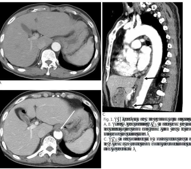
Fig. 1. A 66-year-old man with a history of small cell lung cancer.
A, B. Axial (A) and sagittal (B) CT images show an intraluminal-fill- ing defect of the thrombus (arrow) from the mid thoracic aorta to just above the celiac trunk.
C. A CT image obtained two months before the image presented in (B) shows no aortic thrombosis, atherosclerotic change, dissec- tion, or aneurysm.
boses associated with malignant disease, aortic occlu- sion was seen in pancreatic adenocarcinoma, T-cell lym- phoma and acute myelomonocytic leukemia. Cases of aortic thrombosis in cancer patients occurred in the ab- sence of atherosclerosis, dissection, an aneurysm or any predisposing factors causing the formation of clots. In the cancer patients, the neoplastic lesions were in imme- diate proximity to the aorta (4).
In a previous study, a close correlation of the throm- boembolic events and the location of the cancer for col- orectal, prostate, pancreas, lung, and ovary cancers was reported (5). In the present cases of lung cancer, the aor- tic thrombosis developed mainly in the abdominal aorta and was not related to the tumor location. Possible con- tributing factors such as inappropriate intravascular co- agulation and fibrinolysis, tumor production of procoag- ulants, and the physical characteristics of the tumor
cells leading to the interaction with components of the clotting and inflammatory cascades may have a role in the thrombotic condition in cancer patients.
Furthermore, a poor physical status, decreased activity and compressive effects of the tumor on the vasculature may also increase the risk of thromboembolic events (6, 7).
Czaykowski et al. (7) reported the incidence of throm- boembolic events in patients receiving multi-agent chemotherapy for urothelial cancer. In 271 patients that received multi-agent cisplatin based chemotherapy for transitional cell carcinomas, 35 patients (12.9%) showed vascular events and seven patients showed arterial thromboses. The investigators explained the relation- ship between the vascular events and systemic chemotherapy, and emphasized the necessity of the use of prophylactic anticoagulation in patients with risk fac-
J Korean Radiol Soc 2007;57:337-340
─ 339 ─
A B
C
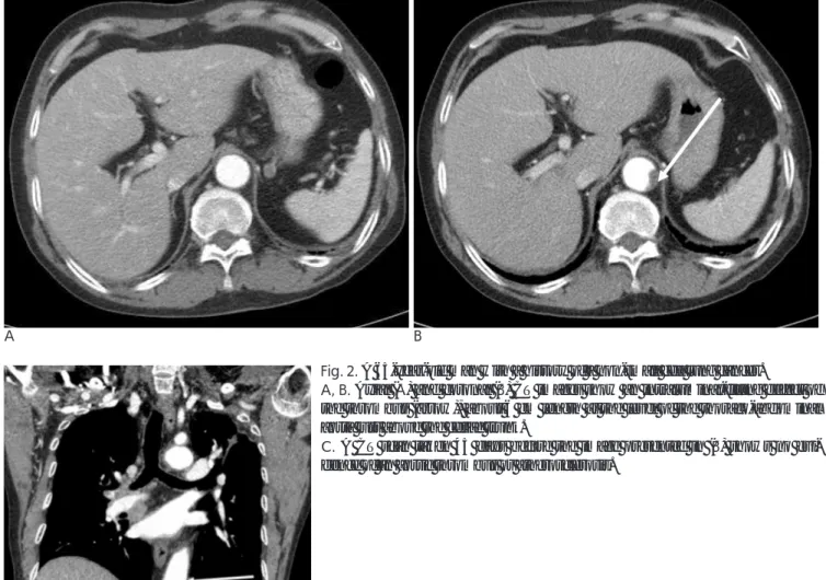
Fig. 2. A 65-year-old man with a history of a non-small cell lung cancer.
A, B. Axial (A) and coronal (B) CT images show an intraluminal-filling defect of the thrombus (arrow), about 6 cm length at the level of the thoraco-abdominal aorta just above the celiac trunk.
C. A CT scan taken 45 days before the image presented in (B) shows no evi- dence of an aortic thrombus or atherosclerosis.
tors for thromboembolic disease. The investigators em- phasized the cisplatin-related thromboembolic events;
there is some evidence that cisplatin can induce platelet activation and aggregation and alter endothelial cell in- tegrity (7). In addition, Apiyasawat et al. (8) reported a case related to a cisplatin-induced localized aortic thrombus. These investigators reported on a patient with a localized mobile aortic thrombus that was treated with cisplatin and 5-fluorouracil and the patient present- ed with embolic events to the right lower extremity. The patient did not have any contributing factors related to the hypercoagulable state. In the present cases, the pa- tients were treated with carboplatin. Carboplatin is an analogue of cisplatin that has been used since the 1990s and has fewer side effects but a decreased anti-cancer drug effect than the use of cisplatin. The use of carbo- platin may be related to the formation of an aortic thrombus.
Reported cases of an acute aortic or arterial thrombo- sis were treated successfully by anticoagulation therapy or by performing a surgical thrombectomy (4, 7, 8). As both patients of the present cases had thrombocytopenia and did not show any complications associated with the aortic thromboses, the physicians decided to proceed with close follow-up and no anticoagulation therapy.
In conclusion, an acute aortic thrombus may occur during chemotherapy of lung cancer, especially with the
use of carboplatin, it should be recognized carefully, and proper therapy must be performed. We have presented two cases of acute aortic thrombi during chemotherapy of lung cancer patients with a review of the literature.
References
1. Machleder HI, Takiff H, Lois JF, Holburt E. Aortic mural throm- bus: an occult source of arterial thromboembolism. J Vasc Surg 1986;4:473-478
2. Khorana AA, Francis CW, Culakova E, Fisher RI, Kuderer NM, Lyman GH. Thromboembolism in hospitalized neutropenic can- cer patients. J Clin Oncol 2006;24:484-490
3. Stein PD, Beemath A, Meyers FA, Skaf E, Sanchez J, Olson RE.
Incidence of venous thromboembolism in patients hospitalized with cancer. Am J Med 2006;119:60-68
4. Poiree S. Monnier-Cholley L, Tubiana JM, Arrive L. Acute abdom- inal aortic thrombosis in cancer patients. Abdom Imaging 2004;29:
511-513
5. Gouin-Thibaut I, Samama MM. Venous thrombosis and cancer.
Ann Biol Clin 2000;58:675-682
6. Winter PC. The pathogenesis of venous thromboembolism in can- cer: emerging links with tumour biology. Hematol Oncol 2006;24:
126-133
7. Czaykowski PM, Moore MJ, Tannock IF. High risk of vascular events in patients with urothelial transitional cell carcinoma treat- ed with cisplatin based chemotheraphy. J Urol 1998;160:2021- 2024
8. Apiyasawat S, Wongpraparut N, Jacobson L, Berkowitz H, Jacobs LE, Kotler MN. Cisplatin induced localized aortic thrombus.
Echocardiography 2003;20:199-200
Kyung-Ryeol Lee, et al: Acute Aortic Thromboses Occurring in Cancer Patients Treated with Chemotherapy
─ 340 ─
대한영상의학회지 2007;57:337-340
항암화학치료를 받은 환자에서 발생한 급성 대동맥 혈전증1
1경희대학교 의과대학 영상의학과교실
이 경 렬・성 동 욱
동맥경화, 대동맥 박리, 동맥류와 같은 인자를 동반하지 않은 급성 대동맥 혈전증은 매우 드문 질환이며 문헌에 서도 찾아보기 어렵다. 이러한 급성 대동맥 혈전증은 동맥색전증 등의 심각한 후유증을 가져와 환자에게 치명적인 위험이 될 수 있어 급성 대동맥 혈전증의 발생 가능성에 대해 항상 주의해야 한다. 저자들은 폐암으로 항암화학치 료를 받고 있던 환자에서 발생한 급성 대동맥 혈전증 2 예를 문헌 조사와 함께 보고하는 바이다.