자기공명영상은 골수를 침범하는 다양한 질환들의 진단과 평 가에 유용한 검사방법으로 다발성 골수종의 척추 침범여부를 평가하는데 가장 좋은 영상진단법으로 알려져 있다 (1-3). 그 러나 자기공명영상에서 다발성 병변을 보이는 환자가 임상적 진단이나 정보 없이 단순히 목과 허리 등의 통증이 주소일 때 척추전이에 의한 병변을 감별해야 한다. 이에 척추를 침범한 다발성 골수종과 척추전이에서 자기공명영상소견의 차이점이 있는지 알아보고자 하였다.
대상과 방법
다발성 골수종 환자 23명의 25예와 척추전이 환자 37명의 37예를 대상으로 후향적으로 자기공명영상을 분석하였다. 다 발성 골수종 환자 중 남자는 14명, 여자는 9명이었으며 척추 전이 환자는 남자가 19명, 여자가 18명이었다. 다발성 골수종 환자의 나이는 38-79세 (평균 나이, 58세)였고 척추전이 환 자의 나이는 29-78세 (평균 나이, 55세)였다. 다발성 골수종 환자는 모두 혈액 및 소변의 생화학적 검사에서 단일클론성 감 마그로불린장애가 증명되었고, 이 중에서 18예는 장골의 골수 생검으로 확진되었다. 척추전이 환자에서 5예는 병리조직학적 으로 확진되었고, 32예는 1개월에서 6개월사이에 임상적 및 골스캔과 같은 방사선학적 추적검사를 종합하여 진단하였다.
척추의 다발성 골수종과 전이암: 자기공명영상 소견의 차이점1
이영준・지원희・하기용2・이배영・김연실・김범수・서경진3・최규호
목적:척추를 침범하는 다발성 골수종과 척추전이의 자기공명영상의 차이점을 알아보고자 하였다.
대상과 방법: 25예의 다발성 골수종과 37예의 척추전이의 자기공명영상을 후향적으로 분석하
였다. 침범유형, 신호강도, 조영증강 유형, 3개의 연속된 척추체의 침범여부, 하나의 척추체내 의 병변의 개수, 그리고 경막외와 척추주위 종괴 유무에 대해 분석하였다. 시상면과 축상면 조 영전 그리고 조영후 T1-강조영상과 고속 스핀에코 T2-강조영상을 얻었다. 통계분석은 Fisher exact test로 검증하였다.
결과: 모든 다발성 골수종과 척추전이는 T1-강조영상에서 저 신호강도를 보였고, 신호강도나 조영증강에 있어서 두 질환간에 통계적으로 유의한 차이는 없었다. 다발성 골수종 환자 중 국 소형이 13예 (52%), 미만형이 8예 (32%), 소금과 후추형이 4예 (16%) 였고, 척추전이 환자 에서는 국소형이 25예 (68%), 미만형이 12예 (32%) 였으며 소금과 후추형은 없었다. 소금과 후추형은 양질환의 감별에 유의한 차이를 보였으나 (p < 0.05) 국소형과 미만형은 유의한 차 이를 보이지 않았다. 하나의 척추내에 5개 이상의 병변을 보인 예는 다발성 골수종에서 10예 (58.8%)였고 척추전이에서는 2예 (8.3%)로 다발성 골수종에서 유의하게 많았다 (p < 0.01).
다발성 골수종에서는 3개 이상의 연속된 척추체를 침범한 경우가 20예 (80%)였고 척추전이 에서는 16예 (43%)로, 통계학적으로 유의한 차이를 보였다 (p < 0.01). 고속스핀에코 T2- 강조영상에서 신호강도는 다발성 골수종과 척추전이에서 각각 고신호강도는 12%와 32% (p
> 0.05), 등신호강도는 36%와 3% (p < 0.05), 저신호강도는 52%와 65% (p > 0.05)였다.
경막외 종괴와 척추주위 종괴는 각각 다발성 골수종에서 12예 (48%), 9예 (36%)이고 척추전 이에서 18예 (49%), 19예 (51%)를 보여서 차이가 없었다.
결론: 양 질환간의 감별은 대부분에서 어려우나 자기공명영상에서 소금과 후추형의 골수 침범 유형, 하나의 척추를 침범하는 병변의 개수가 5개 이상일 때, 그리고 연속된 3개 이상의 척추 체를 침범했을 때 전이암보다는 다발성 골수종을 시사하였다.
1가톨릭대학교 의과대학 방사선과학교실
2가톨릭대학교 의과대학 정형외과학교실
3경북대학교 의과대학 방사선과학교실
이 논문은 2000년도 가톨릭 중앙의료원 학술 보조비로 이루어졌음.
이 논문은 2000년 5월 3일 접수하여 2000년 12월 6일에 채택되었음.
자기공명영상은 1.5-T 자기공명영상장치 (GE Signa Advantage; General Electric Medical Systems, Milwaukee, WI, U.S.A.)를 사용하였다. 절편두께는 3 mm, 절편간격은 1 mm, echo train length는 16으로 하였다. 모든 환자에서 시상 면, 축상면 고식적 스핀에코 T1-강조영상 (350-450 msec/11-17 msec [TR/TE])과 고속스핀에코 T2-강조영상 (3000-3500 msec/108-144 msec [TR/TE])을 얻었다. 또 한 Gadolinium-DTPA (Magnevist, Schering, Berlin, Germany)를 정맥주사한 후 시상면과 축상면 T1-강조영상을 얻었으며 6예를 제외하고는 지방억제 T1강조영상을 얻었다.
T1- 및 T2-강조영상에서 신호강도, 골수 침범 유형, 조영 증강 정도와 유형, 하나의 척추내의 병변의 수, 3개 이상의 연 속된 척추체의 침범여부, 그리고 척추주위 종괴와 경막외 종괴 형성 유무에 대하여 분석하였다. 다발성 골수종환자에서는 치 료 전후의 구분없이 시행한 자기공명영상을 분석하였다.
골수침범의 유형은 Moulopoulos 등 (4)과 stabler등 (7)이 분류했던 형태를 바탕으로 국소형, 미만형, ‘소금과 후추’
(salt-and-pepper)형으로 분류하였다. 국소형은 T1-강조영 상에서 저 신호강도를 보이며 조영증강이 국소적으로 있는 병 변으로 하였고, 미만형은 하나의 척추체 전체를 침범하여 조영 강도 전반적으로 균일하게 되는 것으로 분류하였다. ‘소금과 후추’형은 척추체에 셀 수 없이 많은 작은 불규칙한 형태의 병 변들이 T1- 및 T2-강조영상에서 불균일한 신호강도를 보이 고, 이들 병변이 불균일하게 조영증강이 되는 것으로 분류하였
다. 미만형과‘소금과 후추’형을 제외한 국소형만을 대상으로 하나의 척추내 병변의 개수가 5개 이상인 척추체가 하나라도 있으면 5개 이상인 예로 하였다.
자기공명영상의 분석은 세 명의 방사선과 의사가 상호 합의 에 의해 일치되는 소견으로 하였다. 통계적 분석은 Fisher exact test를 통해 유의수준 0.05로 검증하였다.
결 과
다발성 골수종과 척추전이의 병변들은 모든 환자에서 T1- 강조영상에서 골수 신호강도에 비해 저 신호강도를 보였으며 두 질환에서 모두 조영증강을 보여 T1-강조영상에서의 신호 강도차이와 조영증강의 차이는 유의하지 않았다. 고속 스핀에 코 T2-강조영상에서 고신호강도를 보인 예는 다발성 골수종 과 척추전이에서 각각 3예 (12%) (Fig. 1)와 12예 (32%) (Fig.
2), 등신호강도를 보인 예는 9예 (36%) (Figs.3, 4)와 1예 (3%), 저신호강도를 보인 예는 13예 (52%)와 24예 (65%) (Fig. 5)로 T2- 강조영상에서 등신호강도를 보인 예가 다발 성 골수종에서 유의하게 많았다 (p < 0.05) (Table).
골수 침범 유형은 다발성 골수종 환자 중 국소형이 13예 (52%) (Fig. 1), 미만형이 8예 (32%) (Fig. 4), 소금과 후추 형이 4예 (16%) (Fig. 3)였고 척추전이 환자에서는 국소형이 25예 (68%)(Fig. 2), 미만형이 12예 (32%)였으며 소금과 후 추형은 없어, 소금과 후추형은 감별에 유의한 차이를 보였으나
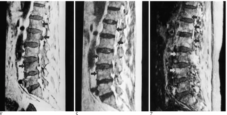
A B C
Fig. 1. Multiple myeloma with focal pattern in a 60-year-old man.
A. Sagittal T1-weighted image shows multiple variable sized hypointense lesions (arrows) at lumbar spine.
B. On sagittal T2-weighted image, these lesions are hyperintense (arrows).
C. Gadolinium-enhanced fat suppressed T1-weighted image shows multiple well enhancing masses (arrows). There are more than five lesions in one vertebra and three consecutive vertebrae are involved.
(p < 0.05) 국소형과 미만형은 유의한 차이를 보이지 않았다.
하나의 척추내에 5개 이상의 병변을 보인 예는 다발성 골수 종중 국소형 13예중 6예로 46%였고 (Fig. 1) 척추전이에서는 25예의 국소형 중 2예에서만 5개 이상의 병변을 보여 8% 밖 에 되지 않아 다발성 골수종에서 하나의 척추체내에 5개 이상 의 병변을 보인 경우가 유의하게 많았다 (p < 0.05).
다발성 골수종에서는 3개 이상의 연속된 척추체를 침범한
경우가 20예로 80%였고 (Figs.1, 3) 척추전이에서는 16예로 43%를 보여 통계적으로 유의한 차이를 보였다 (p < 0.01).
경막외 종괴는 다발성 골수종에서 12예로 48%, 척추전이에서 18예로 49%였고, 척추주위 종괴도 다발성 골수종에서 9예로 36%, 척추전이에서 19예로 51%를 보여 두 질환간에 유의한 차이가 없었다.
Table. Multiple Myeloma versus Metastasis on MR Findings
Multiple myeloma (n=25) Metastasis (n=37) Signal Intensity on FSE T2WI
High-SI (p > 0.05) 12% (n = 3) 32% (n = 12)
Iso-SI (p < 0.05) 36% (n = 9) 3% (n = 1)
Low-SI (p > 0.05) 52% (n = 13) 65% (n = 24)
Infiltration and enhancement patterns
Focal (p > 0.05) 52% (n = 13) 68% (n = 25)
Diffuse (p > 0.05) 32% (n = 8) 32% (n = 12)
Salt and pepper (p < 0.05) 16% (n = 4) 0%
*More than five lesions within one
vertebra in focal pattern (n’) (p < 0.05) 46% (n = 6/n’= 13) 8% (n = 2/n’=25) Involvement of three consecutive
vertebrae (p < 0.01) 80% (n = 20) 43% (n = 16)
Paraspinal mass (p > 0.05) 36% (n = 9) 51% (n = 19)
Epidural mass (p > 0.05) 48% (n = 12) 49% (n = 18)
* n’: each number of focal pattern in multiple myeloma and metastasis FSE T2WI: Fast Spin Echo T2-weighted Image
SI: signal intensity
A B C
Fig. 2. Vertebral metastasis with focal pattern in a 67-years-old man.
A. Sagittal T1-weighted image shows focal hypointense lesions (arrows) at anterior portion of L1 and L2 vertebral bodies.
B. On sagittal T2-weighted image, the lesions reveal hyperintense signal intensity (arrows).
C. Gadolinium-enhanced fat-suppressed T1-weighted image shows intense enhancement of lesions (arrows).
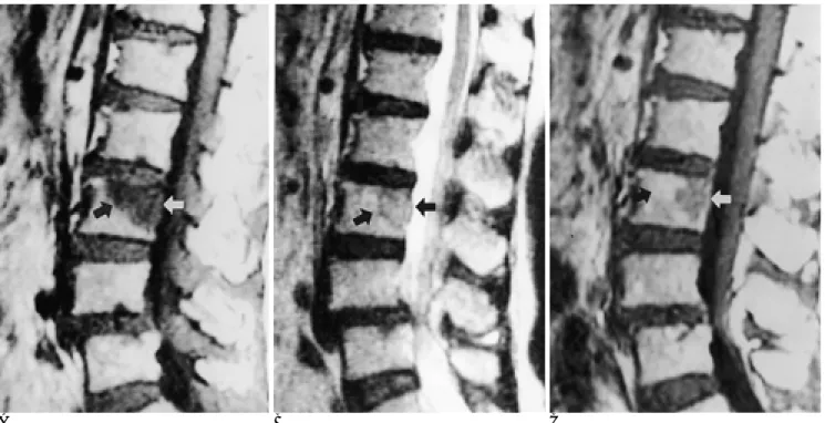
A B C Fig. 4. Multiple myeloma with diffuse pattern in a 69-years-old man.
A. On sagittal T1-weighted image T7 and T8 vertebrae show diffusely hypointense signal intensity (arrows).
B. On sagittal T2-weighted image T7 and T8 vertebrae are isointense (arrows) to the signal of adjacent vertebrae.
C. On contrast-enhanced fat-suppressed T1-weighted image T7 and T8 vertebrae show diffuse, relatively homogeneous contrast enhancement (arrows).
A B C
Fig. 3. Multiple myeloma with ‘salt & pepper’pattern in a 59-years-old man.
A. Sagittal T1-weighted image shows innumerable decreased signal intensity in lumbar spine.
B. Sagittal T2-weighted image shows heterogeneous signal intensity lesions.
C. Gadolinium-enhanced fat-suppressed T1-weighted image reveals heterogeneous enhancement.
고 찰
다발성 골수종은 형질세포의 악성증식의 결과로 골수 형질 세포의 증가와 단일클론성 감마그로불린 장애가 소변이나 혈 중에 나타나며 주로 척추를 비롯한 몸통 골격을 다발성으로 침 범하는 질환이다.
자기공명영상에서 다발성 골수종의 신호강도에 대해 Rahmouni 등 (5)은 T2-강조영상에서 주위 골조직보다 증가 된 균일한 고신호강도를 보이고, T1-강조영상에서는 일부가 등신호 또는 고신호강도를 보이나 대부분 저신호강도를 보였 다고 하였다. Fruehwald 등 (6)은 T2-강조영상에서 대부분 보통 주위 근육이나 침범되지 않은 골수보다 고신호강도의 병 변으로 보이며 일부 저신호강도 병변은 방사선치료를 받은 환 자에서 보였다고 하였다. 본 연구에서는 T2-강조영상에서 고 신호강도를 보인 예는 3예로 12%밖에 되지 않았고, 88%인 22예에서 등신호 또는 저신호강도의 병변으로 관찰되었다. 척 추전이에서도 T2-강조영상에서 역시 비슷한 신호강도를 보여 서 척추전이는 T2-강조영상에서 고신호강도를 보인다는 이전 의 보고 (7, 8)와 상이하였다. 이는 본 연구가 고식적 T2-강 조영상이 아닌 고속스핀에코 T2-강조영상으로 이루어져, 침 범되지 않은 황색골수의 지방의 신호강도가 상대적으로 낮아 지지 않은 결과로 생각된다 (9, 10).
다발성 골수종과 척추전이에서 모두 T1-강조영상에서 저신 호강도를 보이고 조영증강이 상당히 잘되어 T1-강조영상과
조영증강영상은 구별되는 점이 없었다. Breger등 (11)은 정상 적으로 성인의 골수도 조영증강이 될 수 있지만 아주 미미한 정도이기 때문에 조영증강영상이 적색골수에서 황색골수로의 불 균일한 전환과의 감별에는 도움이 될 수 있다고 하였고 본 연구에서도 병변 자체를 찾는데는 조영증강영상이 유용하였다 (Fig. 4C).
다발성 골수종의 골수 침범유형은 다양하여 보고자들마다 분 류양식이 조금씩 다르다 (4, 12, 13). Moulopoulos 등 (4)은 국소형, 미만형, 얼룩덜룩형(variegated pattern)으로 나누었는 데 얼룩덜룩형이 14%였다. 얼룩덜룩형과 같은 형태인 본 연 구의‘소금과 후추’형도 다발성 골수종중 16%를 차지하여 비 슷한 결과를 보였고 척추전이에서는 이러한 형태의 골수침범 유형이 없어 다발성 골수종의 특이소견으로 볼 수 있었다.
Stabler 등 (12)은 국소형, 미만형, 혼합형, ‘소금과 후추’형으 로 나누어‘소금과 후추’형이 골수침범 했던 66예 중 5예로 약 7.6%였고 이들은 모두 임상적 병기가 낮았다고 보고하였 다. 또한 허 등 (13)이 골수침범유형을 국소형, 미만형, 혼합 형의 3형태로 분류했을 때 다발성 골수종에서는 미만형이 약 50%를 보였고 미만형일 경우 다발성 골수종의 가능성이 높다 고 보고하였다. 본 연구의 미만형과‘소금과 후추’형을 종합하 면 48%로 허 등 (13)과 비슷한 결과를 보였다고 할 수 있다.
Kim 등 (14)은 Monoupolous 등 (4)과 같은 분류로 17예의 다발성 골수종과 5예의 림프종, 2예의 백혈병, 모두 24예의 조 혈성 악성종양과 38예의 척추전이를 분석하였는데 조혈성 악 성종양에서 척추전이보다 미만형이 유의하게 많았다고 하였다.
A B C
Fig. 5. Metastasis with focal pattern in a 74-years-old woman.
A. Sagittal T1-weighted image shows a focal hypointense lesion (arrows) at L2 vertebral body.
B. Sagittal T2-weighted image shows a focal heterogeneously hypointense lesion (arrows) at the L2 vertebral body.
C. The mass at L2 vertebral body shows heterogeneous contranst enhancement (arrows) on sagittal contrast-enhanced T1-weight- ed image.
또한 미만형중 균일한 침범양상과 비균일한 침범양상의 분류 에서는 균일한 침범양상이 조혈성 악성종양에서 많았고 본 연 구의‘소금과 후추’형과 같은 비균일한 침범양상은 오히려 척 추전이에서 많았으나 두 질환간에 통계적으로 유의한 차이는 없었다고 하여 본 연구와는 다른 결과를 보였다.
하나의 척추체를 침범한 병변의 개수는 다발성 골수종과 척 추전이의 경우에 모두 하나이상의 병변을 보인 경우가 대부분 이었다. 병변의 수를 5개를 기준으로 분류했을 때 다발성 골수 종에서도 척추전이보다 국소병변의 수가 유의하게 더 많아, 두 개골을 침범한 다발성 골수종이 전이암보다는 국소병변의 개 수가 더 많고 균일한 병변을 보이는 양상과 유사하였다 (15).
2개의 연속된 척추체의 침범은 두 질환간에 차이점이 없었으 나 연속된 3개 이상의 척추체 침범으로 분류했을 때 다발성 골 수종에서 척추전이보다 더 흔하였으나 그 원인은 알 수 없었 다.
다발성 골수종도 척추를 침범했을 때 흔히 주위 연부 조직 종괴를 형성하여 척추주위 종괴나 경막외 침범이 특징적이라 고 하였다 (15). 본 연구도 비슷한 양상을 보여 척추주위 종괴 형성과 경막외 종괴형성에서 척추전이가 다소 높은 백분율을 보였으나 두 질환간의 유의한 차이는 없었다. Kim 등 (14)도 조혈성 악성종양과 척추전이에서 척추주위 종괴형성과 경막외 종괴형성의 유의한 차이는 없었으나 후경막외 종괴형성은 조 혈성 악성종양에서 유의하게 많아 특징적이라고 하였다. 그러 나 허 등 (13)은 경막외, 혹은 척추주위 종괴가 형성된 경우 척추전이를 더욱 시사하는 소견이라 하여 저자들과 다른 소견 을 보였다.
본연구는 여러 제한점을 갖고 있다. 비교적 적은 환자를 대 상으로 하여 통계적으로는 유의하나 증례수가 적다는 점과 후 향적인 연구로 모든 환자의 자기공명영상이 치료 받기전에 시 행한 것이 아니어서 방사선치료와 같은 치료후의 변화가 포함 되었을 가능성도 있다는 점이다. 다발성 골수종 환자와 척추전 이암 중에도 병리조직학적으로 확진되지 않은 예가 있는 점, 골수침범유형에 있어서 혼합형을 분류하지 않은 점이다. 또한 영상소견을 독립적으로 분석하지 않고 다수결로 상호합의하에 분석을 하여 관찰자간의 차이를 비교하지 않았다는 점등이 제 한점이다.
척추를 침범한 다발성 골수종과 척추전이의 자기공명영상 소 견은 대부분의 증례에서 감별이 어려우나 소금과 후추형, 한개 의 추체내에 5개이상의 병변을 보이거나, 3개이상의 연속된 척 추체를 침범한 소견을 보이는 적은 수의 증례에서는 전이암보 다 다발성 골수종을 시사한다. 따라서 확진을 위한 조직생검이 필요하다고 생각한다.
참 고 문 헌
1. Daffner RN, Lupetin AR, Dash N, Deeb ZL, Sefczek RJ, Schapiro RL. MRI in the detection of malignant infiltration of bone marrow.
AJR Am J Roentgenol 1986;146:353-358
2. Ludwig H, Scholakoff D,Neuhold A, Fruhwald F, Radsoul S, Fritz E. Mangentic resonance imaging of the spine in multiple myeloma.
Lancet 1987;15:364-366
3. Edward Gosfield,III, Abass Alavi and Bruce Kneeland.
Comparison of radionuclide bone scans and magnetic resonance imaging in detecting spinal metastases. J Nucl Med 1993;34:2191- 2198
4. Moulopoulos LA, Varma DGK, Dimopoulos MA, et al. Multiple myeloma: spinal MR imaging in patients with untreated newly di- agnosed disease. Radiology 1992;185:833-840
5. Rahmouni A, Divine M, Mathieu D, et al. Detection of multiple myeloma involving the spine: Efficacy of fat suppression and con- trast-enhanced MR imaging. AJR Am J Roentgenol 1993;160:1049- 1052
6. Fruehwald FXJ, Tscholakoff D, Schwaighofer B, et al. Magnetic resonance imaging of the lower vertebral column in patients with multiple myeloma. Invest Radiol 1988;23:193-199
7. Baker LL, Goodman SB, Perkash I, Barton L, Enzmann DR.
Benign versus pathologic compression fracture of vertebral bodies:
assessment with conventional spin-echo, chemical-shift, and STIR MR imagin. Radiology 1990;174:495-502
8. Melki PS, Mulkern RV, Panych LS, Jolesz FA. Comparing the FAISE method with conventional dual-echo sequences. J Magn Reson Imaging 1991;1:319-326
9. Henkelman RM, Hardy PA, Bishop JE, Poon CS, Plewes DB. Why fat is bright in RARE and fast spin-echo imaging. J Magn Reson Imaging 1992;16:41-47
10. Crenod CA, Laredo JD, Chevret S, et al. Acute vertebral collapse due to osteoporosis or malignancy: appearance on unenhanced and gadolinium-enhanced MR images. Radiology 1996;199:541- 549.
11. Breger RK, Williams AL, Daniels DL, et al. Contrast enhancement in spinal cord imaging. AJR Am J Roentgenol 1989;153:387-391 12. Stabler A, Baur A, Bartl R, Munker R, Lamerz R, Reiser MF.
Contrast enhancement and quantitative signal analysis in MR imaging of multiple myeloma. AJR Am J Roentgenol 1996;167:1029- 1036
13. 허서구,윤정희,김창수 등. 척추의 다발성 골수종과 전이암의 감별:
시상면 자기공명영상에서 척추의 골수침범양상을 위주로. 대한방 사선의학회지 1999;40:769-775
14. Kim HJ, Ryu KN, Choi WS, Choi BK, Choi JM, Yoon Y. Spinal in- volvement of hematopoietic malignancies and metastasis:
Differentiation using MR imaging. Clin Imaging 1999;23:125-133 15. Resnick D. Diagnosis of bone and joint disorders. 3rd ed. Philadel-
phia:Saunders, 1995:2148-2162
J Korean Radiol Soc 2001;44:229-235
Address reprint requests to : Won-Hee Jee, M.D., The Catholic University of Korea, Department of Radiology, Kangnam St. Mary’s Hospital, 505 Banpo-dong, Seocho-gu, Seoul 137-701, Korea.
Tel. 82-2-590-2784 Fax. 82-2-599-6771 E-mail: [email protected]
MR Distinction between Multiple Myeloma and Metastasis Involving the Spine
1Young-Joon Lee, M.D., Won-Hee Jee, M.D., Kee-Yong Ha, M.D.2, Bae-Young Lee, M.D., Yeon-Shil Kim, M.D., Bum-Soo Kim, M.D., Kyung-Jin Suh, M.D.3, Kyu-Ho Choi, M.D.
1Department of Radiology, The Catholic University of Korea
2Department of Orthopedic Surgery, The Catholic University of Korea
3Department of Radiology, Kyungpook National University.
Purpose: To differentiate multiple myeloma from metastasis involving the spine at MR imaging.
Materials and Methods: Twenty-five patients with multiple myeloma and 37 with vertebral metastasis were in- cluded in this study. MR images were retrospectively analyzed with regard to infiltration and enhancement patterns, signal intensity, the involvement of three consecutive vertebrae, the number of lesions within one vertebra, and paraspinal and epidural masses. Using a 1.5-T imager, we obtained sagittal and axial, unen- hanced and enhanced T1-weighted images, and fast spin-echo images. For statistical analysis, Fisher’s exact test was used.
Results: All cases of multiple myeloma and metastasis showed low signal intensity on T1-weighted images, and there were no significant differences in signal intensities or enhancement patterns. Infiltration and en- hancement patterns were classified as focal (52% in multiple myeloma vs 68% in metastasis, p> 0.05), diffuse (32% vs 32%, p > 0.05) or salt and pepper (16% vs 0%, p < 0.05) pattern. Differentiation between multiple myeloma and metastasis was based on two criteria: the involvement of three consecutive vertebrae (80% vs 43%, p < 0.01), and the presence of more than five lesions within one vertebra (59% vs 8%, p < 0.05). On fast spin-echo T2-weighted images, signal intensity was as follows: hyperintensity (12% vs 32%, p > 0.05), isoin- tensity (36% vs 3%, p < 0.05), and hypointensity (52% vs 65%, p > 0.05). Paraspinal and epidural masses played little part.
Conclusion: The salt and pepper infiltration pattern, the presence of more than five lesions within one verte- bra, and the involvement of more than three consecutive vertebrae were useful MR findings for differentiation between multiple myeloma and metastasis involving the spine. In most cases, however, it is difficult to distin- guish between the two conditions.
Index words :Spine, MR
Spine, primary neoplasms Spine, secondary neoplasms
2001 / 2 / 3 - 6 1st Annual Penn Vascular Symposium
•Subject: Medicine Vascular Diseases Imaging X-Ray
•Location: Las Croabas: El Conquistador Resort Puerto Rico
•Contact: Hospital of the Univ. of Pennsylvania, Radiology Dept., J. Ford Benner, 3400 Spruce St., MRI B., 1 Founders, Philadelphia, PA 19104, USA
•Phone: (215) 662-6904 Countryphone: +1
•Fax: (215) 349-5925
•E-Mail: [email protected]
•Internet: http://oasis.rad.upenn.edu/radmac/
CME/CMECAL.html
2001 / 2 / 7 - 9 JSNR, Japanese Society of Neuroradiology 30th Annual Meeting
•Subject: Imaging X-Ray Medicine Neuroscience/Neurology
•Location: Osaka Japan
•Contact: Natl. Osaka Hosp., Dept. of Radiology, Dr.
Masanori Mitomo, Osaka, Japan
•Phone: (06) 6942-1331 Countryphone: +81
•Fax: (06) 6943-6467
•E-Mail: [email protected]
2001 / 3 / 2 - 6 ECR 2001: 13. European congress of Radiology
•Subject: Imaging X-Ray
•Location: Vienna Wien Austria
•Contact: European Radiology Association, ECR Office, Neutorgasse 9/2A, A-1010 Vienna, Austria
•Fax: +43 1 533 40 64 9
•E-Mail: [email protected]
•Internet: http://www.ecr.org/
2001 / 3 / 3 - 8 SCVIR, Society of Cardiovascular & Interventional Radiology Annual Scientific Meeting
•Subject: Medicine Cardiology Vascular Diseases Imaging X-Ray
•Location: San Antonio Texas: San Antonio Convention Center USA
•Contact: SCVIR, 10201 Lee Hwy, Ste 500, Fairfax, VA 22030, USA
•Phone: (703) 691-1805 Countryphone: +1
•Fax: (703) 691-1855
•E-Mail: [email protected]
•Internet: http://www.scvir.org
2001 / 3 / 11 - 14 AIUM, American Institute of Ultrasound in Medicine 45th Annual Convention
•Subject: Imaging Ultrasound Medicine
•Location: Orlando Florida USA
•Contact: Amer. Inst. of Ultrasound in Medicine, Stephanie Reisberg, 14750 Sweitzer Lane, Suite 100, Laurel, MD 20707-5906, USA
•Phone: (301) 498-4100 Countryphone: +1
•Fax: (301) 498-4450
•E-Mail: [email protected]
•Internet: http://www.aium.org
2001 / 3 / 14 - 18 ASER, 12th Annual Scientific Meeting of the American Society of Emergency Radiology
•Subject: Imaging X-Ray
•Location: San Francisco California: Grand Hyatt Hotel Union Square USA
•Contact: Amer. Soc. of Emergency Radiologists, 820 Joric Boulevard, Oak Brook, IL 60523, USA
•Phone: (630) 368-3765 Countryphone: +1
•Fax: (630) 571-7837
•E-Mail: [email protected]
•Internet: http://www.erad.org
2001 / 3 / 19 - 23 SCBT-MR, 24th Annual Meeting of the Society for Computed Body Tomography and Magnetic Resonance
•Subject: Imaging X-Ray Magnetic Resonance Imaging
•Location: South Beach Florida: Loews Miami Beach Hotel USA
•Contact: Soc. f. Computed Body Tomography, Matrix Meetings, P.O. Box 1026, Rochester, MN 55903-1026, USA
•Phone: (507) 288-5620 Countryphone: +1
•Fax: (507) 288-0014
2001 / 3 / 25 - 30 SGR, 30th Annual Meeting and Postgraduate Course of the Society of Gastrointestinal Radiologists
•Subject: Imaging X-Ray Medicine Gastroenterology
•Location: Scottsdale Arizona: Marriott Camelback USA
•Contact: Soc. of Gastrointestinal Radiologists, Intl. Meeting Mgrs., K. Schmitt, 4550 Post Oak Place, Suite 342, Houston, TX 77027, USA
•Phone: (713) 965-0566 Countryphone: +1
•Fax: (713) 960-0488
•E-Mail: [email protected]
•Internet: http://www.sgr.org
2001 / 4 / 4 - 8 STR, Society of Thoracic Radiology Meeting
•Subject: Imaging X-Ray Medicine Internal Medicine
•Location: Boca Raton Florida: Boca Raton Resort &
Club USA
•Contact: Soc. of Thoracic Radiology, c-o Ryals &
Associates, P.O. Box 1925, Roswell GA 30077, USA
•Phone: (770) 641-9773 Countryphone: +1
•Fax: (770) 552-9859
•E-Mail: [email protected]
•Internet: http://www.ryalsmeet.com
2001 / 4 / 21 - 27 ISMRM, 9th Annual Scientific Meeting and Exhibition of the International Society of Magnetic Resonance in Medicine
•Subject: Medicine Imaging Magnetic Resonance Imaging
•Location: Glasgow: Scottish Exhibition & Conf.
Centre UK
•Contact: Intl. Soc. f. Magnetic Resonance in, Medicine, 2118 Milvia St, Ste 201, Berkeley, CA 94704, USA
•Phone: (510) 841-1899 Countryphone: +1
•Fax: (510) 841-2340
•E-Mail: [email protected]
•Internet: http://www.ismrm.org