47(1) : 29∼ 37 (2016)
29
참두릅 잎의 Protein Tyrosine Phosphatase 1B와 α-Glucosidase 저해 활성
조윤숙1·성수희2·바크타 히만수 쿠마르2·정희진2·문경호1·최재수2*
1경성대학교 약학대학, 2부경대학교 식품생명과학과
Inhibitory Activity of Aralia elata Leaves on Protein Tyrosine Phosphatase 1B and α-Glucosidase
Yoon Sook Cho1, Su Hui Seong 2, Himanshu Kumar Bhakta 2, Hee Jin Jung2, Kyung Ho Moon1, and Jae Sue Choi2*
1College of Pharmacy, Kyung Sung University, Busan 608-736, Korea
2Department of Food and Life Science, Pukyong National University, Busan 608-737, Korea
Abstract − Anti-diabetic potential of the leaves of A. elata through the inhibitory activity on PTP1B and α-glucosidase has not been reported. In this study, the EtOAc fraction of methanolic extract from the leaves of A. elata showed potent inhibitory activ- ity against the PTP1B and α-glucosidase with IC50 value of 96.29±0.3 and 264.71±14.87μg/mL, respectively. Three known triterpenoids, oleanolic acid, oleanolic acid-28-O-β-D-glucopyranoside and oleanolic acid-3-O-β-D-glucopyranoside were iso- lated from the most active EtOAc fraction. We determined the chemical structure of these triterpenoids through comparisons of published nuclear magnetic resonance (NMR) spectroscopic data. Furthermore, we screened these triterpenoids for their abil- ity to inhibit PTP1B and α-glucosidase over a range of concentrations (12.5-50 μM). All three terpenoids significantly inhibited PTP1B in a concentration dependent manner and oleanolic acid effectively inhibited α-glucosidase. In addition, these com- pounds revealed potent inhibitory activity with negative binding energies toward PTP1B, showing high affinity and tight bind- ing capacity in the molecular docking studies. Therefore, the results of the present study clearly demonstrate that A. elata leaves and its triterpenoid constituents might be beneficial in the prevention or treatment of diabetic disease.
Key words − Aralia elata, PTP1B, α-Glucosidase, Triterpenoid, Molecular docking
제2형 당뇨병 증상 중의 하나인 인슐린 저항성에 관련된 효소 중에는 protein tyrosine phosphatase 1B(PTP1B)가 있 다. PTP1B는 단백질의 인산화된 tyrosine 잔기로부터 인산 기를 제거하는 protein tyrosine phosphatases(PTPs)이다.
PTP에는 PTP-α, leukocyte antigen-related tyrosine phosphatase(LAR), SH2-domaincontaining phosphotyrosine phosphatase(SHP2) 등이 있는데, 이것들은 세포 내 신호와 신진대사에 있어 중요한 역할을 하고, 또한 인슐린 신호전 달 조절에 관련된다.1)주요한 PTP인 PTP1B는 전형적으로 인슐린이 작용하는 간, 근육, 지방세포 등과 같은 조직 내 의 소포체의 세포질의 표면에서, 인슐린의 세포 내 신호전 달을 차단하여 인슐린 저항성을 유발한다.2-5)포유류의 α-
glucosidase는 소장 점막 융모 내에 존재하는 소화 효소로 서6), 체내에 들어온 올리고당류, 다당류 형태의 탄수화물의 단당류로의 가수분해를 촉진하여, 당이 체내로 흡수되도록 한다.7)α-Glucosidase의 작용이 증가하여 분해된 포도당이 증가하게 되면, 혈당 수치가 높아져 고혈당 상태가 야기된 다.8) α-Glucosidase 억제제는 소장 내에서 탄수화물의 소화 를 지연시켜, 식후의 혈당 수치의 증가를 약화시키고9,10), 고 혈당으로 인한 인슐린 분비를 지연시키는데 효과적이다.11) 상업적으로 이용 가능한 α-glucosidase 억제제로는 acarbose, miglitol, voglibose 등이 있으며 이들은 제 2형 당뇨병을 치 료하는데 사용되고 있다.12)
Aralia 속은 초본과 목본으로 구성되며 세계적으로 30여 종이 주로 동남아시아, 말레이지아, 호주, 북미에 분포하고 있다. 땃두릅(Aralia continentalis Kitagawa) (syn.=Aralia
*교신저자(E-mail):[email protected] (Tel): +82-51-629-5845
cordata Thunb.)은 두릅나무과(Araliaceae)에 속하는 다년생 초본식물로13,14) 땃두릅의 뿌리는 한약명으로 독활이라 한 다. 한국에는 목본인 Aralia elata (Miq.) Seem. 두릅나무(일 명 참두릅이라고 부른다)와 초본인 땃두릅이 자생 또는 재 배되고 있으며, 특산으로 A. elata var. rotundata(Nakai) W.
Lee 둥근 잎 두릅나무와, A. elata for. canescens(Siebold &
Zucc.) T.Yamaz. 애기 두릅나무가 분포되어 있다.15) 약용부 위는 뿌리로 정유 1-2%, 스테아린산 0.07%, 수지, 살리실산 과 디테르펜산 I & II, 미량요소로서 구리, 망간, 니켈 등이 들어 있다. 대한약전외 한약 규격집에16) 수재되어 있는 뿌 리에서는 항염증 작용 성분17-19), 혈소판 응집 억제 작용과
그 성분20,21) 에 관한 보고가 있었고 뿌리의 알코올 추출액
은 중추신경계통에 대한 흥분작용이 있고 혈압강하 작용이 알려져 있으며22) 류마티스, 요통등 진통, 부종, 해열, 치통, 관절염 등의 치료에 사용되어 온 생약제이다.13)두릅나무의 수피 및 근피도 민간에서 기침, 위장병 및 당뇨병 치료의 목 적으로, 일본에서는 당뇨 및 위장병 치료의 목적으로23) 중 국에서는 간염, 당뇨병, 변비, 출혈 등의 치료 목적으로24)사 용되어 온 생약이다. 두릅나무나 땃두릅의 어린 순은 모두 식용하여 샐러드로 이용하고 있으며 어린 잎과 줄기는 특 유의 향이 있어 해외에 수출되기도 한다. 땃두릅의 줄기와 잎은 민간에서 열 내림약, 기침약, 염증약 등으로 이용되며 각종 풍을 다스리고 신경쇠약, 성기능저하, 신장병, 당뇨병 등에 쓰기도 하며 땃두릅의 주성분은 flavonoids, diterpene 계인 pimarane 및 kaurane계 화합물들이며, 두릅나무의 주 성분은 saponin으로 보고되어 있으며25) 어린잎으로부터는 정 유 성분에 관한 보고가 있었다.26) 저자 등27,28)은 독활에서 PTP1B와 rat lens aldose reductase 억제 활성과 그 성분들 을 분리한 바 있으며, 또한 어린 잎으로부터 항산화 작용과 그 활성 성분을 분리하였다.22,29) 비록 땃두릅의 뿌리인 독 활에서 PTP1B와 RLAR을 통한 항당뇨 효과를 나타내었지 만 참두릅의 항당뇨 효능을 분석한 연구는 드물기에 참두 릅 잎의 PTP1B와 α-glucosidase 억제 활성을 통한 항당뇨 활성을 검토함으로서 유용 대체 자원으로 활용하고자 한다.
재료 및 방법
실험재료 − 본 실험에서 사용한 참두릅 잎은 2014년 9월 부산 광역시 기장군에서 채취하여 건조한 것을 잎만 분리 하여 실험에 사용하였고, 식물표본은 부경대학교 최재수 교 수가 감별하였으며 실험실에 보관하였다(NO. 201409).
시약 및 기기 − p-Nitrophenyl phosphate(p-NPP)와 ethyl- enediaminetetraacetic acid(EDTA)는 Sigma-Aldrich로부터 구입 하였으며(St. Louis, Mo, USA), PTP1B(human recom- binant)는 Biomol International LP(Plymouth Meeting, PA, USA), dithiothreitol(DTT)는 Bio-Rad Laboratories(Hercules,
CA, USA)에서 구입 하였으며, 그 외 다른 시약은 Merck, Fluka, Sigma-Aldrich에서 구입하였다. 1H-NMR과 13C- NMR은 JEOL JNM-ECP 400 spectrometer(1H-NMR 400 MHz 그리고 13C-NMR 100 MHz, JEOL, Tokyo, Japan)로 측정하였고, EI-MS는 GC-MS QP-5050A spectrometer (Shimadzu, Kyoto, Japan)로 측정하였으며, PTP1B와 α- glucosidase 억제활성 측정은 microplate reader spectro- photometer(Molecular Devices, VERSA max, CA, USA)를 사용하였다.
분획 및 분리 − 참두릅 잎을 분말 1.5 kg를 메탄올로 추 출하여 여과한 후 40oC에서 감압 농축하여 메탄올 추출물 (383.74 g)을 얻었다. MeOH 추출물을 극성에 따른 용매 별 로 분획하여 n-hexane(68 g), CH2Cl2(2.9 g), EtOAc(70.41 g), n-BuOH(132.5 g) 그리고 H2O 분획물(99.69 g)을 얻었다.
EtOAc 분획물(70.41 g)을 silica gel column chromatography (CH2Cl2:MeOH:H2O=10:1:0.1→1:1:0.1→MeOH)하여 12개 의 subfractions(Fr. 1-12)으로 나누었다. 이들 중 Fr. 1(1.5 g) 을 silica gel column chromatography(n-hexane:EtOAc = 5:1)하여 oleanolic acid(30 mg)를 분리하였다. Fr. 4(1.97 g) 를 silica gel column chromatography(CH2Cl2:MeOH:H2O
=10:1:0.1)하여 7개의 fractions(Fr 4-1~4-7)로 나누고, Fr. 4- 5(0.9 g)를 Si gel column chromatography(EtOAc:MeOH:
H2O=500:1:1)하여 3개의 fractions(Fr. 4-5-1~4-5-3)로 나눈 후 Fr. 4-5-2를 100% MeOH 용매로 Sephadex LH-20 gel column chromatography하여 oleanolic acid-28-O-β-D- glucopyranoside(15 mg)를 분리하였으며 Fr. 4-5-3을 70%
MeOH 용매로 RP-18 column chromatography하여 oleanolic acid-3-O-β-D-glucopyranoside(150 mg)를 분리하였다. 화합 물들은 분광학적 기기분석에 의하여 구조를 확인하였다.
분리한 화합물의 성상
Compound I− 무색 침상 결정; mp. 303-306oC; 1H-NMR (CDCl3, 400 MHz) δ:5.28 (1H, m, H-12), 3.22 (1H, dd, J=4.0 and 9.5 Hz, H-3), 1.12 (3H, s, CH3), 0.99 (3H, s, CH3), 0.93 (3H, s, CH3), 0.92 (3H, s, CH3), 0.90 (3H, s, CH3), 0.79 (3H, s, CH3), 0.74 (3H, s, CH3); 13C-NMR (CDCl3, 100 MHz) δ:184.2 (C-28), 144.2 (C-13), 122.8 (C-12), 79.8 (C-3), 56.0 (C-5), 48.4 (C-9), 46.5 (C-17, 19), 42.0 (C-14, 18), 40.0 (C-8), 39.6 (C-4), 38.9 (C-1), 37.5 (C-10), 34.5 (C-21), 33.3 (C-7), 33.2 (C-22, 29), 31.1 (C-20), 28.7 (C-23), 28.4 (C-2, 15), 26.0 (C-27), 23.8 (C-11, 30), 23.7 (C-16), 19.0 (C-6), 18.0 (C-26), 16.5 (C-24), 16.0 (C-25).
Compound II− 무색 침상 결정; mp 226-229oC; EI-MS (m/z, %): 618 (M+, 0.01), 456 (M+-C6H12O6-H2O, 1.88), 248 (100), 203 (75.27); 1H-NMR (CD3OD, 400 MHz) δ:
5.36 (1H, d, J=8.1 Hz, H-1'), 5.24 (1H, t, J=3.0 Hz, H- 12), 3.65 (1H, dd, J=4.2, 9.8 Hz, H-3), 3.13 (1H, dd, J=4.0, 12.0 Hz, H-18), 1.15 (3H, s, CH3), 0.96 (3H, s, CH3), 0.94 (3H, s, CH3), 0.92 (3H, s, CH3), 0.90 (3H, s, CH3), 0.76 (3H, s, CH3), 0.79 (3H, s, CH3); 13C-NMR (CD3OD, 100 MHz) δ: 178.04 (C-28), 144.87 (C-13), 123.79 (C-12), 95.71 (C-1'), 79.71 (C-5'), 78.71 (C-3'), 78.30 (C-3), 73.91 (C-2'), 71.08 (C-4'), 62.38 (C-6'), 56.77 (C-5), 48.01 (C-9), 47.22 (C-17), 42.91 (C-14), 42.58 (C- 18), 40.69 (C-8), 39.84 (C-4), 39.83 (C-1), 38.16 (C-10), 34.88 (C-21), 33.94 (C-7), 33.48 (C-29), 31.13 (C-22), 30.79 (C-20), 28.89 (C-23), 28.73 (C-2), 27.86 (C-15), 26.28 (C-27), 24.54 (C-30, C-16), 23.95 (C-11), 19.50 (C- 6), 17.71 (C-26), 16.31 (C-24), 15.97 (C-25).
Compound III− 무색 침상 결정; mp 248-250oC; EI-MS (m/z, %): 618 (M+, 0.07), 456 (M+-C6H12O6-H2O, 0.73), 248 (100), 203 (69.66); 1H-NMR (CD3OD, 400 MHz) δ:
5.23 (1H, t, J=3.0 Hz, H-12), 4.31 (1H, d, J=7.80 Hz, H- 1'), 3.65 (1H, dd, J=4.2, 9.8 Hz, H-3), 2.83 (1H, dd, J=4.0, 12.0 Hz, H-18), 1.15 (3H, s, CH3), 1.05 (3H, s, CH3), 0.95 (3H, s, CH3), 0.93 (3H, s, CH3), 0.90 (3H, s, CH3), 0.84 (3H, s, CH3), 0.81 (3H, s, CH3); 13C-NMR (CD3OD, 100 MHz) δ: 181.87 (C-28), 145.20 (C-13), 123.66 (C-12), 106.74 (C-1'), 90.80 (C-3), 78.27 (C-5'), 77.67 (C-3'), 75.66 (C-2'), 71.66 (C-4'), 62.80 (C-6'), 57.05 (C-5), 49.85 (C-9), 47.65 (C-17), 47.26 (C-19), 42.89 (C-14), 42.74 (C-18), 40.58 (C-8), 40.15 (C-4), 39.79 (C-1), 37.90 (C-10), 34.91 (C-21), 34.03 (C-7), 33.83 (C-22), 33.58 (C-29), 31.62 (C-20), 28.84 (C-15), 28.55 (C-23), 27.02 (C-2), 26.43 (C-27), 24.55 (C-16), 24.07 (C-11), 23.99 (C-30), 19.34 (C-6), 17.73 (C-26), 17.01 (C-24), 15.96 (C-25).
Protein Tyrosine Phosphatase 1B 억제 활성 실험30)– 효소 10 μL, PTP 1B buffer[0.1 M NaCl, 1 μM EDTA, 1 mM DTT(dithiothreitol)] 30μL, PTP1B buffer를 이용하 여 다양한 농도로 희석한 시료(추출물과 분획물: 400, 200, 100 그리고 50 μg/mL, 화합물: 100, 20 그리고 10 μM) 10μL 그리고 기질(p-NPP) 50 μL을 첨가한 후, 35oC 20분간 incubation한 후 10 M NaOH 10 μL를 넣어 반응을 종결시켰 다. 이후 microplate reader spectrophotometer(Molecular Devices, VERSA max, CA, USA)로 405 nm에서 흡광도를 측정하였으며 positive control로 ursolic acid 를 사용하였다.
α-Glucosidase 억제 활성 실험31)− 100 mM phosphate buffer(pH 6.8) 20μL와 10% DMSO로 다양한 농도로 희석 한 시료(추출물과 분획물: 800, 400, 250, 125 그리고 62.5μg/mL, 화합물: 1000, 400, 200, 40 그리고 8 μM)
20μL를 넣은 후 효소인 0.2 unit/mL α-glucosidase를 10 mM phosphate buffer(pH 6.8)에 녹여 20 µL를 넣고, 기질로서 100 mM phosphate buffer(pH 6.8)에 녹인 2.5 mM pNPG를 20 µL 넣었다. 그리고 37oC에서 15분간 incubation 시킨 후, 반응을 종결시키기 위하여 0.2 M NaCO3 80μL를 넣었다.
이후 405 nm에서 microplate reader spectrophotometer (Molecular Devices, VERSA max, CA, USA)를 사용하여 흡광도를 측정하였으며 acarbose를 positive control로 사용 하였다.
분자적 도킹 시뮬레이션 연구32-34)− PTP1B 효소와 분리 된 화합물 간의 분자 간 도킹 시뮬레이션을 위해서, PTP1B 효소와 알려진 경쟁적 억제제인 compound 23의 결정 구조 는 Protein Data Bank(PDB ID: 1NNY for human PTP1B) 에서 제공하는 자료를 이용하였다. 그리고 이 단백질 구조 는 X-선 회절법(X-ray diffraction method)을 이용하여 분석 하였다. Automated docking simulation은 Autodock tools (ADT)을 이용하여 수행하였고, Autodock 4.2 프로그램에서 Lamarkian genetic algorithm method를 사용하였다. 결과는 UCSF Chimera program과 Ligplot program에 의해 나타내 었다.
통계 분석 − 통계 자료의 값은 평균값±표준오차(mean±
S.E.M.)로 표시 하였다. 그룹 간의 통계적 유의성 검정은 Student's t-test를 통해서 분석하였다.
결과 및 고찰
참두릅 잎의 MeOH 추출물로부터 얻어진 용매별 분획 물의 PTP1B와 α-Glucosidase 억제활성 − 참두릅 잎의 MeOH 추출물로부터 분획한 n-hexane, CH2Cl2, EtOAc, n- BuOH과 H2O 분획물들에 대한 PTP1B와 α-glucosidase 억 제 활성을 측정한 결과를 Table I에 나타내었다. PTP1B 억 Table I. Protein tyrosine phosphatase 1B and α-glucosidase inhibitory activities of the soluble fractions from leaves of A.
elata
Samples IC50 (μg/mL), Mean±S.E.M.
PTP1Ba α-Glucosidaseb n-Hexane fraction 128.97±5.92 367.49±31.67 CH2Cl2 fraction 171.22±3.51 342.08±17.67 EtOAc fraction 96.29±0.30 264.71±14.87 n-BuOH fraction 186.16±5.70 351.96±3.27 H2O fraction 263.56±2.06 563.70±7.13 Ursolic acidc 3.86±0.24
Acarbosed 42.11±11.08
a,b
The IC
50is expressed as the mean±S.E.M. of triplicate
experiments.
c,dUrsolic acid and acarbose were used as a
positive control.
제 활성에서 EtOAc 분획물의 IC50 값이 96.29±0.3 μg/mL로 다른 분획물들에 비하여 가장 높은 억제 활성을 나타내었 으며 또한 α-glucosidase 억제 활성 역시 EtOAc 분획물에서 가장 높은 활성이 나타났으며, IC50 값은 264.71±14.87 μg/
mL로 나타났으며, Fig. 1에서와 같이 PTP1B와 α-glucosidase 를 농도 의존적으로 억제하였다.
참두릅의 EtOAc 분획물 유래 Subfraction들의 PTP1B 와 α-Glucosidase 저해 활성 − Table II에서 보는 바와 같 이 PTP1B와 α-glucosidase 억제 활성이 가장 높은 EtOAc 분획물을 column chromatography하여 모두 12개의 subfraction(Fr. 1-12)을 얻고 500 μg/mL 농도에서 각 subfraction들에 대하여 PTP1B와 α-glucosidase 억제 활성을 측정하여 억제율을 % 백분율로서 나타내었다. 모든 subfraction에서 다양한 억제 활성이 나타났지만 Fr. 1(71.1%) 과 Fr. 4(78.2%)는 PTP1B 억제활성이 높게 나타난 반면에 Fr. 8(65.99%)과 Fr. 11(79.38%)은 α-glucosidase 억제 활성 이 높게 나타났다.
EtOAc 분획물 유래 Subfraction Fr. 1과 Fr. 4로부터 활성 성분의 분리 및 동정 − Fr. 1과 Fr. 4를 각각 column chromatography하여 Fr. 1로부터 compound I(oleanolic acid)와 Fr. 4로부터 compound II(oleanolic acid-28-O-β-D- glucopyranoside)와 compound III(oleanolic acid-3-O-β-D- glucopyranoside)를 분리하였으며 1H-및 13C-NMR과 이미 보고된 자료와의 비교 분석을 통하여 그 구조를 동정하였 다.35-38)
분리된 화합물의 구조 − Compound I은 무색침상결정으 로 표준품 oleanolic acid와 TLC 및 NMR 비교를 통하여 oleanolic acid로 동정하였다.38) Compound II는 무색침상결 정으로 IR spectrum에서 hydroxyl(3,420 cm-1)과 ester(1,735 cm-1) group들의 흡수 band를 나타내었다. Compound II를 산 가수분해하면 당부로서 D-glucose(EtOAc:MeOH:H2O:HAc=
65:20:15:15)와 비당부로서 oleanolic acid를 얻었으며 1H- NMR spectrum에서 한 개의 anomeric proton(δ 5.36 (1H, d, J=8.1 Hz, anomeric) signal로부터 glucose의 한 분자가 compound I인 oleanolic acid에 에스테르 결합하고 있으며 anomeric proton의 J치가 8.1 Hz인 것으로 β-배위로서 결합 하고 있음을 알 수 있었다. 13C-NMR spectrum은 oleanolic acid에 기인하는 30개의 탄소 signal 이 외에 D-glucose에 기 Fig. 1. Concentration-dependent inhibitory activity of the EtOAc fraction of A. elata on PTP1B (A) and α-glucosidase (B). Ursolic acid and acarbose were used as a positive control. Values were expressed as the mean±S.E.M. in triplicate.
Table II. Effect of various subfractions derived from the EtOAc fraction of A. elata MeOH extract on PTP1B and α- glucosidase
Sample Yield (g)
500μg/mL
(Inhibition %, mean±S.E.M.)a PTP1B α-Glucosidase Fr.1 1.50 71.10±2.52 59.52±0.57 Fr. 2 1.88 58.15±2.59 40.15±9.13
Fr. 3 0.52 – –
Fr. 4 1.97 78.21±4.31 41.12±6.70 Fr. 5 2.64 15.42±5.87 26.40±8.70 Fr. 6 8.33 33.66±1.91 37.19±4.15 Fr. 7 3.68 35.13±2.84 42.91±2.70 Fr. 8 2.94 43.16±1.30 65.99±4.06
Fr. 9 28.81 – 47.91±1.35
Fr. 10 6.16 – 35.37±2.70
Fr. 11 6.32 56.81±3.37 79.38±2.36 Fr. 12 5.66 20.58±8.88 20.70±1.12 Ursolic
acidb
95.73±0.96
Acarbosec 83.84±1.91
a
Values are expressed as the mean±S.E.M. of triplicate experiments.
bUrsolic acid (10 µg/mL) and
cacarbose (250 µg/
mL) were used as a positive control.
인하는 δ 62.38에서 95.71 (δ 95.71 (28-Glc-C1'), 79.71 (Glc-C5'), 78.71 (Glc-C3'), 78.30 (C-3), 73.91 (Glc-C2'), 71.08 (Glc-C4'), 62.38 (Glc-C6'))까지 범위에 6개의 peak들 이 나타났으며 carboxyl carbon signal이 δ 178.04로 높은 화 학적 이동값을 보여 D-glucose는 oleanolic acid의 28번 탄 소에 결합되어 있음으로 측정하였다. D-Glucose가 3번 탄 소에 결합하면 약 δ 90 ppm 근처에서 낮은 화학적 이동값 을 보이는 3번 탄소의 signal이 나타나지만 compound II는 δ 78.30로서 3번 탄소는 유리 상태로 존재한다. 또한 HMBC spectrum에서 D-glucose의 anomeric proton signal δ 5.36이 oleanolic acid의 28번 탄소인 δ 178.04에 long range correlation함으로써 D-glucose는 oleanolic acid의 탄소 28에 위치함을 알 수 있었다. 따라서, compound II의 구조는 oleanolic acid 28-O-β-D-glucopyranosyl ester로서 Aralia elata의 rootbark35), Panax japonicus36), Hemsleya gracil- iflora37) 등에서 분리 보고된 바 있지만 땃두릅 잎에서 분리 는 처음이다. Compound III은 무색침상결정으로 IR spectrum에서 hydroxyl(3,420 cm-1)과 acid(1,700 cm-1) group 들의 흡수 band를 나타내었다. Compound III은 compound II와 마찬가지로 산 가수분해하면 당부로서 D-glucose(EtOAc:
MeOH:H2O:HAc=65:20:15:15)와 비당부로서 oleanolic acid 를 얻었으며 1H-NMR spectrum에서 한 개의 anomeric proton(δ 4.31 (1H, d, J=7.80 Hz, anomeric) signal로부터 glucose의 한 분자가 compound I인 oleanolic acid에 결합하 고 있으며 anomeric proton의 J치가 7.80 Hz인 것으로 β-배 위로서 결합하고 있음을 알 수 있었다. 13C-NMR spectrum 은 compound II와 마찬가지로 oleanolic acid에 기인하는 30 개의 탄소 signal이 외에 D-glucose에 기인하는 δ 62.80에서 106.74 (δ 106.74 (3-Glc-C1'), 78.27 (Glc-C5'), 77.67 (Glc- C3'), 90.80 (C-3), 75.66 (Glc-C2'), 71.66 (Glc-C4'), 62.80 (Glc-C6')까지 범위에 6개의 peak들이 나타났으나 compound II와 달리 3번 탄소가 저자장 이동되어 약 δ 90.80 ppm에 나타나는 것으로 D-glucose는 oleanolic acid의 3번 탄소에 결합되어 있음을 알 수 있었다. 또한 HMBC spectrum에서 D-glucose의 anomeric proton signal δ 4.31이 oleanolic acid의 3번 탄소인 δ 90.80에 long range correlation함으로 써 D-glucose는 oleanolic acid의 탄소 3에 위치함을 알 수 있었다. 따라서 compound III의 구조는 oleanolic acid 3-O- β-D-glucopyranoside로서 참두릅 잎에서 분리는 처음이다.
참두릅 잎의 EtOAc 분획물에서 분리된 화합물들에 대 한 PTP1B 저해 활성 − 참두릅 잎의 MeOH 추출물의 극 성에 따른 분획물들 중 가장 높은 PTP1B 억제 활성을 보 여준 EtOAc 분획물에서 분리된 화합물들에 대하여 PTP1B 억제 활성을 측정하여 50% PTP1B 억제 활성을 나타내는 IC50 값(μM)으로 나타내었으며, 그 결과는 Table III와 Fig.
2에 나타내었다. Fig. 2에 나타난 바와 같이, 세 화합물 모
두 농도 의존적으로 높은 PTP1B 억제 활성을 나타내었으 며, 특히 compound I은 10.79±3.89 μM의 IC50값을 가짐으 로써 양성 대조군인 ursolic acid(IC50=8.46±0.53μM)와 유 사한 PTP1B 억제 활성을 보였다. 그리고 oleanolic acid 배 당체인 compound II와 III 는 각각 16.11±1.51 그리고 56.82
±5.96μM의 농도에서 PTP1B의 활성을 50% 억제하였다. 따 라서, oleanolic acid의 3번 탄소 위치에 당이 결합하는 것보 다 28번 탄소 위치에 결합하는 것이 PTP1B 저해에 있어서 더욱 효과적이라고 볼 수 있다.
참두릅 잎의 EtOAc 분획물에서 분리된 화합물들에 대 한 α-Glucosidase 저해 활성 − 참두릅 잎의 EtOAc 분획 물에서 분리된 화합물들에 대하여 α-glucosidase 억제 활성 을 측정하여 IC50 값(μM)으로 나타내었으며, 그 결과는 Table III에 나타내었다. Compound I은 37.69±2.25 μM의 IC50 값을 가짐으로써 양성 대조군인 acarbose(IC50=65.22±17.16μM) 와 비교해서 약 3배 더 높은 α-glucosidase 억제 활성을 보 였다. 그리고 oleanolic acid 배당체인 compound II는 Table III. Protein tyrosine phosphatase 1B and α- glucosidase inhibitory activities of compounds isolated from the EtOAc fraction of A. elata MeOH extract
IC50 (μM) Mean±S.E.M.
PTP1Ba α-Glucosidaseb Compound I 10.79±3.89 37.69±2.25 Compound II 16.11±1.51 530.76±9.42 Compound III 56.82±5.96 > 500 Ursolic acid c 8.46±0.53
Acarbosed 65.22±17.16
a,b
The 50% inhibitory concentration (IC
50) values (µM) were calculated from a log dose inhibition curve and are expressed as mean±S.E.M of triplicate experiments.
c,dPositive control
Fig. 2. PTP1B inhibitory activities of compounds isolated from the EtOAc fraction of A.elata MeOH extract.
530.76±9.42μM의 농도에서 α-glucosidase의 활성을 50%
억제하였으며, compound III는 500 μM의 농도 하에서 α- glucosidase 억제 활성이 나타나지 않았다. 따라서, oleanolic acid는 α-glucosidase 억제 활성이 상당히 높지만 oleanolic acid에 당이 결합함으로써 α-glucosidase 억제 활성이 감소 하였음을 알 수 있다.
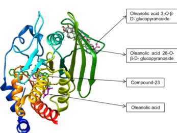
PTP1B에 대한 분자적 도킹 시뮬레이션 연구 − Autodock 4.2 program을 사용하여 PTP1B와 참두릅에서 분리된 활성 화합물인 compound I-III 사이의 결합을 3D 구조를 통해 시 뮬레이션하였고, 이 실험을 통해서 활성 화합물이 PTP1B에 결합하여 화합물-PTP1B 복합체가 형성됨으로써 PTP1B에 결합되는 위치와 상호작용하는 잔기를 알아볼 수 있었다 (Table IV). Compound 23(3-({5-[(N-acetyl-3-{4-[(carboxy- carbonyl)(2-carboxyphenyl)amino]-1-naphthyl}-L-alanyl) amino]pentyl}oxy)-2-naphthoic acid)은 강력한 비 펩티드성 (non-peptide)의 PTP1B 억제제로 보고되어있다.34) 따라서, compound 23과 PTP1B의 결합 시뮬레이션을 통하여 분리 된 화합물들과 비교분석 하였다.
Compound I과 PTP1B 잔기와의 상호작용을 살펴보면, PTP1B와 -8.09 kcal/mol의 결합에너지를 가지고 oleanolic acid의 수산기의 산소가 ASP48 잔기의 산소와 2.55 Å의 결 합길이로 수소결합을 형성하고, 에스터기의 산소가 ARG221 의 질소와 2.79 Å의 결합길이로 수소결합을 형성하였으며, 이 외에도 VAL49, ALA217, TYR46, GLN266, VALL84, TRP179, ASP265, THR263, GLY183, GLY220 그리고 GLN262 잔기와 소수성 상호작용을 형성하였으며, 이러한 소수성 상호작용은 compound I이 PTP1B의 pocket에 단단 하게 결합할 수 있도록 도와준다(Fig. 4B). Compound I과 상호작용하는 대부분의 잔기가 compound 23이 PTP1B와 수소결합을 형성하는 잔기와 거의 일치하였으며, 이 결과를 바탕으로 compound I이 compound 23(Fig. 4A)과 같이
PTP1B에 경쟁적 억제제로서 작용함을 알 수 있다. Compound II의 경우, -7.58 kcal/mol의 결합 에너지를 가지고, 28번 위 치의 탄소에 결합된 당의 수산기 3개가 GLN127 그리고 LYS131 잔기와 3개의 수소결합을 형성하였으며, ASP137, ASN90, PRO89, MET133, GLU132, PRO126, TRP125, GLN123, CYS92, PHE135, ILE134, GLY93, 그리고 THR91 잔기와 소수성 결합을 형성하였다(Fig. 4C). Compound III 는 -8.56 kcal/mol의 결합 에너지를 가지고, 3번 위치의 탄 소에 결합된 당의 수산기 3개가 ILE134, ASP137, GLU136, GLY93 잔기와 6개의 수소결합을 형성하였으며, 알데히드 기의 산소가 PRO89 잔기와 하나의 수소결합을 형성하였다.
그 밖에도, CYS92, LYS131, PRO126, GLN127, LYS128, GLU132, MET133, 그리고 PHE135 잔기와 소수성 결합을 형성하였다(Fig. 4D). Compound II와 III의 분자적 도킹 시 뮬레이션 결과, oleanolic acid의 3번과 28번 탄소에 결합된 Table IV. Molecular interaction of PTP1B active site with known inhibitor compound 23, and three unknown compounds
Compound Binding energya (kcal/mol)
No. of
H-bondb H-bond interacting residuesc Van der Waals bond interacting residuesd Compound
23
- 10.18 11 ARG221, SER216, ASP48, GLY220, ARG254, ALA217, ILE219
TYR46, LYS120, THR263, CYS215, GLN266, GLN262, ARG24, SER28, ASP29, MET258, VAL49.
Compound I
-8.09 2 ASP48, ARG221 VAL49, ALA217, TYR46, GLN266, VALL84, TRP179, ASP265, THR263, GLY183, GLY220, GLN262
Compound II
-7.58 3 LYS131, GLN127 ASP137, ASN90, PRO89, MET133, GLU132, PRO126, TRP125, GLN123, CYS92, PHE135, ILE134, GLY93, THR91
Compound III
-8.56 7 ASP137, GLY93, PRO89, ILE134, GLU136
CYS92, LYS131, PRO126, GLN127, LYS128, GLU132, MET133,PHE135
a
Binding energy, which indicate binding affinity and capacity for the active site of PTP1B enzyme.
b,c,dThe number of hydrogen bonds, and all amino acid residues from the enzyme-inhibitor complex were determined by autodock 4.2 program.
Fig. 3. Molecular docking models of the PTP1B inhibition of compound 23, oleanolic acid, oleanolic acid 28-O-β-D-gluco- pyranoside, and oleanolic acid 3-O-β-D-glucopyranoside.
당이 각각 PTP1B와 수소결합을 형성하는 것이 관찰되었고, 경쟁적 억제제인 compound 23과 유사한 위치에 결합하는 compound I과는 달리 활성자리와 떨어진 곳에 결합하는 것 을 통해서 두 화합물이 비경쟁적 억제를 함을 확인할 수 있 었다(Fig. 3). 또한, in vitro에서 compound II가 III 보다 약 3.5배 활성이 높았음을 고려했을 때, compound II가 결합 에너지는 더 낮지만 PTP1B와 결합하는 위치가 compound III가 PTP1B에 결합하는 위치에 비해 PTP1B 저해에 더 적 합하다는 것을 알 수 있다.
결 론
본 연구는 참두릅(Aralia elata (Miq.) Seem.) 잎의 protein
tyrosine phosphatase 1B(PTP1B)와 α-glucosidase 억제 활성 을 통한 항당뇨 활성을 검토하였다. 참두릅 잎의 메탄올 추 출물로부터 얻어진 용매 별 분획물의 PTP1B와 α- glucosidase 억제활성에서 EtOAc 분획물의 PTP1B 억제 활 성의 IC50값은 96.29±0.3 μg/mL로 다른 분획물들에 비하여 가장 높은 억제 활성을 나타내었으며 α-glucosidase 억제 활 성 또한 264.71±14.87 μmg/mL의 IC50 값을 가짐으로써 여 러 가지 다른 분획물들 중에서 가장 높은 억제 활성을 나타 내었다. 그리고 EtOAc 분획물에서 분리한 triterpene 및 triterpenoid glucosides 즉, oleanolic acid, oleanolic acid- 28-O-β-D-glucopyranoside와 oleanolic acid-3-O-β-D-glucopy- ranoside들이 활성 성분임을 밝혔다. 뿐만 아니라 이들 화합 물들은 분자 도킹 시뮬레이션을 통하여 PTP1B에 대하여 음 Fig. 4. Ligand interaction diagram of PTP1B inhibition of (A) compound 23, (B) oleanolic acid, (C) oleanolic acid 28-O-β-D-glu- copyranoside, and (D) oleanolic acid 3-O-β-D-glucopyranoside.
의 결합 에너지를 지닌 억제활성을 나타내므로 높은 친화 력과 결합력을 가진 것으로 나타났다. 이러한 결과로 볼 때, 참두릅 잎 EtOAc 분획물의 PTP1B와 α-glucosidase 억제 활성은 이러한 triterpene 및 triterpenoid glycosides 성분에 의한 것으로 생각된다. 이상에서 살펴본 바와 같이 참두릅 잎에서 PTP1B와 α-glucosidase 억제 활성을 나타내므로 인 슐린 저항성을 개선하여 당뇨병 치료에 도움이 되리라 생 각된다.
사 사
이 논문은 부경대학교 자율창의학술연구비(2016년)에 의 하여 연구되었음.
인용문헌
1. Kwon, J. H., Chang, M, J., Seo, H, W., Lee, J. H., Min, B. S., Na, M., Kim, J. C., Woo, M, H., Choi, J, S., Lee, H. K., and Bae, K. (2008) Triterpenoids and a sterol from the stem-bark of Styrax japonica and their protein tyrosine phosphate 1B inhibitory activities. Phytother. Res. 22: 1303-1306.
2. Tonks, N. K. (2003) PTP1B: From the sidelines to the front lines. FEBS. Lett. 546: 140-148.
3. Ahmad, F., Azevedo, J. L., Cortright, R., Dohm, G. and Gold- stein, B. J. (1997) Alterations in skeletal muscle protein-tyro- sine phosphatase activity and expression in insulin-resistant human obesity and diabetes. J. Clin. Invest. 100: 449-458.
4. Byon, J. C., Kusari, A. B. and Kusari, J. (1998) Protein-tyro- sine phosphatase-1B acts as a negative regulator of insulin signal transduction. Mol. Cell. Biochem. 182: 101-108.
5. Goldstein, B. J., Ahmad, F., Ding, W., Li, P. M. and Zhang, W. R. (1998) Regulation of the insulin signaling pathway by cellular protein tyrosine phosphatases. Mol. Cell. Biochem.
182: 91-99.
6. Mohamed Sham Shihabudeen, H., Hansi Priscilla, D. and Thirumurugan, K. (2011) Cinnamon extract inhibits α-glu- cosidase activity and dampens postprandial glucose excursion in diabetic rats. Nutr. Metab. (Lond.) 8: 46.
7. Toshiyuki, T. and Mitsuo, M. (2011) Potent α-Glucosidase Inhibitors from Safflower (Carthamus tinctorius L.) Seed.
Phytother. Res. 26: 722-726.
8. Caspary, W. F. (1978) Sucrose malabsorption in man after ingestion of α-glucoside hydrolase inhibitor, Lancet 1: 1231- 1233.
9. Hillebrand, I., Boehme, K., Frank, G., Fink, H. and Berchtold, P. (1979) The effects of the alpha-glucosidase inhibitor BAY g 5421 (Acarbose) on meal-stimulated elevations of circu- lating glucose, insulin, and triglyceride levels in man. Res.
Exp. Med. (Berl.) 175: 81-86.
10. van de Laar, F. A. (2008) Alpha-glucosidase inhibitors in the
early treatment of type 2 diabetes. Vasc. Health Risk Manag.
4: 1189-1195.
11. Sato, Y. and Rifkin, D. B. (1989) Inhibition of endoethelial cell movement by pericytes and smooth muscle cells: Acti- vation of a latent transformation growth factor B1-like mol- ecule by plasmin during coculture. J. Cell. Biol. 109: 309- 315.
12. van de Laar, F. A., Lucassen, P. L., Akkermans, R. P., van de Lisdonk, E. H., Rutten, G. E. and van Weel, C. (2005) Alpha- glucosidase inhibitors for patients with type 2 diabetes:
results from a Cochrane systematic review and meta-analysis.
Diabetes Care 28: 154-163.
13.이창복 (1989) 대한 식물도감, 575. 향문사, 서울.
14. Bae, K. W., Medicinal Plants of Korea, Kyohak Publishing Co. Ltd., Seoul, p. 363, 2000.
15.이우철 (1997) 한국식물명고, 771-772. 아카데미 서적, 서울.
16.지형준, 이상인 (1988) 대한 약전외 한약(생약) 규격집 주해서, 120. 한국 메디칼 인덱스사, 서울.
17. Han, B. H., Han, Y, N., Han, K. A., Park, M. H. and Lee, E.
O. (1983) Studies on the anti-inflammatory activity of Aralia continentalis (I). Arch. Pharm. Res. 6: 17-23.
18. Han, B. H., Park, M. H., Han, Y. N. and Manalo, J. B. (1983) Studies on the anti-inflammatory activity of Aralia conti- nentalis (II). Arch. Pharm. Res. 6: 75-77.
19. Han, B. H., Woo, E. R., Park, M. H. and Han, Y. N. (1985) Studies on the anti-inflammatory activity of Aralia conti- nentalis (III). Arch. Pharm. Res. 8: 59-65.
20. Yun-Choi, H. S., Kim, J. H. and Lee, J. R. (1986) Screening of potential inhibitors of platelet aggregation from plant sources (II). Kor. J. Pharmacogn. 17: 19-22.
21. Kosela, S., Rasad, A., Achmad, S. A., Wicaksonon, W., Baik, S. K., Han, Y. N. and Han, B. H. (1986) Effects of diterpene acids on malon-dialdehyde generation during thrombin induced aggregation of rat platelets. Arch. Pharm. Res. 9:
189-191.
22. Kim, J. S., Kang, S. S., Lee, M. W. and Kim, O.K. (1995) Isolation of flavonoids from the leaves of Aralia continen- talis. Kor. J. Pharmacogn. 26: 239-243.
23. Perry, L. M. (1980) Medicinal plants of east and southeast asia, Attributed properties and uses, 41. The MIT Press, Lon- don.
24. Jiangsu xin yi xue yuan (1977) Zhong-yao-ci-dian, 1268.
Shanghai ke xue ji shu chu ban she, Shanghai.
25. Kang, S. S. (1997) Chemistry and biological activity of the constitiuents from Aralia species. Ann. Rept. Nat. Prod. Sci.
5: 1-26.
26. Sawamura, M., Lee-Kim, M.-S., Shichiri, K.-I., Tsuji, T. and Machida, K. (1989) Volatile constituents of Japanese and Korean Udo (Aralia cordata Thunb.) and Butterbur (Pet- asites japonica Miq.). Research Reports of the Kochi Uni- versity 38: 1-12.
27. Jung, H. J., Jung, H. A., Kang, S. S., Lee, J. H., Cho, Y. S., Moon, K. H., and Choi, J. S. (2012) Inhibitory activity of Aralia continentalis roots on protein tyrosine phosphatase 1B and rat lens aldose reductase. Arch, Pharm Res. 135: 1771- 1777.
28. Jung, H. A., Cho, Y. S., Oh, S. H., Lee, S. H., Min, B. S., Moon, K. H. and Choi, J. S. (2013) Kinetic and molecular docking studies of pimaran type diterpenes as protein tyrosine phosphatase (PTP1B) inhibitors from Aralia continentalis roots. Arch. Pharm. Res. 36: 957-965.
29. Kim, J. S., Kang, S. S., Choi, J. S., Lee, M. W., and Lee, T.
S. (1998) Antioxidant components from Aralia continentalis.
Kor. J. Pharmacogn. 29: 13-17.
30. Na, M., Jang, J., Njamen, D., Mbafor, J. T., Fomum, Z. T., Kim, B. Y., Oh, W. K. and Ahn, J. S. (2006) Protein tyrosine phosphatase-1B inhibitory activity of isoprenylated flavo- noids isolated from Erythrina mildraedii. J. Nat. Prod. 69:
1572-1576.
31. Li, T., Zhang, X. D., Song, Y. W. and Liu, J. W. (2005) A microplate-based screening method for α-glucosidase inhib- itors. Chin. J. Clin. Pharmacol. Ther. 10: 1128-1134.
32. Berman, H. M., Battistuz, T., Bhat, T. N., Bluhm, W. F., Bourne, P. E., Burkhardt, K., Feng, Z., Gilliland, G. L., Iype, L., Jain, S., Fagan, P., Marvin, J., Padilla, D., Ravichandran, V., Schneider, B., Thanki, N., Weissig, H., Westbrook, J. D., and Zardecki, C. (2002) The protein data bank. Acta. Cryst.
58: 899-907.
33. Bernstein, F. C., Koetzle, T. F., Williams, G. J., Meyer, E. F.
Jr., Brice, M. D., Rodgers, J. R., Kennard, O., Shimanouchi, T., and Tasumi, M. (1977) The protein data bank: a com- puterbased archival file for macromolecular structures. J.
Mol. Biol. 112: 535-542.
34. Szczepankiewicz, B. G., Liu, G., Hajduk, P. J., Abad-Zapa- tero, C., Pei, Z., Xin, Z., Lubben, T., Trevillyan, J. M., Stas- hko, M. A., Ballaron, S. J., Liang, H., Huang, F., Hutchins, C.
W., Fesik, S. W., and Jirousek, M. R. (2003) Discovery of a potent, selective protein tyrosine phosphatase 1B inhibitor using a linked-fragment strategy. J. Am. Chem. Soc. 125:
4087-4096.
35. Kang, S. S., Kim, J. S., Kim, O. K. and Lee, E. B. (1993) Tri- terpenoid saponins from the root barks of Aralia elata. Arch.
Pharm. Res. 16: 104-108.
36. Cai, P., Xiao, Z. and Wei, J. (1982) Studies on the chemical constituents of Zhu Jie Shen (Panax japonicas) Chin Trad Herb Drugs, 13: 1-2.
37. Kasai, R., Tanaka, T., Nie, R.-L., Miyakoshi, M., Zhou, J. and Tanaka, O. (1990) Saponins from Chinese medicinal plants, Hemsleya graciliflora (Curcubitaceae). Chem. Pham. Bull.
38: 1320-1322.
38. Lee, W. H., Yang, E. J., Ku, S. K., Song, K. S. and Bae, J. S.
(2012). Anticoagulant activities of oleanolic acid via inhi- bition of tissue factor expressions. BMB reports, 45: 390-395.
((2016. 1. 21 접수; 2016. 2. 24 심사; 2016. 3. 10 게재확정)