대한내과학회지 : 제 66 권 제 3 호 2004
Ebstein 기형과 WPW 증후군이 동반된 환자의 전기생리학적 특성과 전극도자절제술 결과
계명대학교 의과대학 내과학교실
현대우·김윤년·이영수·한성욱·허승호
=Abstract=
Electrophysiologic characteristics and result of radiofrequency catheter ablation of WPW syndrome in patients with Ebstein's anomaly
Dae Woo Hyun, M.D., Yoon Nyun Kim, M.D., Young Soo Lee, MD., Seong Wook Han, M.D. and Seung Ho Hur, M.D.
Department of Internal Medicine, Keimyung University School of Medicine, Daegu, Korea
Background : The purpose of this study was to investigate the electrophysiologic characteristics and result of radiofrequency catheter ablation of patients with Wolff-Parkinson-White (WPW) synd- rome associated with Ebstein's anomaly.
Methods : After performed radiofrequency catheter ablation in five (1 male, 4 females, average age 40.6) patients, we then evaluated their clinical manifestation, echocardiography and electrophysio- logic characteristics.
Results : All patients had palpitation and two patients showed dizziness. In transthoracic echo- cardiography, all of the patients had severe tricuspid regurgitation and the mean distance of the septal tricuspid valve leaflet from the right atrioventricular annulus was 2.3 cm. In electrophysiologic study, eight accessary pathways were found in the 5 patients and all pathways were located on the right-side of the heart. Five manifest accessary pathways were found (one on the anterolateral, two on the lateral, one on the posterior, one on the posteroseptal wall) and three concealed pathways were found at posteroseptal wall. Three patients (60%) had multiple accessary pathways. The most common combination pattern was the manifest right lateral wall and concealed right posteroseptal wall accessary pathway. Catheter ablation of the concealed right posteroseptal accessory pathway was not successful in one patient with multiple accessary pathway. The average time of radiation was 56.0 minutes and the average time of procedure was 141.3 minute.
Conclusion : WPW syndrome associated with Ebstein's anomaly had a high frequency of multiple accessary pathways and there was much difficulty in catheter ablation because morphologic and anatomical change of the heart structure. Further study will be necessary on the role of arrhythmia in atrialized right ventricle.(Korean J Med 66:250-258, 2004)
Key Words : Ebstein's Anomaly, Wolff-Parkinson-White Syndrome
∙접 수 : 2003년 6월 30일
∙통 과 : 2004년 1월 2일
∙교신저자 : 김윤년, 대구시 중구 동산동 194, 계명대학교 동산의료원 순환기내과(700-712) E-mail : [email protected]
- Dae Woo Hyun, et al : Electrophysiologic characteristics and result of radiofrequency catheter ablation of WPW syndrome in patients with Ebstein's anomaly -
서 론
엡스타인 기형은 1866년 엡스타인에 의해 처음 기술되 었으며 삼첨판의 후첨과 중격첨이 판첨 하방으로 변위를 보이는 구조적 기형이다. 대개 삼첨판막의 폐쇄부전이 동 반되며 심방중격결손 및 다른 기형의 동반이 흔하여 다 양한 범위의 형태적, 생리적 변화를 보인다1, 2). 이러한 심 장내 해부학적인 위치 이상으로 발작성 방실회귀빈맥이 엡스타인 기형 환자의 25∼30%에서 발생하며 표면 심전 도상 심실 조기흥분은 5∼25%에서 관찰된다1-4). 또한 엡 스타인 기형 환자에서 발작성 방실회귀빈맥 환자의 50%
까지 다발성 부전도로를 가진다1, 5).
도자 절제술(Catheter ablation)은 발작성 방실회귀빈 맥의 기본적인 치료로 인정되고 있고 엡스타인 기형과 같은 선천성 심질환과 동반된 부정맥의 치료도 성공적으 로 이루어지고 있다. 이에 연구자들은 엡스타인 기형 중 Wolff-Parkinson-White (WPW) 증후군으로 도자 절제 술을 시행한 5예에 대해 전기 생리학적 특성과 전극도자 절제술 결과에 대해 알아보고자 하였다.
대상 및 방법 1. 대상
1993년 2월부터 2003년 4월까지 계명대학교 동산의료 원에서 엡스타인 기형으로 진단받고 심계항진으로 전기 생리학적 검사와 도자 절제술을 시행한 5명을 대상으로 하였고, 이들은 모두 표면 심전도상 WPW 증후군을 보 였다. 엡스타인 기형은 삼첨판막 중격첨이 우측 방실륜보 다 1.2 cm 이상 심첨부로 이동하여 부착한 것으로 정의 하였다6). 진단은 경흉부 심초음파 검사로 하였고, 삼첨판 막 중엽의 이동은 중벽륜 돌출부부터 심실 중벽에서 삼 첨판막이 나오는 부위 사이의 거리를 측정하였다. 삼첨판 막 폐쇄부전의 정도는 도플러 혈류 영상으로 정량화 하 였다.
2. 전기생리학적 검사
전기생리학적 검사는 표준 심내지도 방법으로 시행하 였다. 우측 대퇴정맥을 통해 6F 4극 도자를 우심방과 우 심실에 각각 위치하였고, 6F 6극 도자를 히스속 부위에 위치하였다. 좌측 쇄골하정맥을 통해 6F 10극 도자를 관
상정맥동에 위치한 후 기본 전기생리학적 검사와 iso- proterenol을 분당 2 ug씩 투여하면서 전기생리학적 검사 를 시행하였으며 도자 절제술 후 반복하여 시행하였다.
심장내 심전도는 multichannel oscilloscope recorder (Car- dioLabTM, Amplifier, Prucka Engineering Inc., U.S.A.
혹은 Electronics for Medicine, PPG, Midas 2500, U.S.A.) 를 이용하였고, 한 환자(patient 5)는 3차원 전자기 지도 시스템(Carto, BiosenseWebster, Diamond Bar, U.S.A) 을 이용하였다. 프로그래밍 전기자극계(Bloom DTU 215 A, USA)를 이용하여 프로그램밍 전기자극을 시행하였 다. 계획된 심실이나 심방자극으로 빈맥을 유도하였고, 빈맥이 유도되지 않는 경우는 isoproterenol을 분당 2 ug 씩 정주하면서 다시 빈맥을 유도하였다.
현성 우회로인 경우 동조율시, 불현성 우회로시는 빈 맥이 지속되는 동안이나 심실 자극 동안에 관상정맥동으 로 삽입한 전극도자와 우심방의 도자를 이용하여 좌심방 및 우심방의 판막륜 주위의 활성순서를 관찰하였다.
다발성 부전도의 유무는 부전도로들 사이의 불일치 거 리가 2 cm 이상일 경우로 정의하였다.
3. 도자 절제술
도자 절제술을 위한 고주파에너지 생성기는 Radionics RFG-3C generator system (Radionics®, Inc., Burlington, Messachusetts, U.S.A.), EP TechnologiesTM(Boston Sci- entific®, U.S.A.)와 AtakrTM (medtronic® Cardiorhythm, U.S.A)을 사용하였다. 절제 도자는 6-7F quadripolar steera- ble 4mm tip (Mansfiled-Webster catheter, Watertown, Messachusstts 혹은 EPT catheter, Mountain view, Cali- fonia, U.S.A. 혹은 Daig catheter, Minnesota, U.S.A. 혹 은 NaviStarTM, BiosenseWebster, U.S.A.)을 사용하였다.
도자 절제술시 고주파에너지는 30∼60볼트 정도로 30
∼60초 동안 가하거나 온도 도자를 이용시는 60℃로 하 였다. 도자 절제 부위는 (1) 동리듬시 가장 빠른 심실활성 부위 또는 (2) 심실 자극시 심방이 후향적으로 가장 빨리 활성 되는 부위로 하였다. 고주파 전극 도자 절제술의 성공 은 우회로를 통한 정방향과 역방향의 차단을 기준으로 하였고 절제부위의 위치는 45도 좌전사위에서 결정하였다.
- 대한내과학회지 : 제 66 권 제 3 호 통권 제 523 호 2004 -
결 과
1. 임상상
남성이 1명 여성이 4명이었으며 이들의 평균 연령은 40.6세(최소 18세, 최대 61세)였다. 모두 심계항진이 있었 고, 2명의 환자에서 심계항진시 어지러움증을 동반하였 다(표 1).
모든 환자에서 안정시 표면 심전도상 심실 조기흥분이 관찰되었고, 3명의 환자에서 도자 절제술전 좁은 QRS빈 맥이 확인되었고, 1명은 도자 절제술시 좁은 QRS빈맥이 유도되었다. 한명의 환자(patient 5)에서는 심전도상 빈맥 이 확진되지는 않았지만 반복되는 심계항진이 있고 표면 심전도상 심실 조기흥분이 있어 도자 절제를 실시하였다.
심초음파 검사상 삼첨판막 중엽의 평균 이동거리는 2.4 cm (최소 1.5 cm, 최대 3.5 cm)였고, 삼첨판막 폐쇄부 전의 정도는 5예 모두 심한 삼첨판막 역류를 보였다. 좌 심실 구혈율은 평균 67.4% (최대 75%, 최소 58%)였다 (표 1).
2. 전기생리학적 특징
네 명의 환자에서 도자 절제전 표면 심전도상 평균 P 파 간격은 80 msec, PR 간격은 117 msec, QRS 간격은 142 msec였다. 도자 절제술 후 평균 PR 간격은 188 msec, QRS 간격은 102 msec, AH 간격은 131 msec, HV 간격 은 55 msec, QRS 간격은 93 msec였다. 빈맥시 평균 실 방전도시간은 93 msec였다. 한명의 환자(patient 1)에서 심내 심전도의 자료의 분실로 심전도 측정을 하지 못하 였다(표 2). 4명의 환자에서 도자 절제술시 빈맥이 유도 되었고 모두 정방향 방실회귀성빈맥이었으며 평균 빈맥 속도는 480 msec였다.
5명의 환자에서 모두 8개의 부전도로가 있었고, 3 (60%) 명에서 각각 2개의 다발성 부전도로를 보였다. 모든 부전 도로는 우측 심장에 위치하였고, 불현성 부전도로가 3개 관찰되었는데 이는 모두 후중벽에 위치하였다. 현성 부전 도로는 4개가 자유벽, 1개가 후중벽에 위치하였다(그림 1, 표 3).
Table 2. Electrophysiologic characteristics of the patients
Patient Surface ECG Intracardiac ECG
Preablation Postablation Preablation Postablation
P PR QRS PR QRS Tachy CL VA AH HV
1 2 3 4 5
90 100
60 80 70
130 115 110 120 110
160 118 120 150 160
160 200 160 240 180
140 110 70 100
90
400, 500 450 571 ND
80 90 120
80
120 105 180 120
55 45 60 60
mean 80 117 142 188 102 480 93 131 55
The basic unit is msec. P, P wave duration; PR, PR interval; QRS, QRS duration; Tachy CL, tachycardia cycle length; ND, not documented; AH, AH interval; HV, HV interval; QRS, QRS duration; VA, VA conduction time;
ECG, electrocardiography; *, right bundle block after ablation Table 1. Clinical characteristics of the patients
Patient Age Year Palpitation CTR (%) LVEF TR TV disp 1
2 3 4 5
18 61 40 43 41
95, 96 95 99 01 03
+ + + + +
61%
55%
61%
57%
47%
58%
65%
65%
75%
74%
severe severe severe severe severe
2.3 cm 3.0 cm 1.8 cm 3.5 cm 1.5 cm Year, ablation year; CTR, cardiothoracic ratio; TR, tricuspid regurgitation; TV disp, tircuspid valve displacement
- 현대우 외 4 인 : Ebstein 기형과 WPW 증후군이 동반된 환자의 전기생리학적 특성과 전극도자절제술 결과 -
Figure 2. Right anterior oblique view of right ventriculo- gram shows downward displacement of the tricuspid val- ve into the right ventricle and ablation catheter located at anatomical atrioventricular groove (arrow, ablation catheter;
arrow head, tricupid valve)
현성 부전도로는 모두 성공적으로 제거하였으나 불현 성 부전도로 중 1개(patient 2)는 도자 절제를 실패하였 다. 또 한명의 환자(patient 1)에서는 첫 번째 도자 절제 시 우측벽 현성 부전도로는 성공적으로 시술 후 퇴원하 였으나 외래 추적기간 중 상심실성 빈맥이 확인되어 두 번째 도자 절제시 불현성 우후중벽 부전도를 확인하여 성공적으로 도자 절제하였다.
우후측에 현성 부전도로를 가진 환자(patient 4)의 우 심실 조영사진을 보면 삼첨판막의 심첨으로 하방변위가
관찰되고 실제 도자 절제부위는 해부학적 방실륜이 있는 부위에 있었다(그림 2). 2003년에 도자 절제를 시행한 환 자(patient 5)의 경우는 3차원 전자기 지도 시스템을 이용 해 도자 절제를 시행하였는데(그림 3, 4) 동조율시 가장 빨리 활성화를 보이는 곳은(적색) 우전측벽이었다(그 림 4). 평균 방사선 시간은 56.0분이었고, 평균시술시간은 141.3분이었다.
도자 절제술에 의한 합병증은 없었다.
3. 추적관찰
도자 절제술 후 4명의 환자에서 심계항진과 표면 심전 도상 심실 조기흥분은 없어졌다. 다발성 부전도로를 가지 고 있고 우후중벽 불현성 부전도로 절제를 실패한 환자 (patient 2)에서는 표면 심전도상 심실 조기흥분은 보이지 않았으나 심계항진이 있고, 표면 심전도상 발작성 상심실 성빈맥이 있어 현재 amiodarone으로 약물 치료 중이다.
3명의 환자(patient 1, 2, 4)에서 추적 심초음파를 시행 하였으며 모두 좌심실 구혈율은 정상이었고, 한명의 환자 (patient 2)에서 중등도 삼첨판 폐쇄부전으로 호전 보였 고 나머지 두명의 환자에서는 심한 삼첨판 폐쇄부전을 보여 변화는 관찰되지 않았다. 이들 두명의 환자(patient 1, 4)에서 전신부종 등 우심부전의 증상이 있어 현재 심 부전 치료 중이다. 모든 환자에서 개심술은 시행하지 않 았다.
고 찰
엡스타인 기형은 선천성 기형의 1% 미만을 차지하는 드문 질환으로 유아기에 진단된 환자의 경우는 예후가 좋지 않고 청장년기에 진단된 경우는 대개 우연히 발견 되고 부정맥이 흔하며 예후는 상대적으로 좋다2). 엡스타 인 기형과 같이 선천성 심질환과 동반된 발작성 상심실 성빈맥은 빈맥과 연관된 실신과 심장마비의 빈도가 많다
7)고 하였으나 본원의 증례에서는 실신 또는 심장마비 등 의 증상은 관찰되지 않았고, 심계항진시 어지러움증과 같 은 뇌 저관류에 의한 증상이 2예에서 관찰되었다.
Kastor 등8)은 엡스타인 기형에서 커진 우심방에 의해 PA 간격이 길어지고 심방화된 우심실에 의해 전도 시스 템의 신전에 의해 HV 간격이 연장된다고 하였는데 본원 의 예에서는 2명의 환자에서 HV 간격이 연장(각각 60 msec)되어 있었다. Smith 등5)은 정방향성 빈맥시 부전도 Figure 1. Schematic of the tricuspid and mitral valve
annuli, as viewed in the 45°left anterior olblique view, illustrates the locations of the 8 accessory pathways.
There were 4 posteroseptal pathways, 1 posterior pathway, 2 lateral pathways, 1 anterolateral pathway (TV, tricuspid valve; MV, mitral vavle)
- Korean Journal of Medicine : Vol. 66, No. 3, 2004 -
로가 있는 국소 심실과 심방의 지연된 활성화로 실방전 도시간이 길어진다고 하였고, 표면 심전도상 우측 후측벽 또는 우측 후중벽에 심실 조기흥분이 보이고, 빈맥시 긴 실방간격을 가지는 것이 엡스타인 기형의 심전도 특징이
라고 하였다. 이들의 연구에 의하면 기형이 없는 WPW 증후군 환자의 평균 실방전도 시간은 110 msec였고, 엡 스타인 기형환자 20명의 환자에서 평균 실방전도 시간은 192 msec였다. 그러나 일부 연구에 의하면 부전도 자체
Figure 3. Serial changes on the surface ECGs in a patient 5 who had WPW syndrome with manifest right anterolateral and concealed right posteroseptal pathways. (A) The baseline ECG shows delta wave. (B) After RF ablation, the delta wave is disappeared
Table 3. Electrophysiologic characteristics of the patients
Patient Clinical arrhythmia Ablation site Result 1
2
3 4 5
AVRT, orthodromic
AVRT, orthodromic
AVRT, orthodromic AVRT, orthodromic Not documented
Right lateral (M) Right posteroseptal (C) Right lateral (M) Right posterosetal (C) Right posteroseptal (M) Right posterior (M) Right anterolateral (M) Right posteroseptal (C)
Success Success Success Fail Success Success Success Success ECG, electrocardiography; AVRT, atrioventricular reentrant tachycardia; M, manifest; C, concealed
A
B
- Dae Woo Hyun, et al : Electrophysiologic characteristics and result of radiofrequency catheter ablation of WPW syndrome in patients with Ebstein's anomaly -
의 늦은 전도 또는 복잡하고 긴 부전도로에 의해 실방간 격이 연장된다고 하였다. 본원의 증례에서는 실방전도 시 간의 연장은 관찰되지 않았다.
Chiou 등7)과 Hare 등9)은 엡스타인 기형에서 정확하게 방실륜의 위치를 찾기가 어렵고 절제 도자로 부터의 전 기도 판독의 어려움이 있는데, 심방화된 심실에 불필요한 에너지 투여를 피하기 위해 절제 도자위치에서 정확하게 동일한 심방과 심실파의 폭을 확인하는 것이 중요하다.
엡스타인 기형에서 방실결절은 해부학적인 삼첨판막 륜을 따라 Koch 삼각형의 후하방으로 변형된 위치에 놓 이므로 해부학적인 접근방법으로 도자 절제를 시도하면 방실 결절의 손상을 초래할 수 있다11). 또한 정확한 방실 륜의 위치와 확장된 심방으로 인해 심방 전기도의 분석 의 어려움이 있다7). 상기의 어려움으로 평균 방사선 시간 및 시술 시간이 선천성 기형이 없는 경우보다 길었다21). 3차원 전자기 지도 시스템의 장점은 방사선 노출시간과 시술 시간을 줄이고, 3차원적인 높은 공간해상도를 보여 부정맥 발생기전에 대한 이해가 쉽고 해부학적으로 정확 한 위치에 에너지 투여가 가능하여 불필요한 에너지 투 여를 줄여준다12-14). 특히 선천성 심질환에 의한 심장구조
의 해부학적 변형이 있는 경우 정상 전도로의 위치와 부 정맥 활성 순서를 좀 더 정확히 알 수 있다15). 본원에서 시행한 3차원 전자기 지도 시스템을 이용시 정확한 방실 륜과 His속 위치 지정이 가능함으로 방실 결절과 같은 정상적인 전도 시스템의 손상을 피할 수 있다.
엡스타인 기형에 동반될 수 있는 부정맥은 심방세동, 발작성 방실회귀성빈맥, 방실 결절회귀성빈맥, 심실성 빈 맥 등1, 4, 11, 17)
이 있으나 부전도로에 의한 방실 회귀성 빈 맥이 특징적이다. 이 중 WPW 증후군은 약 5∼25% 정도 에서 동반된다. 부전도로는 우측에 위치하고 다발성인 것 이 특징이다. Smith 등5)은 부전도로를 가진 21명의 엡스 타인 기형 환자에서 32개의 부전도로가 발견되었으며 38% 환자에서 다발성 부전도로가 있었다. Cappato 등10) 은 21명의 환자에서 34개의 부전도로가 있었는데 다발성 부전로를 가진 환자가 52%였다. 또한 현성 부전도로가 25개, 불현성 부전도로가 9개 있었으며 위치를 보면 현성 부전도로는 중벽에 8개, 자유벽에 17개가 있었고, 불현성 부전도로는 중벽에 4개 자유벽에 5개가 있었다. 본원에서 는 현성 부전도로는 80%가 우측 자유벽에 위치하였고, 불현성 부전도로는 모두 우후중벽에 위치하였으며 다발 I
aVF V1 Abl-d Abl-p His-p His-m His-d CS-p CS-m CS-d RV
Figure 4. Serial changes on the intracardiac ECGs in a patient 5 who had WPW syndrome with manifest right anterolateral and concealed right posteroseptal pathways. (A) during ablation at manifest right lateral pathway. After RF ablation, the fusion site at ablation catheter became separated. (B) during ablation at concealed right posteroseptal pathway, After RF ablation, the retrograde VA conduction time is more prolonged during right ventricle pacing (Abl, albation; d, distal; m, midle; p, proximal; cs, coronary sinus; RV, right ventricle)
A B
- 대한내과학회지 : 제 66 권 제 3 호 통권 제 523 호 2004 -
성 부전도로의 조합을 보면 우측벽의 현성 부전도와 우 후측벽의 불현성 부전도로가 많았다. 본원에서 시행한 전 기 생리학적검사에서 다발성 부전도로의 빈도 4.2%였으 며21)엡스타인 기형에서 높은 빈도를 보였다.
발작성 상심실성빈맥이 있거나 심계항진 등의 증상이 있는 엡스타인 기형 환자에서 수술을 계획할 때에는 수 술 전에 심장 전기생리검사를 시행하여 도자 절제로 빈 맥성 부정맥을 해결하고 수술 또는 부전도로의 정확한 위치 및 기전을 알고 수술시에 빈맥성 부정맥에 대한 수 술을 같이 해 준다. 여러 연구17-19)에 의하면 빈맥성 부정 맥에 대한 수술을 어려움없이 같이 할 수 있었다는 보고 가 있다.
부정맥에 의한 급사가 엡스타인 기형에서 보고 되고 있는데 이는 주로 우측 방실 회귀성빈맥, 심방세동 및 심 방조동에 의하며, 또한 심실성 부정맥이 심방화된 우심실 에서 생기는 것과 서맥도 급사의 원인이 될 수 있다22-24). 심방화된 심실이 흥분하기 쉬워 도자 절제시 카테터 조
작에 의해 발작성 빈맥이 25%에서 생기며 이 중 사망과 심장마비가 21%에 발생하여1) 심도자술 또는 도자 절제 술시 유의하여야 하겠다.
엡스타인 기형은 부전도로가 흔하며 또한 심방화된 좌 심실의 부정맥 유발성이 있어 이의 기질적 원인 및 전기 생리학적 특징에 대한 좀 더 많은 연구가 있어야 하겠다.
요 약
목적 : Wolff-Parkinson-White (WPW) 증후군을 보 인 엡스타인 기형환자의 전기 생리학적 특성과 전극도자 절제술 결과에 대해 알아보고자 하였다.
방법 : 계명대학교 동산의료원에서 전기 생리학적 검 사와 고주파 도자 절제술을 시행한 WPW 증후군을 보인 엡스타인 기형 환자 5명(남 1, 여 4, 평균연령 40.6세)을 대상으로 임상 증상, 심초음파 소견, 전기 생리학적 특성 에 대해 알아보았다.
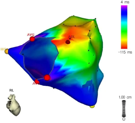
Figure 5. Three dimensional mapping system (Carto, BiosenseWebster) image of the right-sided chambers showing the successful site of ablation of the manifest right anterior accessory pathway in a patient 5. His bundle (His), atrioven- tricular groove (AVG) and ablation site (ABL) are noted (RL, right lateral view)
4 ms
-115 ms
1.00 cm
- 현대우 외 4 인 : Ebstein 기형과 WPW 증후군이 동반된 환자의 전기생리학적 특성과 전극도자절제술 결과 -
결과 : 증상으로는 심계항진 5명, 어지러움증이 2명의 환자에서 있었고, 심초음파 소견상 모두 심한 삼첨판 역 류가 관찰되었으며 삼첨판막 중격첨의 평균이동거리는 2.4 cm였다. 전기 생리학적 특징으로는 부전도로는 모두 우측에 있었고, 5명의 환자에서 모두 8개의 부전도로가 있었다. 현성 부전도로는 전측벽 1명, 측벽 2명, 후벽 1명, 후중벽에 1명 있었고, 불현성 부전도로는 3명에서 관찰되 었고, 모두 후중벽에 위치하였다. 다발성 부전도로는 3명 (60%)의 환자에서 있었고, 우측벽의 현성 부전도로와 우 후중벽의 불현성 부전도로 조합이 많았다. 이 중 1명에서 우후중벽의 불현성 부전도로의 절제를 실패하였다. 평균 방사선 시간은 56.0분이었고, 평균시술 시간은 141.3분이 었다
결론 : WPW 증후군을 보이는 엡스타인 기형 환자는 다발성 부전도로가 많고 해부학적 형태 및 위치의 변화 로 도자 절제의 어려움이 있을 수 있고, 향후 심방화된 심실이 부정맥을 일으키는 기질적 원인에 대한 연구가 있어야 할 것으로 생각된다.
REFERENC ES
1) Watson H. Natural history of Ebstein's anomaly of tricuspid valve in childhood and aldolescense: an international co-operative study of 505 cases. Br Heart J 36:417-427, 1974
2) Celermajer DS, Bull C, Till JA, Cullen S, Vassillikos VP, Sullivan ID, Allan L, Nihoyannopoulos P, Somer- ville J, Deanfield JE. Ebstein's anomaly: presentation and outcome from fetus to adult. J Am Coll Cardiol 23:170-176, 1994
3) Mair DD. Ebstein's anomaly: natural history and management. J Am Coll Cardiol 19:1047-1048, 1992 4) Radford DJ, Graff RF, Neilson GH. Diagnosis and
natural history of Ebstein's anomaly. Br Heart J 54:517-522, 1985
5) Smith WM, Gallagher JJ, Kerr CR, Sealy WC, Kasell JH, Benson DW, Reiter MJ, Sterba R, Grant AO. The electrophysiologic basis and management of sympto- matic reccurrent tachycardia in patients with Ebs- tein's anomaly of the tricuspid valve. Am J Cardiol 49:1223-1234, 1982
6) Feigenbaum H. Echocardiography. 4th ed. p. 652, Phila- delphia, Lea & Febiger, 1986
7) Chiou CW, Chen SA, Chiang CE, Wu TJ, Tai CT, Lee SH, Cheng CC, Ueng KC, Chen CY, Wang SP, Chiang BN, Chang MS. Radiofrequency catheter ablation of
paroxysmal supraventricular tachycardia in patients with congenital heart disease. Int J of Cardiol 50:
143-151, 1995
8) Kastor JA, Goldreyer BN, Josephson ME, Perloff JK, Scharf DL, Manchester JH, Shelburne JC, Hirshfeld JW. Electrophysiologic characteristics of Ebstein's ano- maly of the tricuspid valve. Circulation 52:987-995, 1975
9) van Hare GF, Lesh MD, Stanger P. Radiofrequency catheter ablation of supraventricular arrythmias in patients with congenital heart disease: results and technical consideration. J Am Coll Cardiol 22:883- 890, 1993
10) Cappato R, Schlüter M, Weiß C, Antz M, Koschyk DH, Hofmann T, Kuck KH. Radiofrequency current catheter ablation of accessary atrioventricular path- ways in Ebstein's anomaly. Circulation 94:376-383, 1996 11) Okishige K, Azegami K, Goseki Y, Ohira H, Sasano T, Yamashita K, Satake S. Radiofrequency ablation of tachyarrhythmia in patients with Ebstein's anomaly.
Int J Cardiol 60:171-180, 1997
12) Kopelman HA, Prater SP, Tondato F, Chronos NA, Peters NS. Slow pathway catheter ablation of atrio- ventricular nodal re-entrant tachycardia guided by electroantomical mapping: a randomized comparison to the conventional approach. Europace 5:171-174, 2003 13) Gepstein L, Evans SJ. Elecroantomical mapping of the heart: basic concepts and implications for the treat- ment of cardiac arrhythmias. Pacing Clin Electro- physiol 21:1268-1278, 1998
14) Cooke PA, Wilber DJ. Radiofrequency catheter abla- tion of atrioventricular nodal reentry tachycardia uili- zing nonfluoroscopic electroanatomical mapping. Pacing Clin Electrophysiol 21:1802-1809, 1998
15) Dorostkar PC, Cheng J, Scheinman MM. Electroan- tomical mapping and ablation of the substrate su- pporting intraatrial reentrant tachycardia after pallia- tion for complex congenital heart disease. Pacing Clin Electrophysiol 21:1810-1819, 1998
16) Kocheril AG, Losenfeld LE. Radiofrequency ablation of an accessory pathway in a patient with corrected Eb- stein's anomaly. Pacing Clin Electrophysiol 17:986- 990, 1994
17) Chauvaud SM, Brancaccio G, Carpentier AF. Cardiac arrythmia in patients undergoing surgical repair of Ebstein's anomaly. Ann Thorac Surg 71:1547-1552, 2001
18) Pass RH, Williams MR, Quaegebeur JM, Liberman L, Hordof AJ. Intraoperative radiofrequency linear ca- theter ablation of accessary pathways in children with Ebstein's anomaly undergoing tricuspid annuloplasty.
- Korean Journal of Medicine : Vol. 66, No. 3, 2004 -
Am J Cardiol 90:817-819, 2002
19) Lazorishinets VV, Glagola MD, Stychinsky AS, Ru- denko MN, Knyshov GV. Surgical treatment of Wolf- Parkinson-White syndrome during plastic operations in patients with Ebstein's anomaly. Eur J Cardio- thorac Surg 18:487-490, 2000
20) Iturralde P, Guevara-Valdivia M, Rodriguez-Chávez L, Medeiros A, Colin L. Radiofrequency ablation of mul- tiple accessary pathways. Europace 4:273-280, 2002 21) 이영수, 권택근, 김성열, 손봉준, 조봉기, 한성욱, 허승호,
김윤년. 다발성 부전도로의 심장 전기생리적 특성. 대한 내과학회지 63:394-401, 2002
22) Lo HM, Lin FY, Jong YS, Tseng YZ, Wu TL. Ebs- tein's anomaly with ventricular tachycardia: evidence for the arrythmogenic role of the atrialized ventricle.
Am Heart J 117:959-962, 1989
23) Obidha-Ngwu O, Milliez P, Richardson A, Pittaro M, Josephson ME. Ventricular tachycardia in Ebstein's anomaly. Circulation 104:e92-e94, 2001
24) Carballal J, Asensio E, Hernández R, Narváez R, Gómez M, Dorantes J, Orea A, Rebollar V, Oseguera J. Ebstein's anomaly, atrial paralysis and atrio-ven- tricular block: an uncommon association. Eropace 4:
451-454, 2002