서 론
유방암의 일반적인 방사선치료는 림프절 1문조사와 유방 의 접선방향조사로 이루어진다. 유방의 접선방향조사는 초 기 암의 유방보존술(Breast conservative surgery, BCS) 후 잠재적인 병을 예방하기 위해 사용하였다.1,2)치료기법의 발 전에 따라 최근에는 접선방향 치료방법의 큰 틀은 유지하면 서 세기조절방사선치료(Intensity modulated radiation therapy, IMRT)나 Field-in-Field technique 등이 시도되고
있다. 본원에서 유방의 방사선치료계획은 주로 3차원 입체 조형치료계획(3D Conformal Radiation Therapy, 3D-CRT) 을 수립하며 쐐기필터(wedge filter)를 이용한 technique, Field-in-Field technique, 그리고 Dose Fluence를 이용한 Irregular compensation 방법 등을 사용한다. 방사선치료에 있어서 치료부위 내 균등한 선량분포는 환자의 치료효과를 증대시키고 장애발생을 최소화하는 중요한 인자이며, 특히 유방의 방사선치료에 있어 조직병리검사결과에 따라 Scar 또는 Drain site 부위에 원하고자 하는 선량을 충분히 전달 하고 반대편 유방(Contralateral breast dose)의 방사선노출 선량에 대해 아는 것이 중요하다.3)반대편 유방의 방사선노 출 요인은 주로 Housing leakage와 쐐기필터로 인한 이차
본 논문은 2015년 11월 20일 접수하여 2015년 12월 14일 채택되었음.
책임저자 : 김선명, 고려대학교구로병원 방사선종양학과 서울시 구로구 구로동로 148
Tel : 02) 2626-1387
E-mail : [email protected]
열형광선량계(TLD)와 MOSFET을 이용한 유방암 방사선치료계획에 대한 피부선량 평가
고대안산병원 방사선종양학과
목 적 :유방암 치료에 있어 피부선량의 측정은 매우 중요하다. 치료계획시에는 처방선량에 비해 초과선량이나 부족선량이 생길 수 있으므로 이에 대한 유방암의 여러 가지 치료계획간 피부선량 평가가 필요하다. 이에 대해 본원에서는 다양한 선량계를 이용하여 선량을 분석하여 유방암치료에 적용하고자 한다.
대상 및 방법 :유방암은 기본적으로 접선방향 치료계획 시 일어나는 skin dose(Drain site, Scar)의 선량차이를 알아보기 위하여 인체모형팬텀을 이용하였다. 인체모형팬텀을 전산화단층촬영하고 전산화치료계획에서 open과 쐐기필터(Wedge filter)를 이용한 치료계획, Field-in-Field를 이용한 치료계획, 그리고 Dose fluence를 이용한 Irregular compensation 치료계획을 세우고 컴퓨터치료계획 프로그램(Eclipse)으로 선량관심점과 측정점 의 선량을 비교하였다. 치료실에서 인체모형팬텀을 위치시키고 선량비교를 위하여 각 치료계획 측정점에 열형광선량계(themoluminesence dosimeter, TLD)와 MOSFET(Metal oxide-silicon field effect transistor)을 이용하여 선량을 측정하여 비교평가 하였다.
결 과 :피부선량은 치료계획 중심점을 기준으로 위와 아래는 Dose fluence를 이용한 Irregular compensation 치료계획 사용 시 MOSFET을 이용 한 선량측정에서 가장 많은 선량이 들어가는 것으로 나타났다. 내측과 외측의 측정선량은 open과 쐐기필터 치료계획에서 TLD와 MOSFET을 이용 하여 측정시 5.7%에서 10.3%까지 낮게 나타났다. 반대쪽 유방의 선량은 open 치료계획이 가장 적었고, Dose fluence를 이용한 Irregular compensation 치료계획을 사용 시 가장 많은 선량이 측정되었다. 치료종별 치료계획상에서는 내측과 외측의 선량편차가 가장 컸으며, TLD와 MOSFET 측정시에도 같은 경향을 보였다. 외측은 TLD , 내측은 MOSFET이 가장 편차가 컸다.
결 론 :치료계획에 따른 피부선량은 전반적으로 Dose fluence를 이용한 Irregular compensation의 치료계획을 사용 시 가장 많이 들어가는 것으 로 나타났으며, 이는 많은 MLC의 움직임에 의한 산란선 영향으로 생각된다. 모든 치료계획에서 피부의 위치에 따라 약간의 차이는 있으나 부족선 량이 생기는 부분에서는 내측의 내유임파절(Intramammary lymph nodes)선량이나 Scar, Drain site등에서 세심한 주의가 필요하다. 부족선량을 높 이기위해서는 Dose fluence를 이용한 Irregular compensation의 치료계획을 사용하는 것이 좋겠으나, 전체적인 선량을 높이기보다는 선택적인 범 위내에서 선량을 높이게 되므로 환자의 연령이나 움짐임 등을 고려하여 치료기술을 선택하는 것이 바람직할 것으로 사료된다.
핵심용어 :열형광선량계(TLD)와 MOSFET을 이용한 유방암 방사선치료계획에 대한 피부선량 평가
김선명・김영범・이상록・백상윤・정세영
법이 많이 사용되고 있다. MOSFET은 에너지 의존성과 방 향의존성, 에너지스펙트럼의 따른 오차 등의 단점은 있지만 방사선치료분야에 있어 재현성은 열형광선량계보다 우수 하며 6MV 영역에서 에너지 스펙트럼의 따른 단점은 미미하 다.6)
이를 위해 본원에서는 방사선치료계획에 따른 피부선량 의 측정을 평가하기 위해 인체모형팬텀에 전산화단층 모의 치료계획촬영(CT simulation)을 실시하고 관심선량 측정 점의 선량계산을 하였다. 다양한 치료계획에 열형광선량계
- Humanoid phantom(Anderson Rando phantom, USA)
- CT simulator(Brightspeed Elite,GE,USA)
- TLD(Harshaw 5500, Thermo Fisher Scientific Inc.) - MOSFET(Best Medical Canada,Ottawa,ON) - Clinac-iX(Varian medical system, USA) - Eclipse(version 8.6, Varian, USA)
2. 실험방법
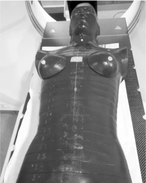
본 원 에 서 사 용 하 고 있 는 모 의 치 료 촬 영 장 치 인 CT simulator(BrightSpeed Elite, GE, USA)를 이용하여 인체모 형팬톰(Anthropomorphic phantom)을 스캔하였다.(Fig.
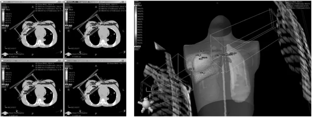
1). 획득한CT 영상을 이용하여 가상의 임상표적체적 (clinical target volume, CTV)을 설정하고 전산화치료계획 시스템 (Eclipse, version 8.6, Varian, USA)에서 Open beam을 이용한 치료계획, 쐐기필터를 이용한 technique, Field-in-Field technique, Dose Fluence를 이용한 irregular compensator technique을 계획하였다.(Fig.2)
전산화치료계획상 유방의 관심선량 지점을 표시하여 일 일선량 200 cGy로 포인트선량(Point dose)을 계산하였다.
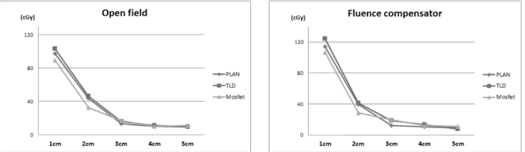
측정점은 중심점(Normalization point)을 기준으로 상방 6 cm, 하방 6 cm, 내측 8 cm, 외측 4.5 cm에 위치하였고, 반 대편 Breast의 측정점은 치료중심점에서 1 cm, 2 cm, 3 cm, 4 cm, 5 cm지점에 표시하여 선량을 계산하였다(Fig. 3).
CL-iX(Varian medical system, USA) 6MV 광자선으로 각 각의 치료계획에 따라 치료계획 선량을 조사하였고 열형광 선량계와 MOSFET를 이용하여 관심선량 지점에서의 선량 을 비교, 평가하였다.(Fig. 4)
Fig 1. The image of CT scan for Anthropomorphic phantom
결 과
전산화치료계획시 유방의 측정점 선량을 측정한 결과, 중 심점(Normalization point)을 기준으로 상방과 하방의 부분 에서는 각각 8.06%, 8.7%로 높게 나타났고, 이는 Dose fluence를 이용한 irregular compensation technique 치료 계획 시 MOSFET으로 측정하였을 때 가장 높은 선량이 측 정되었다.(Fig. 5) 중심점에서 내측과 외측의 측정선량을 비 교했을 경우, 특히 Open beam을 이용한 technique과 쐐기 필터를 이용한 치료계획에서 TLD와 MOSFET을 이용하여 측정하였을 때 작게는 5.7%에서 크게는 10.3%까지 낮게 측 정되었다. (Fig. 6)
반 대 쪽 유 방 의 측 정 선 량 은 Open beam을 이 용 한 technique의 경우에서 치료중심에서 1cm단위로 멀어질수 록 각각 97.4cGy, 43.5cGy, 12.8cGy, 9.7cGy, 9cGy로 가장 적은 선량이 들어감을 알 수 있었고, Dose Fluence를 이용 한 irregular compensation technique에 서 는 각 각 114.3cGy, 39.7cGy, 12.4cGy, 10.4cGy, 9.6cGy로 가장 많 은 선량이 들어감을 알 수 있었다. (Fig. 7) 치료종별 선량은 전산화치료계획시스템을 이용한 치료계획상에서는 내측 75.5cGy과 외측 62.3cGy의 편차가 가장 크게 나타났으며, 실제 TLD(내측 98.93cGy, 외측 81.69cGy)와 MOSFET(내측 100.2cGy, 외측 79cGy) 을 이용한 측정에서도 비슷한 경향 을 보였다. 외측은 TLD, 내측부분의 선량은 MOSFET이 가 Fig 2. Computed planning of Breast
Fig 3. Interest points at RT plan
(A) (B) (C)
Fig 4. Dose measurement of MOSFET(a) and TLD(b) at interest point contralateral breast dose measurement of MOSFET at interest point(c)
Fig 5. Measured dose of RT plan(upper/lower point)
Fig 6. Measured dose of RT plan(inner/lower point)
장 편차가 큰 것으로 측정되었다. (Fig. 8)
결 론
유방암의 방사선치료계획 시 다양한 방법의 3차원 입체 조형치료계획 뿐만 아니라 치료기법의 발전에 따라 세기조 절방사선치료 등을 통하여 최적의 선량분포를 얻을 수 있
다. 치료부위에 정확한 선량을 충분히 전달하고 확인하기 위해서는 여러 선량측정이 필요하게 되는데, 이를 위해 TLD와 MOSFET을 이용하여 방사선 선량측정의 비교 및 평 가를 하고 있다.
본 실험에서 치료계획에 따른 피부선량은 피부의 위치에 따라 약간의 차이는 있으나 전반적으로 Dose fluence를 이 용한 irregular compensation technique 치료계획 시에 가 장 높게 측정되었다. 이는 MLC leaf의 움직임이 많으므로 이에 따른 산란선의 영향으로 생각된다. 또한 Dose fluence 를 이용한 irregular compensation technique 치료계획 시 상방과 하방 부분의 피부선량에 상대적으로 더 높은 선량이 측정되고 내측이나 외측에는 낮게 측정되는 것으로 나타나 치료계획 시 내측의 내유임파절(Intramammary lymph nodes)의 부분이나 외측부분의 Scar 또는 Drain site에 부족 선량(under dose)이 생기지 않도록 주의하여야 한다. 반대 쪽 유방에 들어가는 선량은 MLC leaf 움직임에 의한 영향이 Fig 7. Measured dose of opposite breast plan(Open/Fluence)
Fig 8. Measured dose at Open field plan
로 치료계획전 반드시 환자의 병력을 숙지하여야하며 특히 수술부위가 양성(positive)으로 판명된 환자는 Scar, Drain site의 선량을 높이기 위해서는 Dose fluence를 이용한 irregular compensation technique 방식의 치료계획이 사용 되는 것이 좋을 것으로 사료된다. 그러나 전체적인 피부선 량을 높이기보다는 선택적인 범위 내에서 선량을 높이게 되 므로 환자의 연령이나 움직임 등을 고려하여 치료계획기술 을 선택하는 것이 바람직할 것으로 사료된다.
이번 연구에서는 세기조절방사선치료의 표면선량측정은 제외하였지만 추후 유방의 고정용구(optimold)를 사용하는 세기조절방사선치료에서의 선량측정과 본 연구에서 측정 된 데이터를 비교해본다면 유의미할 것으로 생각된다.
참고문헌
1. Jemal A, Siegel R, Ward E, Hao Y, Xu j, Thun MJ.
Cancer statistics 2008. CA Cancer j Clin 2008;58:71- 96
2. Holland R, Veling SHJ, Mravunac M, Hendriks JHCL.
Histologic multifocality of Tis, T1-2 breast carcinomas: implications for clinic trialsl of breast- conserving surgery. Cancer 1985;56:979-90
3. Brooks, P.S. Dose to contralateral breast: A comparative study. Med. Dosim. 20:301-7; 1995 4. Warlic, W.B.; O’Rear, J.H; Early, L.; et al. Dose to
the contralateral breast: A comparison of two techniques using the enhanced dynamic wedge versus a standard wedge. Med. Dosim. 22:185-91;
1997
5. Fairbanks, E. J.; DeWerd, L. A. Thermoluminescent characteristics of LiF:Mg, Ti from three
and analysis system for MOSFET radiation detectors.
Med Phys 1991;18;542-8
9. Hughes RC, Huffman D, Snelling JV, Zipperian JE, Ricco AJ, Kelsey A. Miniature radiation dosimeter for in-vivo radiation measurements. Int J Radiat Oncol Biol Phys 1988;14;963-7
10. Ramani R, Russell S, O’Brien P. Clinical dosimetry using MOSFET’s. Int J Radiat Oncol Biol Phys 1997;37(4);959-64
Purpose :The measurement of skin dose is very important that treatment of breast cancer. On account of the cold or hot dose as compared with prescription dose, it is necessary to analyse the skin dose occurring during the various plan of the breast cancer treatment. At our hospital, we want to apply various analyses using a diversity of dosimeters to the breast cancer treatment.
Subjectss and Methods : In the study, the anthropomorphic phantom is used to find out the dose difference of the skin(draining site), scar and others occurring from the tangential treatment plan of breast cancer. We took computed tomography scan of the anthropomorphic phantom and made plans for the treatment planing using open and wedge, Field-in- Field, Dose fluence. Using these, we made a comparative analysis of the dose date points by using the Eclipse. For the dose comparison, we place the anthropomorphic phantom in the treatment room and compared the measurement results by using the TLD and MOSFET on the dose data points.
Results :On the central point of treatment planing basis, the upward and downward skin dose measured by the MOSFET was the highest when the fluence was used. The skin dose of inner and outer was distinguished from the figure(5.7% ~ 10.3%) when the measurements were fulfilled by using TLD and MOSFET. The other side of breast dose was the lowest in the open beam, on the other hand, is highest in the Dose fluence plan. In the different kinds of treatment, the dose deviation of inner and outer was the highest, and so this was the same with the TLD and MOSFET measurement case. The outer deviation was highest in the TLD, and the Inner’was highest in the MOSFET.
Conclusion :Skin dose in relation to the treatment plan was the highest in the planing using the fluence technique in general and it was supposed that the high dose had been caused by the movement of the MLC. There’s some differences among the all the treatment planning, but the sites such as IM node occurring the lack of dose, scar, drain site are needed pay close attention. Using the treatment planning of dose fluence is good to compensate the lack of dose, but It increases the dose of the selective range rather than the overall dose. Therefore, choosing the radiotherapy technique is desirable in the lights of the age and performance of the patient.
Kim seon myeong, Kim young bum, Bak sang yun, Lee sang rok, Jeong se young
Evaluation of the Breast plan using the TLD and Mosfet for the skin dose
Department of Radiation Oncology, Korea University Ansan Hospital
Abstract
Keyword :Mosfet(Metal oxide-silicon field effect transistors), TLD(Thermoluminescence dosimeter), Field-in-Field technique, Dose Fluence.