1. Introduction
Samples for rare cell capture experiments, such as circulating tumor cell (CTC) capture research, may not
be perfectly prepared by current methods because of cell loss during collecting or transferring[1,2]. The number of target cells (5 to 10 CTCs per 7.5 mL for practical test) should be exact to accurately assess the
* This work was supported by Kyungnam University Foundation Grant, 2015.
*Corresponding Author : Taehyun Park ([email protected]) Received February 14, 2018
Accepted March 20, 2018
Revised March 2, 2018 Published March 28, 2018
Fabrication and Application of Micro Polymer Chip Platform for Rare Cell Sample Preparation
Taehyun Park
School of Mechanical Engineering, Kyungnam University
희귀 세포 샘플 준비를 위한 마이크로 폴리머 칩 플랫폼 제작 및 활용
박태현
경남대학교 기계공학부
Abstract In this paper, a new micro polymer chip platform and protocol were developed for rare cell sample preparation. The proposed platform and protocol overcome the current limitation of the dilution method which is based on statistics and the FACS method which expensive and requires fluorescence staining. It allows collecting exact number of target cells simply and selectively because the cells are visually confirmed during the collecting process. The collected cells can be transported or spiked into a desired locations, such as a microchamber, without cell loss. This research may applicable not only to a rare cell sample preparation for Lab on a Chip cancer diagnosis, but also to a single/double/multiple cell sample preparation for a cell analysis field. To verify this platform and protocol, five human breast cancer cells (MCF-7) were collected and transported into a hemocytometer chamber.
Key Words : Cell Collection, Micro Fabrication, Micro Polymer Chip, Rare Cell Sample, Sample Preparation
요 약 본 논문에서는 정확한 수의 희귀 세포 포집 및 이송을 위한 마이크로 폴리머 칩 플랫폼의 디자인과 제작, 그리고
프로토콜을 소개하고 있다. 본 플랫폼과 프로토콜은 기존의 통계학적인 샘플 준비 방법인 희석(Dilution)의 한계와 고가이 며 형광염색이 요구되는 유세포분석기(Fluorescence activated cell sorter)의 단점을 극복하였다. 타켓 세포를 선택적으로 쉽고 간단하게 채집할 수 있으며 채집되는 세포의 수는 시각적으로 검증되므로 매우 정확한 방법이다. 또한, 채집된 세포들 은 마이크로 챔버 등의 원하는 곳으로 세포의 손실 없이 이송 또는 주입 시킬 수 있다. 본 연구는 암진단 등을 목적으로 하는 칩 속의 실험실(Lab on a chip) 등에 필요한 희귀 세포 샘플 준비를 위해 활용 될 수 있을 뿐만 아니라 세포분석을 위한 싱글/더블/다수 세포 샘플의 준비에도 활용 가능하다. 본 논문에서 제시하는 세포 채집 플랫폼과 프로토콜을 검증하기 위해 5개의 인간 암세포(MCF-7)를 채집한 뒤 세포계수기(Hemocytometer) 안으로 주입시켜 세포의 수를 확인하였다.
주제어 : 희귀 세포 샘플 준비, 마이크로 폴리머 칩, 세포 채집, 샘플 준비, 미세 공정
effectiveness of rare cell capture test. If there is a one cell lost or add during the sample preparation procedures, cell collection and transportation, it effects around 20% error that may not acceptable for clinical application[3-5]. Also, in cell research, it is required a single or a pair of cells to study cell properties and communication between cells[6-8].
For clinical diagnostic applications, cells are cultured in large-scale environments by conventional methods with culture dish or microwell existing with contacting with other neighboring cells. In microbiology, the cell information on the response to environment and the interaction with other cells has traditionally been derived by the use of population-level data under assuming the cell samples are identical. However, cells are known to differ between individuals due to their environment such as temperature distribution or irregularity[9].
In contrast to the population-based analysis, the single cell analysis provides insights into the properties of chemical pathways and mechanisms between cellular biochemistry and is able to precisely understand differences between individual cells[10]. The application of single cell technologies to stem cell or rare cancer cell research, for example, will enable to understand how a single stem or cancer cell can give rise to a particular lineage of cells. Therefore, single or multiple cell sample preparation is an essential technology for cell analysis, rare cell research, or stem cell study[11].
However, a suitable protocol for the collection of multiple (five to ten) cells, which is important to test circulating tumor cell capture devices, has not yet been introduced.
Commercial Cell Counter or Fluorescence Activated Cell Sorter may possible to collect single or multiple rare cells. But they require expensive instruments and fluorescence labeling procedures which affect cell properties. Moreover, transportation of the collected cells in a tube or vial to a desired location, usually in a microchannel or microchamber can lead to cell loss during the delivery process.
For single-cell diagnosis, microfluidic devices have been widely used, which enables to isolate cells from a group of cells in cell suspension inside the microchannels[12-15]. The employment of microfluidic devices is beneficial to accurate cell capture or trapping, but it is limited to a specific application only.
Protocols for the isolation of cells from colonies were introduced, but not suitable for microfluidic applications due to the complicated and lengthy process[16,17]. A method for counting small number of cells was introduced by Badders[18]. However, it was still not suitable for less than 1000 cells/mL. Single cell separation methods were introduced in previous research, but were only for a specific application with a complicated device required for each use[19].
In this paper, we present the design of a micro polymer chip platform and introduce an easy and simple protocol for collection of exact numbers of multiple cells using an optical microscope and capillaries.
2. Platform Design and Fabrication
In order to collect multiple cell using an optical microscope, the depth of chamber for cell collection
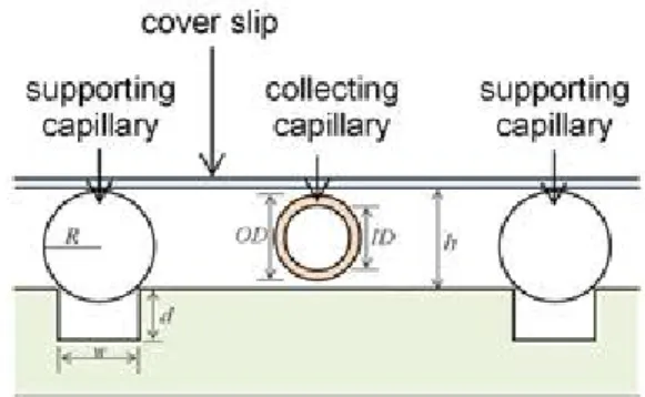
Fig. 1. Schematic of micro-milled PMMA cell collection platform. R: radius of supporting capillary, W:
width of adjusting groove, d: depth of adjusting groove, h: depth of collecting well chamber, ID: inner diameter of collecting capillary, OD: outer diameter of collecting capillary.
should not be deep due to the short focal length of optical microscope, but be deep enough for collecting capillary to move. Fig. 1 shows the schematic of micro-milled PMMA cell collecting chip platform. As shown in Fig. 1, the platform consists of a plate with micro-milled grooves which can be adjusted with any combination of width or depth of grooves and different diameter capillaries, a cover slip and two capillaries;
one for supporting the cover slip and another for collecting cells. The inner diameter of the collecting capillary is larger than the diameter of targeting cells and the outer diameter of the capillary is smaller than the chamber depth to free to move.
The developed technique is to use the visible silica capillary for capturing single or precise number of multiple cell collection. Polymethylmethacrylate (PMMA) plate, transparent and biocompatible, was
employed as cell collection chips and directly milled in micron-scale. Depth of the cell collection well can be adjusted using different diameter capillaries for different sizes of target cell or width of micro-milled grooves.
Micro milling on the PMMA was achieved using a
(a) (b)
(c) (d)
Fig. 2. PMMA cell collection chips with pairs of supporting capillaries in the micro milled grooves to support a cover glass, a cell collection capillary is inserted and movable under the cover glass. (a) Single well cell collection chip, (b) multi well cell collection chip, (c) cover glass supporting capillary area, and (d) cell collecting capillary area.
(a)
(b)
(c)
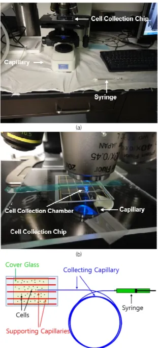
Fig. 3. (a) Pictures of the cell collecting platform on a microscope stage, (b) Picture of the cell collection chip, cell collection chamber, and polyimide removed cell collection capillary under a microscope, and (c) Illustration of the simple cell collection platform.
micromilling machine (MMP, KERN Micro- und Feinwerktechnik GmbH & Co. KG, Eschenlohe, Germany) at Louisiana State University, USA. For micro-milling, a rotational speed was at 30,000 rpm and a feed rate was of 350 mm/min. For finishing, the RPM was of 40,000 and the feed rate of 150 mm/min.
For small width of adjusting groove (W < 2R), the chamber depth is determined by.
and for ≥ ,
Here, h is the depth of chamber, R is the radius of the supporting capillaries, W is the width of adjusting grooves, d is the depth of adjusting grooves, ID is inner diameter of collection capillary and OD is outer diameter of collection capillary.
A single-well cell collection chip and a multi-well cell collection chip are shown in Fig. 2(a) and Fig. 2(b).
The multi well cell collection chip minimized the cover gass bending that occurred due to the fixing tapes on the cover glass, and it offered the number of cell collection wells. Supporting capillaries of 363 μm outer diameter (Polyimide Coated Fused Silica Capillary
#TSP075375, Molex, Lisle, IL, USA) are located in a micro-milled groove to support a cover slip and create a chamber with 168 μm depth shown in Fig. 2(c). A cell collecting capillary of 164 μm of outer diameter (Polyimide Coated Fused Silica Capillary #TSP100170, Molex, Lisle, IL, USA) is located in the chamber shown in Fig. 2(d). The polyimide removed cell collecting capillary can be movable in the chamber because the outer dimeter of the cell collecting capillary decreased to 140 μm.
3. Preparation and Cell Collection
The cell collecting capillaries are polyimide coated silica fused capillary tubes and the inside of the capillary is not clearly visible. The polyimide at the end
of the collecting capillary tubes was removed by flaking off the polyimide over a 600°C flame. Cells are then visible in the collecting capillary and the collecting capillary is more movable in the cell collection chip.
The other end of the collecting capillary are connected to a micro syringe. Fig. 3(a) and Fig. 3(b) show pictures of cell collecting process under a microscope.
Fig. 3(c) shows a simplified cell collection platform.
For precise number of cell collection, a low concentrations cell solution, approximately 103 ~ 104 cells/mL, is injected into the cell collection chip. The polyimide removed collecting capillary is inserted into the well under a microscope. Once the collecting capillary is located near a target cell, the syringe is withdrawn to enable collection of the cell in the collecting capillary. The number of collected cells are visually confirmed during the collection process. The collected cells are temporary stored in the collecting capillary to be transferred to a target location later.
After collection of the target cells, the remaining cells in the chamber and collecting capillary surfaces are washed out several times with a phosphate buffered saline (PBS, pH 7.4). Then, the collected cells in the collecting capillary can be easily and safely transferred or spiked to a desired location, such as a microdevice, microchamber, or micro vial, for a further processing without any cell loss during the transfer process. Prior to applying cells, the cell collection platform and cell collecting capillary are treated with 4% bovine serum albumin(BSA) over 1 hour to avoid nonspecific binding of cells on the PMMA or capillary surfaces during cell collection process. The end of the cell collection capillary should be cut neatly and clearly to avoid cell trap at the edge of the capillary during the collection process.
4. Results and Discussion
Fig. 4 shows the collection procedure of MCF-7 cells in a 168 μm deep cell collection chip using a polyimide
removed cell collecting silica capillary (inner diameter:
100 μm) connected with a 10 μL syringe. Once the polyimide removed cell collection capillary located near the target cell, the syringe was pulled to draw to the target cell in the capillary. The cell moved slowly at the outside of the capillary shown in Fig. 4 (ⅰ), (ⅱ), and (ⅲ). After entering the capillary, the cell flow rapidly into the capillary shown in Fig. 4 (ⅳ), (ⅴ), and (ⅵ).
It is clearly visible and countable the collecting cell in the polyimide removed capillary.
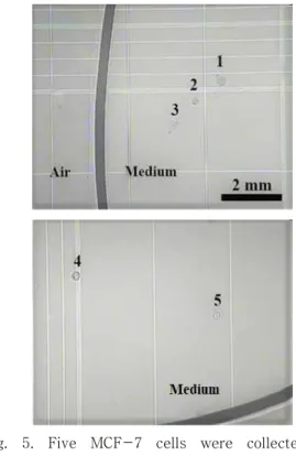
To confirm the protocol, five MCF-7 cells are collected in the cell collecting capillary and safely transported into a hemocytometer. All five collected MCF-7 cells successfully delivered without cell loss or cell trap during the process. Fig. 5 shows the transported five MCF-7 cells in the hemocytometer. It takes less than a minute to collect multiple cells.
5. Conclusions
A simple protocol for single or precise number of multiple target cell collection was introduced. This protocols can collect the exact number of target cells and deliver to a desire location.
The protocol can be used for any single or multiple number of cell manipulation, collection, and transportation for various application.
This protocol is especially suitable for a sample preparation of several target cells for testing a sensitivity or accuracy of cell capture devices.
To collect different size of target cells, the chamber depth can be adjusted with a different combination of the grooves on the cell collection polymer chip and the supporting capillaries.
Further study of automated single or multiple cell manipulation systems based on this platform and protocol will offer a new tools for rare cell or stem cell research fields.
Fig. 4. Microscopic pictures of cell collection using a cell collecting capillary inserted under the cover glass. A 100 μm of inner diameter of polyimide removed cell collecting capillary is inserted in a cell collection chip and located near a target cell and collect the target cell, MCF-7 cell in a cell collection chamber.
Fig. 5. Five MCF-7 cells were collected and delivered into a hemocytometer to confirm the process without cell loss or cell trap.
REFERENCES
[1] L. Zhu, X. L. Peh, H. M. Ji, C. Y. Teo, H. H. Feng, &
W. T. Liu. (2007). Cell loss in integrated microfluidic device,Biomedical Microdevices, 9(5), 745-750.
DOI : 10.1007/s10544-007-9085-z
[2] Y. Kim et al. (2007). Novel platform for minimizing cell loss on separation process: Droplet-based magnetically activated cell separator, Review of Scientific Instruments, 78(7), 074301.
DOI : 10.1063/1.2751414
[3] A. L. Allan & M. Keeney. (2010). Circulating tumor cell analysis: Technical and statistical considerations for application to the clinic,Journal of Oncology, 2010.
DOI : 10.1155/2010/426218
[4] A. G. J. Tibbe, M. C. Miller &, L. W. Terstappen. (2007).
Statistical considerations for enumeration of circulating tumor cells, Cytometry Part A, 71(3), 154-162.
DOI : 10.1002/cyto.a.20369
[5] M. Shackleton. (2010). Normal stem cells and cancer stem cells: Similar and different, Seminars in Cancer Biology, 20(2), 85-92.
DOI : 10.1016/j.semcancer.2010.04.002
[6] F. Guo et al. (2013). Probing cell-cell communication with microfluidic devices, Lab on a Chip, 13(16), 3152-3162.
DOI : 10.1039/c3lc90067c
[7] J. P. Frimat et al. (2011). A microfluidic array with cellular valving for single cell co-culture,Lab on a Chip, 11(2),231-237.
DOI : 10.1039/c0lc00172d
[8] A. Gross, J. Schoendube, S. Zimmermann, M. Steeb, R.
Zengerle, & P. Koltay. (2015). Technologies for single-cell isolation,International Journal of Molecular Sciences, 16(8), 16897-16919.
DOI : 10.3390/ijms160816897
[9] J. Y. Park, S. Takayama, & S. H. Lee. (2010). Regulating microenvironmental stimuli for stem cells and cancer cells using microsystems, Integrative Biology, 2(5-6), 229-240.
DOI : 10.1039/c000442a
[10] K. Eyer, P. Kuhn, C. Hanke, & P. S. Dittrich. (2012). A microchamber array for single cell isolation and analysis of intracellular biomolecules, Lab on a Chip, 12(4), 765-772.
DOI : 10.1039/c2lc20876h
[11] B. F. Brehm-Stecher & E. A. Johnson. (2004).
Single-cell microbiology: Tools, technologies, and
applications, Microbiology and Molecular Biology Reviews, 68(3), 538-559.
DOI : 10.1128/MMBR.68.3.538-559.2004
[12] C. Liberal et al. (2013). Integrated microfluidic device for single-cell trapping and spectroscopy,Scientific reports, 3, 1258.
DOI : 10.1038/srep01258
[13] A. R. Wheeler et al. (2003). Microfluidic device for single-cell analysis, Analytical Chemistry, 75(14), 3581-3586.
DOI : 10.1021/ac0340758
[14] C. H. Lin et al. (2015). A microfluidic dual-well device for high-throughput single-cell capture and culture,Lab on a Chip, 15(14), 2928-2938.
DOI : 10.1039/c5lc00541h
[15] T. Gerhardt, S. Woo, & H. Ma. (2011). Chromatographic behaviour of single cells in a microchannel with dynamic geometry, Lab on a Chip, 11(16), 2731-2737.
DOI : 10.1039/c1lc20092e
[16] L. G. Villa-Diaz et al. (2009). Microfluidic culture of single human embryonic stem cell colonies, Lab on a Chip, 9(12), 1749-1755.
DOI : 10.1039/b820380f
[17] H. Shadpour, J. S. Zawistowski, A. Herman, K. Hahn, &
N. L. Allbritton. (2011). Patterning pallet arrays for cell selection based on high-resolution measurements of fluorescent biosensors, Analytica Chimica Acta, 696(1-2), 101-107.
DOI : 10.1016/j.aca.2011.04.012
[18] N. M. Badders, C. M. Alexander, H. Yu, & D. J. Beebe.
(2008). Quantification of small cell numbers with a microchannel device, BioTechniques, 45(3), 321-325.
DOI : 10.2144/000112906
[19] D. D. Carlo, L. Y. Wu, & L. P. Lee. (2006). Dynamic single cell culture array, Lab on a Chip, 6(11), 1445-1449.
DOI : 10.1039/b605937f
박 태 현(Taehyun Park) [정회원]
▪2011년 5월 : 루이지애나 주립대학 교(미국) 기계공학과 (공학박사)
▪2012년 9월 ~ 현재 : 경남대학교 기계공학부 조교수
▪관심분야 : 마이크로 나노 폴리머, BioMEMS
▪E-Mail : [email protected]