Parkin Interacts with the PDZ Domain of Multi-PDZ Domain Protein MUPP1
Won Hee Jang
1, Young Joo Jeong
1, Sun Hee Choi
1, Won Hee Lee
2, Mooseong Kim
2, Sang-Jin Kim
3, Sang-Hwa Urm
4, Il Soo Moon
5and Dae-Hyun Seog
1*
1
Departments of Biochemistry and u-HARC, Inje University College of Medicine, Busan 614-735, Korea
2
Departments of Neurosurgery, Inje University College of Medicine, Busan 614-735, Korea
3
Departments of Neurology, Inje University College of Medicine, Busan 614-735, Korea
4
Departments of Preventive Medicine, Inje University College of Medicine, Busan 614-735, Korea
5
Departments of Anatomy & Dongguk Medical Institute, College of Medicine, Dongguk University, Gyeongju 780-714, Korea
Received June 23, 2014 /Revised August 14, 2014 /Accepted August 14, 2014The localization to specific subcellular sites and the regulation of cell surface receptors and channels are crucial for proper functioning. Postsynaptic density-95/Disks large/Zonula occludens-1 (PDZ)-domain is involved in recognition of and interaction between various proteins, by which the localization and the regulation are mediated. Multi-PDZ domain protein 1 (MUPP1) contains 13 PDZ domains. MUPP1 serves a scaffolding function for structure proteins and signaling proteins, but the mechanism how MUPP1 is stabilized and signalized has not yet been elucidated. We used the yeast two-hybrid system to identify proteins that interact with PDZ domains of MUPP1. We found an inter- action between MUPP1 and Parkin. Parkin is an E3 ubiquitin ligase. Loss-of-function mutations of Parkin gene are known to cause an autosomal recessive juvenile parkinsonism. Parkin bound to the 12
thPDZ domain, but not to other PDZ domains of MUPP1. The C-terminal end of Parkin has a type II PDZ-association motif, which was essential for the interaction with MUPP1 in the yeast two-hybrid assay. When co-expressed in HEK-293T cells, Parkin co-localized with MUPP1. When co-expressed with ubiquitin in HEK-293T cells, MUPP1 has been strongly ubiquitinated by Parkin. These findings collectively suggest that MUPP1 is a novel substrate of Parkin and its function or stability could be modulated by Parkin-mediated ubiquitination.
Key words : MUPP1, Parkin, PDZ domain, protein-protein interaction, ubiquitination
*Corresponding author
*Tel : +82-51-890-6974, Fax : +82-51-894-5801
*E-mail : [email protected]
This is an Open-Access article distributed under the terms of the Creative Commons Attribution Non-Commercial License (http://creativecommons.org/licenses/by-nc/3.0) which permits unrestricted non-commercial use, distribution, and reproduction in any medium, provided the original work is properly cited.
Journal of Life Science 2014 Vol. 24. No. 8. 820~826 DOI : http://dx.doi.org/10.5352/JLS.2014.24.8.820
Introduction
The localization of membrane receptors at specific sub- cellular site can be crucial for proper function. The cell-cell interactions, such as cell junctions and neuronal synapse are emerging as multimolecular composites whose structure and regulation are governed in part by their associated pro- teins [13]. PSD-95/Dlg/Zo-1 (PDZ)-domain containing pro- teins are modular proteins that act as adaptors, by linking the cell membrane receptors via PDZ domains or other pro- tein modules to cytoskeletal proteins or signaling proteins that include regulators of membrane trafficking, protein kinase and regulators of small GTPase, such as guanine ex-
change factors (GEFs) and GTPase activating proteins (GAPs) [9-11, 13, 22, 24].
PDZ domains are built of 80~100 amino-acid residues and specialized for binding of the carboxyl (C)-terminus in partner proteins, which are most often transmembrane re- ceptors and channel proteins [8, 22, 27]. Such interactions localize membrane proteins to specific subcellular domains, thus enabling assembly of large molecular complexes [24].
The role of PDZ domains in clustering and localization of proteins at the plasma membrane has important biological implications, e.g., in signaling, mediating the adhesive properties of particular cells, ion transport, and formation of the paracellular barriers also known as tight junctions [1, 10, 11, 13, 22].
Multi-PDZ domain protein 1 (MUPP1) was identified as
a protein that interacts with the C-terminus of the serotonin
5-hydroxytryptamine type 2C (5-HT
2C) receptor [29]. It is
highly expressed in the central nervous system, with high-
est levels in all cerebral cortical layers, the hippocampus,
the granular layer of the dentate gyrus, and the choroid
plexus, and enriched in synaptosomes, specifically in post-synaptic density (PSD) [4, 26]. MUPP1, which pos- sesses an L27 domain and 13 PDZ domains has been re- ported to interact with a variety of integral membrane pro- teins, including a synaptic adhesion molecule Cadm1, junc- tional adhesion molecule-A, a sodium channel Nav1.4, a melatonin receptor MT
1, Claudin-1, and γ-aminobutyric acid receptor 2 using different PDZ domains [1, 3, 4, 12, 14, 18].
MUPP1 might play a multifaceted role in assembling and localizing of the integral membrane proteins.
To help define the function of MUPP1, it is necessary to identify the interacting proteins of MUPP1. We screened for proteins that interact with the PDZ domains of MUPP1 through the yeast two-hybrid assay and identified the E3 ubiquitin ligase Parkin, the causal protein responsible for hereditary recessive early-onset parkinsonism [5, 19, 21, 25].
The MUPP1 and Parkin interaction suggests that MUPP1 may be a direct substrate for Parkin-mediated ubiquitination.
Materials and Methods Plasmid constructs
Full-length rat MUPP1 cDNA in the pCMV vector (a gift from Dr. H. Lubbert, Ruhr-Universitat, Denmark) was tag- ged with a FLAG-epitope at the amino (N)-terminus. Trun- cations of MUPP1 corresponding to different PDZ domains were prepared by PCR amplification using the appropriate primers. The amplified fragment was subcloned into T- vector. The fragment was then EcoRI-restricted and subcl- oned into the EcoRI site of pLexA. The correct orientation and in-frame cloning of cDNA inserts were verified by re- striction enzyme analysis and DNA sequencing. EGFP- fused Parkin were constructed and used to visualize the in- tracellular localization in mammalian cells. General re- combinant DNA techniques were performed according to standard protocol [23].
Screening of MUPP1-binding proteins by yeast two- hybrid assay
The Matchmaker LexA two-hybrid system was used for screening according to the manufacturer’s manual (Clon- tech, Palo Alto, CA, USA). In brief, a part of the rat MUPP1 cDNA was fused to the DNA-BD region of the pLexA vec- tor using the PCR and the plasmid DNA was transformed into yeast strain EGY48 carrying the p8op-lacZ gene.
Transformed EGY48 yeast cells containing the MUPP1 bait plasmid were transformed with the mouse brain cDNA li-
brary and grown on synthetic dextrose (SD) plates supple- mented with glucose but with no histidine, tryptophan, or uracil (SD/-His/-Trp/-Ura). The selection of positive clones was performed on an SD/-His/-Trp/-Ura/-Leu plate con- taining galactose, raffinose, X-gal, and BU salts. Plasmids from positive clones were analyzed by restriction digestion.
Unique inserts were sequenced and protein sequence analy- sis was performed with the BLAST algorithm at the National Center for Biotechnology Information (NCBI).
Sequence-verified clones were tested again for interaction with the bait in yeast by retransformation.
β -Galactosidase activity in liquid cultures of yeast The β-galactosidase activity of yeast was assayed as de- scribed previously [28]. Mid-log phase yeast cells were col- lected and permeabilized with 0.1% sodium dodecyl sul- phate (SDS) and chloroform. An excess amount of o-nitro- phenyl-β-D-galactoside (ONPG) was added to yeast lysate, and the mixture was incubated at 30℃, and then the re- action was stopped by increasing pH to 11 by the addition of 1 M Na
2CO
3. The formation of the reaction product, o-ni- trophenol, was determined by measuring absorbance at 420 nm on a spectrophotometer and normalizing for the re- action time. The units of enzyme activity were calculated by the following equation: units =1000× [(OD
420–1.75 × OD
550)] / (reaction time (min) × culture volume (ml) × OD
600) [2]. All experiments were independently performed at least three times.
Cell culture and Transfection
HEK-293T cells were cultured in Dulbecco's modified Eagle's medium supplemented with 10% fetal bovine se- rum, L-glutamine, and antibiotics. Transient transfections were done with the CaPO
4precipitation method.
Immunocytochemistry
Cells grown on poly-D-lysine-coated coverslips were transfected with EGFP-Parkin and MUPP1 constructs.
Twenty-four hours after transfection, cells were washed
with phosphate-buffered saline (PBS), fixed with 4% paraf-
ormaldehyde in PBS for 5 min, and permeabilized with
0.2% Triton X-100 in PBS for 10 min. After blocking with
5% normal goat serum in PBS for 30 min, cells were in-
cubated with anti-MUPP1 antibody (BD science, San Jose,
CA, USA) diluted 1:500 in PBS containing 1% bovine serum
albumin (BSA) and 0.05% Tween-20 overnight at 4℃. After
washing with PBS 3 times, cells were incubated with
A
B
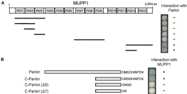
Fig. 1. Identification of the proteins interacting with MUPP1 by yeast two-hybrid screening. (A) Minimal Parkin binding region in MUPP1. Different truncations of MUPP1 were constructed by PCR. Several truncated forms of MUPP1 were tested in the yeast two-hybrid assay for interaction with Parkin. +, interaction with Parkin; -, no interaction with Parkin. PDZ domains are indicated in gray. aa, the amino acid residue number. (B) Specific interaction of MUPP1 with the C-terminus of Parkin.
Several deletion mutants of Parkin were tested in the yeast two-hybrid assay for interaction with MUPP1. +, interaction with MUPP1; -, no interaction with MUPP1.
Dylight 594-conjugated goat anti-rabbit IgG antibody (Jac- kson ImmunoResearch Labs, West Grove, PA, USA) diluted 1:800 for 40 min. After washing with PBS 3 times, the cells were mounted with Fluoromount (DAKO). Fluorescence images were acquired on Zeiss LSM510 META confocal la- ser scanning microscope (Carl Zeiss, Oberkochem, Germany).
Ubiquitination Assay
Twenty-four hours after transfection of myc-Parkin, HA- Ub, and MUPP1 constructs, cells were rinsed with PBS twice and lysed with SDS lysis buffer [PBS containing 1%
SDS and 1X protease inhibitor cocktail set V (Calbiochem, Darmstadt, Germany)]. Lysates were subjected to sonication and then boiled for 10 min. Lysates were centrifuged at 16,000× g for 10 min at room temperature. The supernatant was diluted 1:10 with PBS containing 0.5% NP-40 and in- cubated with anti-FLAG M2 agarose beads (Sigma-Aldrich, St. Louis, MO, USA) for 4 h at room temperature with con- stant rocking. The beads were collected by centrifugation at 2,000× g for 30 sec and washed 5 times with PBS containing 0.5% NP-40. The immunoprecipitated proteins were ana- lyzed by Western blotting.
Results
Identification of MUPP1 interacting proteins by yeast two-hybrid screening
To identify MUPP1-interacting proteins, we screened a mouse brain cDNA library through the yeast two-hybrid as- says using the C-terminal region of MUPP1 containing 12
thand 13
thPDZ domains as bait (Fig. 1A). From 8×10
6colonies screened, we obtained one positive clone. This clone pos- sessed a cDNA fragment of Parkin (Fig. 1B). MUPP1 con- tains 13 homologous protein binding PDZ domains (Fig.
1A) [29]. To determine the binding domain of MUPP1 that
is required for the interaction with Parkin, we constructed
various fragments of MUPP1. Yeast two-hybrid assays with
Parkin showed that the minimal domain required for bind-
ing was critically dependent on the 12
thPDZ domain of
MUPP1 (Fig. 1A). Parkin contains a type II PDZ-association
motif (φXφ), where φ is a hydrophobic residue, at its C-ter-
minus [8, 24]. Next we investigated whether the C-terminal
PDZ-association motif of Parkin mediates protein-protein
interaction. For this purpose, a series of C-terminal deletion
mutants of Parkin were constructed (Fig. 1B), and co-trans-
fected into yeast cells with pLexA-MUPP1. As shown in
Fig. 1B, the C-terminal deletion mutants of Parkin did not
interact with MUPP1. These result indicated that the inter-
A B
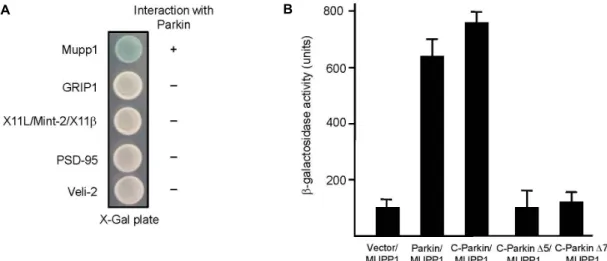
Fig. 2. Interaction between MUPP1 and Parkin. PDZ domain containing proteins were fused to the pLexA DNA binding domain.
(A) Parkin specifically interacted with MUPP1 but not with GRIP1, X11/Mint-2/X11β, PSD-95, or Veli-2. +, interaction with Parkin; -, no interaction with Parkin. (B) The strength of interactions between several deletion mutants of Parkin and MUPP1 were examined quantitatively using β-galactosidase activity in yeast two-hybrid reporter assay.
action between Parkin and MUPP1 is mediated through a PDZ-mediated interaction similar to the previously de- scribed type II PDZ interaction [8, 24].
To determine whether other PDZ domain containing pro- teins interact with Parkin, we tested four different PDZ do- main containing proteins using yeast two-hybrid assay. Fig.
2A shows that Parkin did not interact with other PDZ do- main containing proteins, glutamate receptor-interacting protein 1 (GRIP1), X11L/Mint-2, PSD-95, and Veli-2. A quantitative β-galactosidase assay showed that the C-termi- nus of Parkin is essential in binding MUPP1 and its dele- tion impairs the binding with MUPP1 (Fig. 2B). These data indicated a specific interaction between MUPP1 and Parkin through the 12
thPDZ of MUPP1 and the PDZ-association motif of Parkin.
MUPP1 colocalizes with Parkin in cells and is a substrate for Parkin-mediated ubiquitination
Conjugation of ubiquitin to protein requires the con- serted activity of three enzymes, an E1 ubiquitin activating enzyme, an E2 ubiquitin conjugating enzyme, and an E3 ubiquitin ligase. A previous report has shown that Parkin is an E3 ubiquitin ligase [30]. To determine whether Parkin ubiquitinates MUPP1, we carried out an ubiquitination as- say by co-transfection with FLAG-MUPP1, HA-ubiquitin, and myc-Parkin plasmids in HEK-293T cells. High molec- ular weight, HA-ubiquitinated proteins were observed in the presence of FLAG-MUPP1, HA-ubiquitin, and myc-
Parkin but not in control cultures lacking either myc-Parkin or HA-ubiquitin (Fig. 3A). This result indicates that MUPP1 is a direct substrate for Parkin-mediated ubiquitination.
For a potential interaction between Parkin and MUPP1 to be physiologically relevant, two proteins must co-localize at the same subcellular region in cells. To determine wheth- er Parkin and MUPP1 co-localize, we generated the N-ter- minal EGFP-fused Parkin construct. MUPP1 was co-trans- fected with EGFP-Parkin into HEK-293T cells, and vi- sualized by immunocytochemistry. Confocal microscopic images of EGFP-Parkin (green channel) and MUPP1 (red channel) showed that both proteins formed puncta along cytoplasmic membrane, and that the two proteins ex- tensively overlapped at the same subcellular region in cells (Fig. 3B). These findings indicate that MUPP1 and Parkin interact in cells.
Discussion
In this study, we have shown by yeast two-hybrid assay that Parkin associates with MUPP1 through the C-terminal region. We have further shown that Parkin and MUPP1 co- localizes in HEK-293T cells, and that MUPP1 is a direct sub- strate for Parkin-mediated ubiquitination.
Proteins containing PDZ domains usually form multi-
meric complexes [11, 22, 24]. PDZ domains contain a con-
served peptide-binding groove that associates with the ex-
treme C-terminus of ligands [8]. Interestingly, MUPP1 has
A
B
Fig. 3. Parkin-mediated ubiquitination of MUPP1 and co-localization of MUPP1 and Parkin in mammalian cells. (A) HEK-293T cells were transiently transfected with MUPP1 and HA-Ub plasmids and either control vector or Parkin plasmid as indicated.
Cell lysates prepared with SDS lysis buffer were incubated with anti-FLAG M2 agarose beads to immunoprecipitate MUPP1.
Western blots were subsequently probed with anti-HA antibody. MUPP1 was modified by ubiquitin. (B) Twenty-four hours after transfection with MUPP1 and EGFP-Parkin plasmids, HEK-293T cells were immunostained using anti-MUPP1 antibody.
MUPP1 and EGFP-Parkin are seen at the same subcellular region in cells.
13 PDZ domains that mediate interaction with the other proteins [29]. In this study, we demonstrated through do- main analysis that the 12
thPDZ domain of MUPP1 specifi- cally mediates the interaction with the C-terminal region of Parkin.
What would the association between MUPP1 and Parkin mean? A first possibility is the turn-over of MUPP1 by Parkin. Parkin functions as an E3 ubiquitin ligase [17, 25, 30]. The process of ubiquitination occurs through the trans- fer of an ubiquitin molecule from an activated E1 enzyme to the conjugating E2 enzyme [6, 15]. The E3 enzyme con- fers substrate specificity and acts as a scaffold for the co- valent attachment of ubiquitin. Our data showed that MUPP1 is a direct substrate for Parkin, indicating that Parkin may regulates MUPP1 by ubiquitination. Further study on this possibility may help to understand the patho- genesis of Parkinson’s disease.
A second possibility is subcellular targeting of Parkin to appropriate subcellular localization by MUPP1. Several PDZ proteins, such as GRIP1 and syntenin serve to act as targeting/scaffolding proteins that have potential to bring their interacting proteins to appropriate subcellular local- ization [16, 20]. Therefore, the association of Parkin with MUPP1 could target Parkin to specific subcellular location for appropriate functions. A thorough understanding of the factors that regulate Parkin may help to develop ther- apeutics for the treatment of Parkinson
’s disease. Our find- ings provide insight into the possible regulation of Parkin
by MUPP1 through PDZ-mediated interaction, and could provide a novel therapeutic target [7].
Acknowledgments
The research was supported by the Basic Science Research Program through the National Research Foundation of Korea (NRF) by the Ministry of Science, ICT and Future Planning (NRF-2012R1A1A2020689).
References
1. Adachi, M., Hamazaki, Y., Kobayashi, Y., Itoh, M., Tsukita, S., Furuse, M. and Tsukita, S. 2009. Similar and distinct properties of MUPP1 and Patj, two homologous PDZ do- main-containing tight-junction proteins.
Mol Cell Biol
29, 2372-2389.2. Ausubel, F. M., Brent, R., Kingston, R. E., Moore, D. D., Seidman, J. G., Smith, J. A. and Struhl, K. 1998.
Current Protocols in Molecular Biology
. John Wiley & Sons.3. Balasubramanian, S., Fam, S. R. and Hall, R. A. 2007.
GABAB receptor association with the PDZ Scaffold Mupp1 alters receptor stability and function.
J Biol Chem
282, 4162-4171.4. Becamel, C., Figge, A., Poliak, S., Dumuis, A., Peles, E., Bockaert, J., Lubbert, H. and Ullmer, C. 2001. Interaction of serotonin 5-hydroxytryptamine type 2C receptors with PDZ10 of the multi-PDZ domain protein MUPP1.
J Biol Chem
276, 12974-12982.5. Coelln, R., Valina, L. and Dawson, T. M. 2004. Parkin-asso- ciated Parkinson
’
s disease.Cell Tissue Res
318, 175-184.6. Dev, K. K., van der Putten, H., Sommer, B. and Rovelli, G. 2003. Part I: parkin-associated proteins and Parkinson's disease.
Neuropharmacology
45, 1-13.7. Dev, K. K. 2004. Making protein interactions druggable:
targeting PDZ domains.
Nat Rev Drug Discov
3, 1047-1056.8. Doyle, D. A., Lee, A., Lewis, J., Kim, E., Sheng, M. and MacKinnon, R. 1996. Crystal structures of a complexed and peptide-free membrane protein-binding domain: molecular basis of peptide recognition by PDZ.
Cell
85, 1067-1076.9. Field, C. M. and Kellogg, D. 1999. Septins: cytoskeletal pol- ymers or signaling GTPases?
Trends Cell Biol
9, 387-394.10. Garner, C. C., Nash, J. and Huganir, R. L. 2000. PDZ do- mains in synapse assembly and signalling.
Trends Cell Biol
10, 274-280.11. Gomperts, S. N. 1996. Clustering membrane proteins: It's all coming together with the PSD-95/SAP90 protein family.
Cell
84, 659-662.12. Guillaume, J. L., Daulat, A. M., Maurice, P., Levoye, A., Migaud, M., Brydon, L., Malpaux, B., Borg-Capra, C. and Jockers, R. 2008. The PDZ protein mupp1 promotes Gi cou- pling and signaling of the Mt1 melatonin receptor.
J Biol Chem
283, 16762-16771.13. Guillemot, L., Foglia, A., Paschoud, S., Pulimeno, P. and Citi, S. 2008. The cytoplasmic plaque of tight junctions: a scaffolding and signalling center.
Biochim Biophys Acta
1778, 601-613.14. Hamazaki, Y., Itoh, M., Sasaki, H., Furuse, M. and Tsukita, S. 2002. Multi-PDZ domain protein 1 (MUPP1) is con- centrated at tight junctions through its possible interaction with claudin-1 and junctional adhesion molecule.
J Biol Chem
277, 455-461.15. Hershko, A. and Ciechanover, A. 1992. The ubiquitin sys- tem for protein degradation.
Annu Rev Biochem
61, 761-807.16. Hirbec, H., Perestenko, O., Nishimune, A., Meyer, G., Nakanishi, S. and Henley, J. M. 2002. The PDZ proteins PICK1, GRIP and Syntenin bind multiple glutamate re- ceptor subtypes.
J Biol Chem
277, 15221-15224.17. Imai, Y., Soda, M. and Takahashi, R. 2000. Parkin sup- presses unfolded protein stress-induced cell death through its E3 ubiquitin-protein ligase activity.
J Biol Chem
275, 35661-35664.18. Kimber, W. A., Trinkle-Mulcahy, L., Cheung, P., Deak, M., Marsden, L. J. and Kieloch, A. 2002. Evidence that the tan- dem-pleckstrin-homology-domain-containing protein TAPP1 interacts with Ptd(3,4)P2 and the multi-PDZ-domain-con- taining protein MUPP1
in vivo
.Biochem J
361, 525-536.19. Kitada, T., Asakawa, S., Hattori, N., Matsumine, H., Yamamura, Y. and Minoshima, S. 1998. Mutations in the parkin gene cause autosomal recessive juvenile parkinson- ism.
Nature
392, 605-608.20. Leonoudakis, D., Conti, L. R., Radeke, C. M., McGuire, L.
M. and Vandenberg, C. A. 2004. A multiprotein trafficking complex composed of SAP97, CASK, Veli, and Mint1 is as- sociated with inward rectifier Kir2 potassium channels.
J Biol Chem
279, 19051-19063.21. Lucking, C. B., Durr, A., Bonifati, V., Vaughan, J., De Michele, G. and Gasser, T. 2000. Association between ear- ly-onset Parkinson’s disease and mutations in the parkin gene. French Parkinson’s Disease Genetics Study Group.
N Engl J Med
342, 1560-1567.22. Ponting, C. P., Phillips, C., Davies, K. E. and Blake, D. J.
1997. PDZ domains: targeting signalling molecules to sub- membranous sites.
Bioessays
19, 469-479.23. Sambrook, J., Fritsch, E. F. and Maniatis, T. 1989. Molecular cloning: a laboratory manual.
Cold Spring Habor Laboratory,
Cold Spring Habor, New York.24. Sheng, M. and Sala, C. 2001. PDZ domains and the organ- ization of supramolecular complexes.
Annu Rev Neurosci
24, 1-29.25. Shimura, H., Hattori, N., Kubo, S., Mizuno, Y., Asakawa, S. and Minoshima, S. 2000. Familial Parkinson disease gene product, parkin, is a ubiquitinprotein ligase.
Nat Genet
25, 302-305.26. Sitek, B., Poschmann, G., Schmidtke, K., Ullmer, C., Maskri, L. and Andriske, M. 2003. Expression of MUPP1 protein in mouse brain.
Brain Research
970, 178-187.27. Songyang, Z., Fanning, A. S., Fu, C., Xu, J., Marfatia, S. M.
and Chishti, A. H. 1997. Recognition of unique carbox- yl-terminal motifs by distinct PDZ domains.
Science
275, 73-77.28. Takeda, S., Yamazaki, H., Seog, D. H., Kanai, Y., Terada, S. and Hirokawa, N. 2000. Kinesin superfamily protein 3 (KIF3) motor transports fodrin-associating vesicles im- portant for neurite building.
J Cell Biol
148, 1255-1265.29. Ullmer, C., Schmuck, K., Figge, A. and Luëbbert, H. 1998.
Cloning and characterization of MUPP1, a novel PDZ do- main protein.
FEBS Lett
424, 63-68.30. Zhang, Y., Gao, J., Chung, K. K., Huang, H., Dawson, V.
L. and Dawson, T. M. 2000. Parkin functions as an E2- de- pendent ubiquitin-protein ligase and promotes the degra- dation of the synaptic vesicle associated protein, CDCrel-1.