Fermented Organic Matter as Possible Food for Rearing Anguilla japonica Leptocephali
Hyo-Won Kim1, Jung-Hyun Kim1, Myung-Hee Kim2, Kwang-Hyun Kim3, Jin-Chul Park4, Heum-Gi Park5, Chang-Hee Han2* and Dae-Jung Kim1*
1Jeju Fisheries Research Institute, National Institute of Fisheries Science, Jeju 63610, Korea
2Department of Molecular Biology, Dong-Eui University, Busan 47340, Korea
3Department of Life Science and Biotecnology, Dong-Eui University, Busan 47340, Korea
4East Costal Life Science Institute Gangneung-Wonju National University, Gangneung 25457, Korea
5Department of Marine Bioscience, Gangneung-Wonju National University, Gangneung 25457, Korea Received October 10, 2018 /Revised December 12, 2018 /Accepted December 13, 2018
We prepared flocculated detritus-like organic marine snow originating from various organisms by fer- mentation using microorganisms; this fermented organic material was fed to the leptocephali of the eel (Anguilla japonica) to investigate whether or not such organic matter was an appropriate food source for the larvae. A strain was isolated from a biofloc technology system used to culture fish, and seven types of organic material from hen’s egg, eel muscle, tuna muscle, lugworm, shrimp, manila clam, mussel, and sea squirt were fermented using isolated bacteria (Bacillus sp.). The fermented mat- ter did not show any specific form and was larger than 10-20 μm but no more than 100 μm in size.
Four diets (A-D) were prepared using the various fermented products, and the larvae were fed the prepared food from 20 days after hatching. The leptocephali fed the A, B, and C diets survived until 37, 39, and 37 days after hatching, respectively. However, the leptocephali fed the D diet survived for 60 days after hatching. The protein content of each diet was very similar, but the n-3 HUFA con- centration in the D diet was approximately twice as high as that of the others.
Key words : Anguilla japonica, biofloc, diet, eel, larval rearing
*Corresponding authors
Tel : +82-064-780-5420, Fax : +82-064-780-5409 E-mail : [email protected] (Dae-Jung Kim)
[email protected] (Chang-Hee Han)
This is an Open-Access article distributed under the terms of the Creative Commons Attribution Non-Commercial License (http://creativecommons.org/licenses/by-nc/3.0) which permits unrestricted non-commercial use, distribution, and reproduction in any medium, provided the original work is properly cited.
Journal of Life Science 2018 Vol. 28. No. 12. 1424~1431 DOI : https://doi.org/10.5352/JLS.2018.28.12.1424
서 론
뱀장어(Anguilla japonica) 종자생산을 위한 인위적인 수정 란 생산, 자어 사육 그리고 실뱀장어 생산에 대한 연구들은 한국을 비롯한 일본, 중국 등에서 수행되어지고있다[9, 10, 15, 25, 26, 33, 35]. Yamamoto [38]가 외래성 호르몬을 반복 투여 하여 뱀장어 자어를 최초로 생산하였으나 먹이가 밝혀지지 않아서 장기간 사육에는 실패하였다. 이후 Tanaka [34]가 최초 로 곱상어의 알을 주원료로 하는 슬러리(slurry) 형태의 사료 를 사용하여 부화후 250일에 실뱀장어(glass eel) 생산에 성공 하였고, 사료 첨가물의 개량 및 사육방법의 개선을 통해 실뱀 장어 생산기간을 부화후 131일까지 단축하였다[17]. 또한, 국 내에서도 곱상어 알을 주원료로하는 사료를 개발하였고[13], 개발된 사료로 실뱀장어 생산에 성공하였다[25]. 그러나 슬러
리 형태 사료의 주원료인 곱상어가 IUCN (International Union for Conservation of Nature and Natural Resources) Red List 의 취약종(Vulnerable; VU)으로 선정되어서[6], 최근에는 곱상 어 알을 사용하지 않는 사료에 대한 연구가 활발하게 이루어 지고 있다. Masuda [17]는 뱀상어(tiger shark, Galeocerdo cuv- ier)와 gulper 상어(Centrophorus atromarginatus)의 알을 주원료 로 이용한 사료를 자어에게 공급해서 부화 후 60일까지 사육 하였고, Okamura [27]는 계란을 주원료로 이용한 사료를 공급 해서 부화후 58일까지 사육하였다. 또한, 가수분해된 어분을 주원료로한 사료로 실뱀장어 생산까지 성공하였으나 기존의 곱상어 알을 이용한 사료보다 생존율과 성장률이 낮아 추가적 인 연구가 진행되고 있다[18, 19].
자연에서 뱀장어 자어들은 marine snow를 먹이로 이용한 다고 알려져 있다[20-23]. 그러나, 인공적으로 marine snow를 생산하는 기술은 개발되어있지 않아서 marine snow를 이용 한 뱀장어 자어사육 기술은 연구되지 않았다. Marine snow는 해양의 식물성 플랑크톤(phytoplankton)과 동물성 플랑크톤 (zooplankton)의 사체, 박테리아(bacteria) 그리고 미립자 유기 물(particulate organic matters; POM) 등이 합쳐진 일정한 형 태가 없는 유기물 분해산물의 복합체이다[14]. 해양에서는 다 양한 생명체의 사체들이 미생물들에 의해 분해된 미세한 분해 산물들이 소형 부유성 동물들의 중요한 먹이원이 되고 있으며
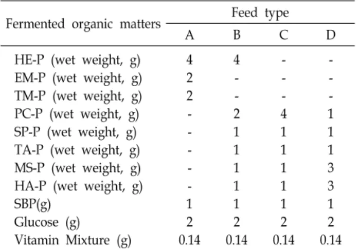
Table 1. Feed formulation table for investigating the survival effect of diets on leptocephalus larvae of eel (A. japon- ica) from day 20 after hatching
Fermented organic matters Feed type
A B C D
HE-P (wet weight, g) EM-P (wet weight, g) TM-P (wet weight, g) PC-P (wet weight, g) SP-P (wet weight, g) TA-P (wet weight, g) MS-P (wet weight, g) HA-P (wet weight, g) SBP(g)
Glucose (g) Vitamin Mixture (g)
4 2 2 - - - - - 1 2 0.14
4 - - 2 1 1 1 1 1 2 0.14
- - - 4 1 1 1 1 1 2 0.14
- - - 1 1 1 3 3 1 2 0.14 HE - hen egg, EM - eel’s muscle, TM - tuna’s muscle, PC - lugworm, SP - shrimp, TA - manila clam, MS - mytilus galloprovincialis, HA - sea squirt, SBP - soybean peptide
[1, 37], 유기물 분해산물 그 자체보다 거기에 함께 서식하는 미생물과 이들이 유기물을 분해하면서 만들어낸 미지의 많은 영양분과 유기물들을 이용한다고 알려져 있다[2]. 최근 BFT (biofloc technology) 양식 방법에서 언급되고 있는 biofloc 도 유기물을 분해하는 미생물과 미생물 산물 그리고 분해상태의 유기물들의 복합체로[3], biofloc과 marine snow는 유사한 구 조적 특징을 가진다고 보고되었다[36]. 따라서 본 연구에서는 BFT의 biofloc 으로부터 분리한 미생물과 해양유래 유기원들 을 이용하여 marine snow와 유사한 유기물 분해산물을 만들 고, 이를 뱀장어 자어에게 공급하여 생존 및 성장효과를 조사 하였다.
재료 및 방법
실험어
뱀장어 친어의 인위적인 성성숙 유도는 Kim [10]과 Kim [12]의 방법에 따라 실시하였으며 인위적으로 성숙된 뱀장어 로부터 얻은 수정란을 부화시켜 실험어로 이용하였다. 예비 실험에서 부화자어의 유구가 모두 흡수되고 입이 열리는 부화 후 7일부터 유기물 분해산물을 공급하였고 최대 19일까지 생 존하였지만 모두 폐사하였다(data not show). 본 연구에서는 부화된 자어를 부화후 7일부터 19일까지 기존의 곱상어 난황 을 주 원료로하는 액상사료[25]로 사육한 후 부화 20일차 자어 를 실험에 이용하였다.
유기물 분해산물의 생산
유기물 분해산물에 이용된 미생물은 동의대학교 생물생산 연구소의 BFT 사육수에서 분리된 타가영양균(heterotrophic bacteria)중에서 단백질 분해능력이 있는 바실러스속(Bacillus sp.) 균을 순수 분리하여 배양하였다. 유기원(organic source) 으로는 계란(hen egg), 참다랑어(Thunnus orientalis), 뱀장어, 담치(mytilus galloprovincialis), 갯지렁이(Perinereis aibuhitensis), 멍게(Halocynthia roretzi) 바지락(Ruditapes philippinarum) 그리 고 새우(Litopenaeus vannamei)의 근육을 이용하였다. 0.2 μm로 필터된 해수에 peptone 0.5%, yeast extracts 0.1%를 첨가하고 멸균(121℃, 15 min)하여 제조한 배지와 마쇄한 유기원을 2.5 : 1 (v/w) 비율로 잘 혼합한 후, 바실러스속(Bacillus sp.) 균주 를 2.2 × 10¹⁴ CFU/ml 농도로 배지 혼합액에 10%(v/v) 접종하 여 발효하였다. 발효기간동안 유기원이 침전되지 않도록 지속 적으로 섞어주었고 온도는 25℃로 유지하였다. 생산된 유기물 들의 분해 상태는 광학현미경(Nikon, Japan)으로 관찰하였고, 유기물들의 크기는 현미경프로그램(NIS-elements, Nikon, Japan)을 사용하여 측정하였다. 유기물의 크기가 100 μm 이하 로 분해될 때까지 발효하였으며, 이때 만들어진 유기물 분해 산물은 원심분리(10,000 rpm, 4℃, 15 min)하여 침전물을 실험 사료의 원료로 사용하였다.
자어 사료 제조 및 영양분석
생산한 유기물 분해산물은 망목이 100 μm인 체(sieve)로 거른 후 homogenizer로 분쇄하여 Table 1의 조성에 따라 사용 하였다. 다양한 유기물 분해산물들을 일정비율로 조합하여 사 료를 제조하였으며, 일정량의 glucose, vitamin mixture를 첨 가하였다. 유기물 분해산물로 제조한 사료의 지방산과 아미노 산 분석은 gas chromatography (HP6890 plus, Agilent, USA) 와 아미노산 분석기(L-8800, Hitachi, Japan)를 이용하여 분석 하였다[27].
자어 사육 실험
부화 7일차 자어를 20 l 유수식 아크릴 사육수조에서 부화후 19일까지 기존 곱상어 알을 주원료로 제조한 액상사료를 이용 하여 사육하였으며, 이때 자어의 전장은 8,692±306 μm이었다.
이후 부화 20일차 자어를 20 l 유수식 사육수조에 각 150마리 씩 수용하여 실험하였으며, 실험기간동안 자어의 사육은 Kim [11]와 Lee [25]의 방법에 따라 진행하였다. 대조구는 곱상어 알이 주원료인 기존 액상사료를 공급하였고, 실험구는 사료에 대한 적응을 위해 실험시작 후 3일 동안(부화후 20일~22일) 제조한 실험 사료(Table 1)와 곱상어 알이 주원료인 액상 사료 의 비율을 1:3, 1:1 그리고 3:1로 단계적으로 전환시켜 공급하였 으며 부화후 23일부터 실험 사료만을 공급하여 사육하였다.
액상 사료 및 실험 사료는 오전 9시부터 오후 5시까지 2시간 간격으로 1일 5회에 걸쳐 1회당 5 ml 씩 공급하였다.
매일 마지막 사료 공급 후 자어들을 새로운 수조로 옮겨주 었으며, 이때 생존개체수를 계수하여 생존율을 조사하였다.
자어의 사료 섭취상태는 광학현미경으로 관찰하였으며 자어 의 전장은 현미경프로그램을 이용하여 측정하였다.
Fig. 1. Artificial organic decomposition products formed by fer- mented organic particles by microbial strain Bacillus sp.
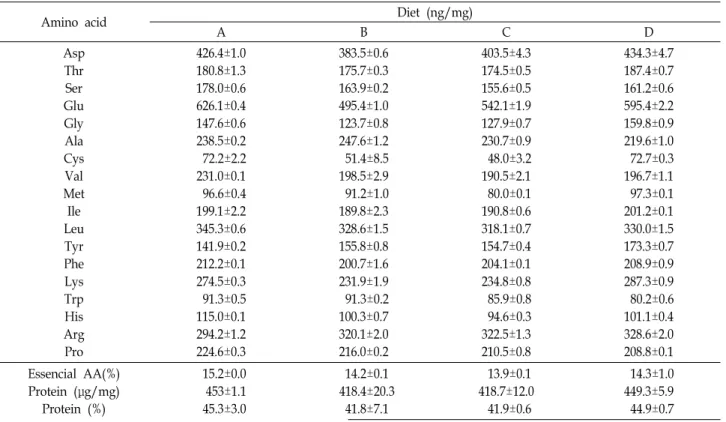
Table 2. Amino acid content and protein concentration of each diet listed in Table 1
Amino acid Diet (ng/mg)
A B C D
Asp Thr Ser Glu Gly Ala Cys Val Met Ile Leu Tyr Phe Lys Trp His Arg Pro
426.4±1.0 180.8±1.3 178.0±0.6 626.1±0.4 147.6±0.6 238.5±0.2 72.2±2.2 231.0±0.1 96.6±0.4 199.1±2.2 345.3±0.6 141.9±0.2 212.2±0.1 274.5±0.3 91.3±0.5 115.0±0.1 294.2±1.2 224.6±0.3
383.5±0.6 175.7±0.3 163.9±0.2 495.4±1.0 123.7±0.8 247.6±1.2 51.4±8.5 198.5±2.9 91.2±1.0 189.8±2.3 328.6±1.5 155.8±0.8 200.7±1.6 231.9±1.9 91.3±0.2 100.3±0.7 320.1±2.0 216.0±0.2
403.5±4.3 174.5±0.5 155.6±0.5 542.1±1.9 127.9±0.7 230.7±0.9 48.0±3.2 190.5±2.1 80.0±0.1 190.8±0.6 318.1±0.7 154.7±0.4 204.1±0.1 234.8±0.8 85.9±0.8 94.6±0.3 322.5±1.3 210.5±0.8
434.3±4.7 187.4±0.7 161.2±0.6 595.4±2.2 159.8±0.9 219.6±1.0 72.7±0.3 196.7±1.1 97.3±0.1 201.2±0.1 330.0±1.5 173.3±0.7 208.9±0.9 287.3±0.9 80.2±0.6 101.1±0.4 328.6±2.0 208.8±0.1 Essencial AA(%)
Protein (μg/mg) Protein (%)
15.2±0.0 453±1.1 45.3±3.0
14.2±0.1 418.4±20.3
41.8±7.1
13.9±0.1 418.7±12.0
41.9±0.6
14.3±1.0 449.3±5.9 44.9±0.7 통계처리
통계처리는 SPSS 프로그램(version 19.0)을 이용하여 Du- can’s new multiple range test에 의해서 유의성 검정을 실시하 였으며(p<0.05), 결과값은 mean±SEM으로 나타내었다.
결 과
미생물 분리 및 유기물 분해산물 생산
바실러스속(Bacillus sp.) 균을 이용하여 계란, 어류 및 무척 추동물들의 근육을 발효하여 생산된 유기물 분해산물은 일정 한 형태를 나타내지 않았으며, 크기는 10-20 μm의 미세한 크 기에서부터 100 μm 이상 되는 다소 큰 크기까지 다양하게 나 타났다(Fig. 1).
유기물 분해산물의 영양학적 분석
유기물 분해산물로 제조한 사료들의 아미노산 조성에서 모 두 glutamic acid, aspartic acid, leucine 등이 나타나 일반적인 다른 단백질의 아미노산 조성과 차이가 없음을 알 수 있었다.
또한, 아미노산 조성과 필수아미노산의 함량에서도 종류에 따 른 차이는 보이지 않았다. 단백질 함량은 실험사료 모두에서 40%이상으로 나타났으며, 단백질 함량은 각각 45.3±3.0%, 41.8
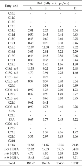
±7.1%, 41.9±0.6%, 44.9±0.7%으로 A, D, C, B-type 순으로 나타 났다(Table 2). 지방산 조성에서는 인공 유기물 분해산물로 제 조한 사료의 종류에 따라 다르게 나타났다. Palmitic acid (C16 :0), oleic acid (C18:1 n-9) 그리고 linoleic acid (C18:2 n-6)의 조성은 A, B, C, D-type순으로 A-type이 가장 높게 나타났으 며, eicosapentaenoic acid (EPA)와 decosahexaenoic acid (DHA)에서는 반대로 D-type이 가장 높게 나타났다. 또한 n-3 HUFA (highly unsaturated fatty acids)에서는 D, C, B, A-type 순으로, n-6 HUFA와 n-9 HUFA는 A, B, C, D-type순으로 나 타났다(Table 3).
자어 성장률 및 생존율
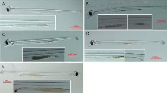
유기물 분해산물을 부화 20일 이후 자어에 대한 사료로 활 용 가능한지를 조사하였다. 부화 19일째 자어의 평균 체장은 8,692±306 μm이었다. 유기물 분해산물로 제조한 사료들을 부 화자어에게 공급한 결과 모든 자어의 장내에 섭취된 모습이 확인되었으며, 사료 섭취 후 배설하는 모습이 확인되어 인공 유기분해산물들을 자어의 사료로 사용할 수 있음을 확인하였
Table 3. Fatty acid content of each diet listed in Table 1
Fatty acid Diet (fatty acid μg/mg)
A B C D
C10:0 C11:0 C12:0 C13:0 C14:0 C14:1 C15:0 C15:1 C16:0 C16:1 C17:0 C17:1 C18:0 C18:1 n-9 C18:2 n-6 C18:3 n-6 C18:3 n-3 C20:0 C20:1 n-9
C20:2 C20:3 n-6
C21:0 C20:3 n-3
ARA C22:0 EPA C22:1 n-9
C22:2 C23:0 C24:0 C24:1 DHA n-3 HUFA n-6 HUFA n-9 HUFA
- - - - 2.01 0.50 0.43 0.46 15.07
3.05 0.46 0.38 1.97 14.53
4.70 - 0.37
- 0.92 0.37 - 0.62 0.90 - - 0.67
- - - 3.33
- 14.88 16.82 4.70 15.45
- - - - 2.23 0.43 0.60 0.26 12.38
2.84 0.45 0.33 1.45 9.22 3.91 0.22 0.37 0.58 1.26 0.90 0.66 0.44 0.73
- - 1.77
- - 1.37 2.97 - 14.16 17.03 4.80 10.48
- - - - 2.62 0.44 0.60 0.30 10.62
3.22 0.58 0.33 1.06 2.89 2.25 - 0.40 0.64 2.00 1.49 0.80 - 0.46
- - 2.45
- - 2.16 3.63 - 16.24 19.55
3.05 4.89
- - - - 3.54 0.63 0.75 0.46 9.02 2.29 0.66 0.44 1.20 2.48 1.60 - 0.54 0.99 1.23 0.77 0.95 - 0.76
- - 3.22
- - 1.72 4.06 - 29.48 34.00
2.55 3.71
Total 183.77 166.66 154.55 187.03
다(Fig. 2). 계란, 뱀장어, 참다랑어로 생산한 인공 유기분해산 물들을 2:1:1의 비율로 제조한 A-type을 공급한 자어는 부화 후 37일까지 생존하였고, 계란, 갯지렁이, 새우, 바지락, 담치, 멍게를 4:2:1:1:1:1의 비율로 제조한 B-type을 공급한 자어는 부화 후 39일, 갯지렁이, 새우, 바지락, 담치, 멍게를 4:1:1:1:1의 비율로 제조한 C-type을 공급한 자어들은 부화 후 37일까지 생존하였다. 담치, 멍게, 갯지렁이, 새우, 바지락를 3:3:1:1:1의 비율로 제조한 D-type을 공급한 자어의 경우 부화 후 60일까 지 생존하였고, 곱상어 알을 주원료로한 액상사료를 공급한 자어는 부화후 60일에 생존율이 12.7% 였다(Fig. 3). 부화 후 33일과 58일에 곱상어 알을 주원료로한 액상사료, A, B, C 그 리고 D-type에 따른 부화자어의 전장을 조사하였다(Fig. 4).
곱상어 알을 주원료로한 액상사료로 사육한 자어의 전장은 부화후 33일에 11,108±343 μm, 부화후 58일에 14,613±237 μm 로 나타났으며, A-type, B-type 그리고 C-type 을 공급한 자어 의 전장은 부화후 33일에 각 10,553±442 μm, 10,935±500 μm 그리고 10,788±365 μm로 나타났다. D-type 을 공급한 자어의 전장은 부화후 33일에 11,074±252 μm 그리고 부화후 58일에 12,714±92 μm로 나타나, 부화후 58일에 대조구보다 전장이 유의하게 낮게 나타났다(p<0.05).
고 찰
본 연구에서는 뱀장어 자어에게 BFT system으로부터 분리 한 미생물을 이용하여 생산된 유기물 분해산물을 공급하여 사료로써의 가능성을 조사하였다. 인공적으로 생산된 유기물 분해산물의 크기는 10-20 μm의 미세한 크기에서 100 μm 이상 되는 다소 큰 크기까지 다양하였고, 일정한 형태를 나타내지 않았다. 부화후 20일부터 담치, 멍게, 갯지렁이, 새우, 바지락 으로 생산된 유기물 분해산물을 3:3:1:1:1의 비율로 제조한 D- type의 사료를 공급하였고 부화후 60일까지 생존하였다.
Biofloc의 영양조성은 분해되는 유기물의 종류, 이를 분해 하는 미생물의 조성 그리고 미생물들이 유기물을 분해할 때 필요로 하는 유기탄소원(organic carbon source)의 종류와 탄 소질소비(C/N ratio) 등에 따라 다르게 나타난다고 알려져 있 으며[5], 본 연구에서도 각 유기물 따라 영양조성 및 형성된 모양이 다르게 나타났다(data not shown).
예비실험에서 부화후 7일차 자어에게 유기물 분해산물들을 각각 단일로 공급하였지만 부화후 19일 이내 모두 폐사하였 다. 뱀장어 부화자어의 소화관 내 trypsin, lipase 그리고 amy- lase 활성은 부화 7일이후 증가하기 시작해서 부화 16~18일이 후로는 부화 7일보다 높은 수치로 안정된다고 알려져 있으며, 다른 어류의 부화자어보다 소화 및 흡수 능력이 저조하여 저 분자 형태의 작은 먹이가 적합하다고 보고되었다[24, 29]. 예비 실험에서 부화후 19일 이내에 모두 폐사한 것은 본 연구의 방법으로 생산된 유기물 분해산물이 낮은 소화 및 흡수 능력 을 가지는 부화후 7일 자어가 먹이로 이용하기 어렵고 각각의 유기물분해산물만으로는 영양이 부족하여서 나타난 결과라 고 생각된다. 따라서 본 연구에서는 부화자어의 소화효소 활 성이 향상 및 안정되어진 부화후 20일의 자어를 이용하였으며 유기물 분해산물들을 배합비를 달리하여 실험을 진행하였다.
부화 초기 뱀장어 자어의 식도에는 muco-polysaccharides 와 같은 점액을 분비하는 점액분비세포가 없으며 식도 구경은 약 50 μm 정도로 알려져 있어서[40], 먹이의 입자 크기는 섭취 가능 여부를 가름하는 중요한 요인으로 작용할 수 있다. 본 연구에서는 생산된 모든 인공 유기물 분해산물들을 망목 100 μm의 체로 거른 후, homogenizer를 이용해 마쇄하여 자어가 섭취에 용이하도록 입자크기를 조절하였다. 이렇게 제조한 유
Fig. 4. Changes in body length of leptocephalus each diet (A, B, C, D-type and dogfish shark egg yolk-based diet) shown in Table 1 from day 20 after hatching. For 20 days after hatching, larvae were fed dogfish shark egg yolk-based diet received from the National Institute of Fisheries Science. Data are expressed as the mean ± SEM (n=5).
* indicates significant differences (p<0.05).
Fig. 2. Photographs of leptocephalus larvae fed each feed type shown in Table 1. Photographs A, B, C, and D show larvae fed each type of feed (feed type A, B, C, and D) listed in Table 1. Photograph E shows larvae fed dogfish shark egg yolk-based feed received from the Nationcal Institute of Fisheries Science.
Fig. 3. Changes in survival rates of leptocephalus fed each diet (A, B, C, D-type and dogfish shark egg yolk-based diet) shown in Table 1 and from day 20 after hatching. For 20 days after hatching, larvae were fed dogfish shark egg yolk-based diet received from the National Institute of Fisheries Science.
기물 분해산물 사료들을 부화자어에게 공급한 결과 모든 자어 의 장 내에 섭취된 모습이 확인되어서 유기물 분해산물들을 자어용 사료로 사용할 수 있음을 확인 할 수 있었다.
본 연구에서 A. B. C-type의 먹이를 공급한 자어들은 39일 이내에 모두 폐사하였으나, D-type의 먹이를 급이한 자어들만 부화 후 60일까지 생존할 수 있었다. 37-39일까지 생존한 A.
B. C-type과 60일까지 생존한 D-type 먹이의 단백질조성과 아 미노산은 차이가 없었으며, D-type 먹이의 단백질 함량은
44.9±0.7%이고 가장 빨리 폐사한 A-type의 단백질 함량은 45.3±3.0%로 단백질 함량의 유의한 차이를 보이지 않았다. 그 러나 뱀장어 자어의 사료 연구들중 계란을 주원료한 사료[31]
와 곱상어 난을 주원료로한 사료[13, 35]의 단백질 함량은 각 26.6%와 21.2%로 본 연구의 유기물 분해산물 사료보다 낮은 단백질 함량을 보였으나 더 오랜기간 생존하였다. 이러한 연 구결과들을 종합해볼 때 뱀장어 자어는 사료내 단백질 함량이
높지 않거나 단백질 함량보다 아미노산 조성이 생존에 영향을 미치는 것으로 생각되지만, 아직까지 뱀장어 자어의 성장단계 별 단백질 요구량에 대한 연구가 수행되지 않아서 향후 이에 대한 연구가 필요하다고 판단된다.
A, B, C 그리고 D-type 먹이의 단백질 조성과는 달리 지방 산 조성은 차이를 나타냈다. 특히 DHA, EPA 그리고 n-3 HUFA의 함량에서 D-type 먹이가 다른 종류의 먹이보다 약 2배이상 많았으며, n-6 HUFA와 n-9 HUFA의 함량에서는 다 른 종류의 먹이보다 2~5배 더 작게 나타났다. 어류들은 주에너 지원으로 지질을 이용한다고 알려져 있으며, 자어 발달시기에 n-3 HUFA 를 주에너지원으로 이용하는 그룹과 palmitic aicd (16:0)와 n-9 HUFA 를 주에너지원으로 먼저 이용하는 그룹 등으로 나누어 진다[8]. DHA, EPA를 포함하는 n-3 HUFA 는 어류에서 합성할 수 없는 필수 지방산(essential fatty acids, EFAs)으로 알려져 있으며, 방어(Seriola quinqueradiata), 감성돔 (Acanthopagrus schlegeli), 귀족도미(Sparus aurata) 및 흰다리새 우(Penaeus vannamei) 등 해양동물의 자·치어기에 정상적인 성 장과 생존을 위해서 꼭 필요하다고 알려져 있다[7, 16, 31, 40].
계란을 주원료로 사료를 만들어 뱀장어 자어를 사육하였을 때 곱상어 난황을 주원료로 사료를 만들어 사육할 때보다 생 존율이 현저하게 떨어지는 원인은 계란 난황의 n-3 HUFA의 농도가 곱상어 난황에 비해 현저하게 낮기 때문이라고 하였다 [27]. 본 연구결과에서도 뱀장어 자어는 지방산 중 n-3 HUFA 를 주에너지원으로 사용하는 것으로 보이며, D-type의 먹이가 다른 종류의 먹이보다 오래 생존할 수 있었던 이유도 D-type 먹이의 n-3 HUFA 함량이 가장 높기 때문이라고 판단된다.
자연에서의 뱀장어 먹이로 알려진 marine snow에는 glu- cose와 galactose와 같은 다양한 단당류와 다당류에 속하는 탄 소화합물들을 많이 함유하고 있다고 알려져 있으며[4, 32], 대 부분의 뱀장어목 자어들의 몸에는 glucuronic acid와 N-ace- tylglucosamine로 구성된 hyaluronan이 많이 존재하고 이들 의 형성에 대한 생리적 기능이 성장과 관련이 있다고 알려져 있다[30]. 그러나 본 연구에서는 사료내 지방산과 아미노산의 성분분석만이 이루어져 탄소화합물 성분에 의한 차이는 비교 할 수 없었으나 액상사료를 공급한 자어보다 유기물분해산물 로 제조한 사료를 공급한 자어의 성장이 저조한 이유는 사료 내 탄소화합물의 조성 또는 함량의 차이가 원인이라고 생각된 다. 향후 뱀장어 자어 사료들의 탄소화합물 조성과 함량에 대 한 비교분석이 필요해 보이며, 뱀장어 자어 사료개발에 있어서 도 탄소화합물의 첨가가 중요한 요소가 될 것으로 생각된다.
감사의 글
본 연구는 국립수산과학원의 지원(R2018035)에 의해 운영 되었습니다.
References
1. Alldredge, A. L. and Youngbluth, M. J. 1985. The sig- nificance of macroscopic aggregates (marine snow) as sites for heterotrophic bacterial production in the mesopelagic zone of the subtropical Atlantic. Deep Sea Research Part A.
Oceanogr. Res. Pap. 32, 1545-1456.
2. Arndt, S., Jørgensen, B. B., LaRowe, D. E., Middelburg, J.
J., Pancost, R. D. and Regnier, P. 2013. Quantifying the deg- radation of organic matter in marine sediments: a review and synthesis. Earth-Science Rev. 123, 53-86.
3. Avnimelech, Y. 2006, Bio-filters: The need for an new com- prehensive approach. Aquac. Eng. 34, 172-178.
4. Cowen, J. P. and Holloway, C. F. 1996. Structural and chem- ical analysis of marine aggregates: in situ macrophotog- raphy and laser confocal and electron microscopy. Mar. Biol.
126, 163-174.
5. Crab, R., Chielens, B., Wille, M., Bossier, P. and Verstraete, W. 2010. The effect of different carbon sources on the nutri- tional value of bioflocs, a feed for Macrobrachium rosenbergii postlarvae. Aquac. Res. 41, 559-567.
6. Fordham, S., Fowler, S. L., Coelho, R., Goldman, K. J. and Francis, M. 2006. Squalus Acanthias. IUCN Red List of Threatened Species. Version 2010.1. Available at http://
www.iucnredlist.org.
7. Furuita, H., Takeuchi, T., Watanabe, T., Fujimoto, H., Sekiya, S. and Imaizumi, K. 1996. Requirements of larval yellowtail for eicosapentaenoic acid, docosahexaenoic acid, and n-3 highly unsaturated fatty acid. Fish. Sci. 62, 372-379.
8. Izquierdo, M. S. 1996. Essential fatty acid requirements of cultured marine fish larvae. Aquac. Nutr. 2, 183-191.
9. Kagawa, H., Tanaka, H., Ohta, H., Unuma, T. and Nomura, K. 2005. The first success of glass eel production in the world: basic biology on fish reproduction advances new ap- plied technology in aquaculture. Fish. Physiol. Biochem. 31, 193-199.
10. Kim, D. J., Kim, E. H., Park, M. W., Cho, Y. C. and Lim, S. G. 2006a. Plasma sex steroid hormone profiles in artifi- cially maturing wild eel, Anguilla japonica. J. Aquac. 19, 267- 274.
11. Kim, D. J., Lee, N. S., Kim, K. K. and Chang, D. S. 2014.
Effects of starvation, water temperature, and water flow on the metamorphosis of leptocephalus of Japanese eel Anguilla japonica. Kor. J. Fish. Aquat. Sci. 47, 597-602.
12. Kim, E. O., Bae, J. Y., Lim, S. G., Son, M. H., Park, M. W., Park, M. S., Cho, Y. C. and Kim, D. J. 2006b. Plasma Sex Steroid Hormone Profiles and Testicular Development in Artificially Maturing Cultured Mille Eel, Anguilla japonica.
Kor. J. Fish. Aquac. Sci. 39, 466-471.
13. Kim, S. K., Lee, B. I., Kim, D. J. and Lee, N. S. 2014.
Development of the slurry type diet for the growing lep- tocephalus, eel larvae (Anguilla japonica). J. Fish. Mar. Sci.
Educ. 26, 1209-1216.
14. KiØrboe, T. 2000. Colonization of marine snow aggregates by invertebrate zooplankton: abundance, scaling, and possi- ble role. Limnol. Oceanogr. 45, 479-484.
15. Lin, H. R., Xie, G., Zhang, L. H., Wang, X. D. and Chen, L. X. 1998. Artificial induction of gonadal maturation and ovulation in the Japanese eel (Anguilla japonica, T ETS.). Bull.
Fr. Pêche. Piscic. 349, 163-176.
16. Ma, J., Shao, Q., Xu, Z. and Zhou, F. 2013. Effect of Dietary n-3 highly unsaturated fatty acids on growth, body compo- sition and fatty acid profiles of juvenile black seabream, Acanthopagrus schlegeli (Bleeker) J. World Aquac. Soc. 44, 311-325.
17. Masuda, Y., Imaizumi, H., Oda, K., Hashimoto, H., Teruya, K. and Usuki, H. 2011. Japanese eel Anguilla japonica larvae can metamorphose into glass eel within 131 days after hatc- ing in captivity. Nippon. Suisan. Gakkaishi. 77, 416-418. (in Japanese with English abstract)
18. Masuda, Y., Jinbo, T., Imaizumi, H., Furuita, H., Matsunari, H., Murashita, K., Fujimoto, H., Nagao, J. and Kawakami, Y. 2013. A step forward in development of fish protein hy- drolysate-based diets for larvae of Japanese eel Anguilla japonica. Fish. Sci. 79, 681-688.
19. Masuda, Y., Yatabe, T., Matsunari, H., Furuita, H., Kamoshi- da, M., Shima, Y. and Kuwada, H. 2016. Rearing of larvae of Japanese eel Anguilla japonica to metamorphosis into glass eel by feeding with fish protein hydrolysate-based diets.
Nippon. Suisan. Gakkaishi. 82, 131-133. (in Japanese with English abstract)
20. Miller, M. J. 2009. Ecology of anguilliform leptocephali : re- markable transparent fish larvae of the ocean surface layer.
Aqua-Bio Sci. Monogr. 2, 1-94.
21. Miller, M. J., Otake, T. Aoyama, J. Wouthuyzen, S., Suhartt, S., Sugeha, H. Y. and Tshkamoto, K. 2011. Observation of gut contents of leptocephali in the north equatorial current and tomini bay, Indonesia. Coast. Mar. Sci. 35, 277-288.
22. Miller, M. J., Chikaraishi, Y., Ogawa, N. O., Yamada, Y., Tsukamoto, K. and Ohkouchi, N. 2013. A low trophic posi- tion of Japanese eel larvae indicates feeding on marine snow. Biol. Lett. 9, 20120826.
23. Mochioka, N. and Iwamizu, M. 1996. Diet of anguilloid lar- vae: leptocephali feed selectively on larvacean houses and fecal pellets. Mar. Biol. 125, 447-452.
24. Murashita, K., Furuita, H., Matsunari, H., Yamamoto, T., Awaji, M., Nomura, K., Nagao, J. and Tanaka, H. 2013.
Partial characterization and ontogenetic development of pancreatic digestive enzymes in Japanese eel Anguilla japon- ica larvae. Fish. Physiol. Biochem. 39, 895-905.
25. Lee, N. S., Kim, D. J., Lee, B. L., Kim, S. K. and Kim, K.
K. 2015. Distribution of ghrelin immunoreactivity in artifi- cially reared Japanese eel, Anguilla japonica, leptocephalus and metamorphosed glass eel. J. Environ. Biol. 36, 521.
26. Ohta, H., Kagawa, H., Tanaka, H., Okuzawa, K., Iinuma, N. and Hirose, K. 1997. Artificial induction of maturation and fertilization in the Japanese eel, Anguilla japonica. Fish Physiol. Biochem. 17, 163-169.
27. Okamura, A., Yamada, Y., Horie, N., Mikawa, N., Tanaka,
S., Kobayashi, H. and Tsukamoto, K. 2013. Hen egg yolk and skinned krill as possible foods for rearing
leptocephalus larvae of Anguilla japonica Temminck & S chlegel. Aquac. Res. 44, 1531-1538.
28. Park, J. C., Kwon, O. N., Hong, S. E., An, H. C., Bae, J.
H., Park, M. S. and Park, H. G. 2013. Changes in the growth and biochemical composition of Nannochloropsis sp. cultures using light-emitting diodes. Kor. J. Fish. Aquac. Sci. 46, 259- 265.
29. Pedersen, B. H., Ueberschär, B. and Kurokawa, T. 2003.
Digestive response and rates of growth in pre-leptocephalus larvae of the Japanese eel Anguilla japonica reared on artifi- cial diets. Aquaculture 215, 321-338.
30. Pfeiler, E. 1999. Developmental physiology of elopomorph leptocephali. Comparative Biochemistry and Physiology.
Part A, Mol. Integr. Physiol. 123, 113-128.
31. Salhi, M., Izquierdo, M. S., Hernandez-Cruz, C. M., Gonzalez, M. and Fernández-Palacios, H. 1994. Effect of lipid and n−
3 HUFA levels in microdiets on growth, survival and fatty acid composition of larval gilthead seabream (Sparus aurata).
Aquaculture 124, 275-282.
32. Skoog, A., Alldredge, A., Passow, U., Dunne, J. and Murray, J. 2008. Neutral aldoses as source indicators for marine snow. Mar. Chem. 108, 195-206.
33. Tanaka, H., Kagawa, H. and Ohta, H. 2001. Production of leptocephali of Japanese eel (Anguilla japonica) in captivity.
Aquaculture 201, 51-60.
34. Tanaka, H. 2003. Techniques for larval rearing. In: Eel Biology (ed. By K. Aida, K. Tsukamoto & K. Yamauchi), pp. 427-434. Springer-Verlag, Tokyo, Japan.
35. Tanaka, H., Kagawa, H., Ohta, H., Unuma, T. and Nomura, K. 2003. The first production of glass eel in captivity: fish reproductive physiology facilitates great progress in aqua- culture. Fish Physiol. Biochem. 28, 493-497.
36. Tansel, B. 2018. Morphology, composition and aggregation mechanisms of soft bioflocs in marine snow and activated sludge: A comparative review. J. Environ. Manage. 205, 231- 243.
37. Tecchio, S., Coll, M., Christensen, V., Ramírez-Llodra, E. and Sardà, F. 2013. Food web structure and vulnerability of a deep-sea ecosystem in the NW Mediterranean Sea. Deep Sea Research Part I: Oceanographic Research Papers 75, 1-15.
38. Yamamoto, K. and Yamauchi, K. 1974. Sexual maturation of Japanese eel and production of eel larvae in the aquarium.
Nature 251, 220.
39. Yoshimatsu, T. 2011. Early development of pre-leptocephalus larvae of the Japanese eel in captivity with special reference to the organs for larval feeding. Bull. Graduate School of Bioresources Mie Univ. 37, 11-18.
40. Wouters, R., Vanhauwaert, A., Naessens, E., Ramos, X., Pedrazzoli, A. and Lavens, P. 1997. The effect of dietary n-3 HUFA and 22:6n-3/20:5n-3 ratio on white shrimp larvae and postlavae. Aquaculture 5, 113-126.
초록:뱀장어(Anguilla japonica) 자어 먹이로 유기물 분해산물의 활용 가능성
김효원1․김정현1․김명희2․김광현3․박진철4․박흠기5․한창희2*․김대중1*
(1국립수산과학원 제주수산연구소, 2동의대학교 분자생물학과, 3동의대학교 생명응용학과, 4강릉원주대학교 동해
안생명과학연구소, 5강릉원주대학교 해양자원육성학과)
본 연구에서는 BFT (biofloc technology) system 수조로부터 분리된 미생물(Bacillus sp.)을 이용하여 Marine snow와 유사한 유기물 분해산물을 만들어 뱀장어 자어먹이로 활용 가능성을 조사하였다. 유기원으로는 계란, 참 다랑어, 뱀장어, 담치, 갯지렁이, 멍게, 바지락 그리고 새우를 이용하여 8 종류의 유기물 분해산물(HE, TM, EM, PC, TA, MS, HA, SP)를 생산했다. 생산된 유기물 분해산물은 특정한 형태는 나타나지 않았으며, 크기도 10-20 μm의 미세한 것부터 100 μm 이상 되는 것까지 다양하였다. 부화 후 20일부터 다양한 유기물 분해산물을 이용하 여 제조한 4 종류의 먹이(A, B, C, D-type)를 급이한 결과 A, B, C-type의 먹이를 급이한 뱀장어 유생은 각각 부화 후 37일, 39일, 37일까지 생존하였으나, D-type의 먹이를 급히한 뱀장어 유생은 부화 후 60일 동안 생존하였다.
각 먹이의 단백질 함량은 비슷하였지만, D-type 먹이의 n-3 HUFA 농도는 다른 type의 n-3 HUFA 농도보다 약 2배 높았다.