An Analysis for Effects of Stain Family Drugs on Osteogenic Differentiation using Human Periosteum-derived Mesenchymal Stem Cells
Dong Kyu Moon1, Jeong-Won Yun2, Bo Gyu Kim3, A Ram Lee3, Sun Young Moon3, June-Ho Byun2, Sun-Chul Hwang1* and Dong Kyun Woo3*
1Department of Orthopedic Surgery and Institute of Health Sciences, School of Medicine and Hospital, Gyeongsang National University, Jinju 52727, Korea
2Department of Oral and Maxillofacial Surgery and Institute of Health Sciences, School of Medicine and Hospital, Gyeongsang National University, Jinju 52727, Korea
3College of Pharmacy and Research Institute of Pharmaceutical Sciences, Gyeongsang National University, Jinju 52828, Korea Received August 28, 2019 /Revised November 10, 2019 /Accepted November 19, 2019
Osteoporosis is characterized by a reduction in bone mass and typically manifests as an increase in fractures. Because this disease is common in elderly populations and lifespans are rapidly increasing, the incidence of osteoporosis has also grown. Most drugs currently used for osteoporosis treatment target osteoclasts in the bone tissue to prevent absorption. However, these medications also cause cer- tain side effects and, furthermore, cannot increase bone mass. Thus, in order to control osteoporosis, regenerative medicine that utilizes adult stem cells and osteoblasts has been extensively studied.
Statins, also known as 3-hydroxy-3-methylglutaryl-coenzyme A (HMG-CoA) reductase inhibitors, are cholesterol-lowering drugs that have been widely prescribed for cardiovascular diseases. Interestingly, recent studies have reported the beneficial effects of various statins on bone formation via the activa- tion of osteoblasts. Thus, the current study investigated the effects of seven statin-family drugs on os- teoblast activity during osteogenic differentiation using adult stem cells from human periosteal tissue.
Specifically, statin effects on alkaline phosphatase activity, an early marker of bone cell differentiation, and on calcium deposit, a late marker of bone cell differentiation, were assessed. The results demon- strate that some statins (for example, pitavastatin and pravastatin) have a weak but positive effect on bone formation, and the findings therefore suggest that statin treatments can be a novel modulator for osteogenic differentiation and regenerative medicine using periosteal stem cells.
Key words : Osteogenesis, osteoporosis, regenerative medicine, statin, stem cell
*Corresponding authors
*Tel : +82-55-772-2428, Fax : +82-55-772-2429
*E-mail : [email protected] (Dong Kyun Woo) [email protected] (Sun-Chul Hwang)
This is an Open-Access article distributed under the terms of the Creative Commons Attribution Non-Commercial License (http://creativecommons.org/licenses/by-nc/3.0) which permits unrestricted non-commercial use, distribution, and reproduction in any medium, provided the original work is properly cited.
Journal of Life Science 2019 Vol. 29. No. 12. 1337~1344 DOI : https://doi.org/10.5352/JLS.2019.29.12.1337
서 론
골다공증은 뼈조직의 다공성이 증가하는 질환이다. 이러한 뼈의 구조적인 변화는 뼈의 밀도와 질을 감소시키며 이로 인 하여 골절의 위험이 현저히 증가한다[5]. 골다공증의 유발 인 자로는 여러 원인이 알려져 있으며, 대표적으로 나이가 증가 함에 따라 골형성과 골흡수의 균형이 깨지면서 발생한다. 전 세계적으로 2억명 이상의 사람들이 골다공증으로 진단 받고 치료 중이고 미국과 유럽에서 폐경 후 여성의 약 30%가 골다 공증을 가지고 있다. 그리고 이러한 골다공증 환자의 15~40%
는 골절을 경험하게 된다[34]. 따라서 골다공증을 치료하기 위
해 많은 연구가 현재 활발히 이루어지고 있다.
Statin은 전세계적으로 가장 광범위하게 사용되는 지질강 하제이다. Statin은 콜레스테롤(cholesterol) 생합성 경로의 속 도조절효소(rate limiting enzyme)인 3-hydroxy-3-methyl- glutaryl-coenzyme A 환원효소(HMG-CoA reductase)의 억제 제로 콜레스테롤의 생합성을 저해한다. 또한 statin은 죽상동 맥경화를 야기하는 염증성 인자들의 활동을 억제한다[20]. 이 를 통해 statin은 지질 대사 이상을 개선하고 심뇌혈관 질환을 예방하여 전체적으로 사망률을 감소시키는 효과를 가진다고 알려져 있다[27, 40]. 최근 들어, statin은 이러한 지질대사 조절 기능 이외에도 항암 및 항산화 효과도 가진 것으로 알려지고 있다[4, 20, 46]. 그리고 흥미롭게도 골형성과 관련하여 긍정적 인 효과를 보인다. Simvastatin을 비롯한 여러 statin 계열 약물 이 지닌 bone metabolism에 이득이 되는 효과가 지속적으로 보고되고 있다[1, 19, 25, 36].
중배엽유래줄기세포(mesenchymal stem cells, MSC)는 지 방세포, 골세포, 그리고 연골세포로 분화가 가능한 다기능 성 체줄기세포다[11]. MSC는 배아줄기세포(embryonic stem cells) 에 비하여 윤리적 문제가 없고 면역학적인 문제가 작다는 장
Table 1. Statin family drugs used in this study
Drug Molecular
weight Solubility References
Atorvastatin Fluvastatin Lovastatin Pitavastatin Pravastatin Rosuvastatin Simvastatin
1209.42 433.45 404.54 440.49 446.51 481.54 418.57
Lipophilic Lipophilic Lipophilic Lipophilic Hydrophilic Hydrophilic Lipophilic
[14, 48]
[16, 28]
[37]
[29, 33]
[17, 22]
[24, 38]
[26, 30, 44]
점이 있다. 재생의학 분야에서는 전통적으로 최근까지 골수 천자를 통하여 획득한 골수유래 성체 중간엽줄기세포(bone marrow-derived mesenchymal stem cells, BMMSCs)를 많이 이용하였다. 하지만 이러한 BMMSCs 획득 과정은 통증 및 감 염과 같은 합병증을 유발 할 수 있다는 단점이 있다. 더욱이 고령인 사람으로부터 획득한 BMMSCs의 경우 세포증식과 분 화능력이 젊은 사람에게서 획득한 BMMSCs보다 감소한다는 단점이 있다[3, 6].
골막(periosteum)은 골의 외변을 둘러 싸고 있는 결합조직 으로 성체줄기세포를 포함하고 있다. 골막유래 성체 중간엽줄 기세포(periosteum-derived mesenchymal stem cells, POMSCs) 는 BMMSCs 와 동일한 성체줄기세포 특이적인 세포막 표지자 (CD73, CD90, and CD105)를 발현하고 다분화 능력을 가진다 [10, 15]. 더욱이 POMSCs는 발치와 같은 간단한 치과적인 처 치과정에서 획득한 골막에서 쉽게 분리가 가능하며, 고령인 사람에게서 획득한 경우에도 분화능력이 유지되는 장점을 가 진다[9]. POMSCs는 또한 임상적으로, 골생성 및 골절치유에 중요한 역할을 한다고 알려져 있다[7, 42]. 이러한 장점으로 인해 골막은 재생의학적인 관점에서 매력적인 MSCs의 공급 처다.
지금까지 statin이 골형성 및 골대사에 미치는 영향에 관한 연구들은 대부분 골수유래 성체줄기세포를 이용하여 개별적 인 statin의 효과에 대한 보고들이다. 따라서 본 연구에서는 최근 재생의학에서 주목받고 있는 골막유래 성체줄기세포를 이용하여, 총 7종류의 다양한 statin 계열 약물이 가지는 조골 세포 및 골세포 활성에 미치는 영향을 분석하였다.
재료 및 방법
POMSCs의 세포배양 및 골세포 분화유도
경상대학교병원 윤리위원회(the Ethics Committee of Gyeongsang National University Hospital, GNUH 2014-05- 012)의 규정에 의거하여 환자 동의 하에 인체유래 골막조직을 획득하여 POMSCs 분리하였다[11]. 세포배양은 10% fetal bo- vine serum과 1% penicillin/streptomycin이 첨가된 Dulbec- co's modified Eagle's medium (DMEM배양액)을 사용하여 통 상적인 37℃, 5% CO2의 배양조건에서 이루어졌다. POMSCs 로부터 골세포로의 분화 유도를 위해서는 DMEM배양액에 50 μg/ml L-ascorbic acid 2-phosphate, 10 nM dexamethasone, 그리고 10 mM β-glycerophosphate를 첨가한 골세포분화유도 배양액(osteogenic differentiation induction medium, OM배 양액)을 사용하여 세포배양하였다. 골세포 분화를 위한 세포 배양에서 OM배양액은 3일을 주기로 교체해주었다.
Statin 계열 약물의 세포독성 분석
본 연구에 사용된 7종류의 statin 계열 약물의 특성은 Table
1에 요약되었으며 Sigma-Aldrich (St. Louis, USA)로부터 구 입하였다. Statin 계열 약물의 세포독성(cytotoxicity)에 대한 영향을 분석하기 위해 2×10⁴ cells/well의 농도로 POMSCs를 24-well plate에 seeding한 후에 각각의 statin을 10 μM, 5 μM, 1 μM, 500 nM, 100 nM, 그리고 50 nM 농도로 처리한 OM배양 액에서 각각 세포배양하였다. 세포배양 14일에 MTT assay를 이용하여 세포독성을 분석하였다.
Alkaline phosphatase (ALP) 활성 분석
Statin 계열 약물의 골세포분화에 대한 영향을 알아보기 위 해 골세포분화의 초기 표지자인 ALP의 활성을 분석하였다.
POMSCs를 2×10⁴ cells/well의 농도로 24-well plate에 seed- ing한 후에 뚜렷한 세포독성을 보이지 않은 농도에서 각각의 statin를 처리한 OM배양액에서 세포배양하였다. 골세포 분화 유도 세포배양 14일에 ALP 활성을 분석하였다. 간략히 기술 하면, 먼저 NP40 Cell Lysis Buffer (Life Technologies, Carls- bad, USA)를 사용하여 세포용해(cell lysis)를 실시하였다. 여기 서 얻어진 일부 cell lysates에 완충액과 ALP의 기질(substrate) 인 p-nitrophenyl phosphate (Thermo Fisher Scientific, Wal- tham, USA)를 첨가한 후에, 37℃에서 20분간 효소반응시켰다.
이 반응을 0.9 N NaOH 용액을 첨가하여 종료시킨 후에 mi- croplate reader (Molecular Devices, San Jose, USA)를 이용하 여 405 nm 파장에 대한 흡광도를 측정하여 ALP 활성을 분석 하였다. 다른 일부 cell lysates에 대해서는 Bradford assay로 단백질 농도를 분석하였다. 세포수에 따른 흡광도 보정을 위 해서, cell lysates에서 같은 양의 단백질 당 흡광도를 계산하여 최종적인 ALP 활성을 결정하였다.
칼슘 침착 분석
Statin 계열 약물의 골세포분화에 대한 영향을 알아보기 위 해 골세포분화의 후기 표지자인 칼슘 침착 정도을 분석하였 다. 먼저, 2×10⁴ cells/well의 농도로 POMSCs를 24-well plate 에 seeding한 후에 앞서 ALP 활성 분석 실험과 동일한 농도로 statin 약물이 처리된 OM배양액으로 각각 세포배양하였다. 다 음으로, 3주일 간의 골세포 분화유도 세포배양후에 세포 밖에 침착된 칼슘을 Calcium C-test kit (Wako Chemicals, Osaka,
Fig. 1. Effects of statin treatments on viabilities of human periosteum-derived mesenchymal stem cells (POMSCs). Overall, statin treatments (1 μM to 50 nM) do not affect POMSC viability in cell cultures with a conventional DMEM (white bars) or an osteogenic differentiation induction medium (OM, black bars). Cell viabilities of 14-day POMSC cultures were assessed by MTT assay. *p<0.05, **p<0.01, and ***p<0.001.
Japan)를 사용하여 정량적으로 분석하였다. 간략하게 기술하 면, 배양된 세포 밖에 침착된 칼슘을 0.6 N HCl 용액을 처리하 여 24 시간 동안 상온에서 탈회하였다. 다음으로 탈회된 용액 에 칼슘과 결합하여 발색하는 o-cresolphthalein를 처리하여 상온에서 10분간 반응시킨 후에 microplate reader (Molecular Devices, San Jose, USA)를 이용하여 575 nm 파장에 대한 흡광 도를 측정하여 분석하였다. 배양용기에 남은 세포를 용해하고 총 단백질 농도를 Bradford assay로 정량하였다. ALP 활성 분석 실험과 동일하게 각 조건에 대한 세포의 총 단백질 농도 를 이용하여 세포수에 대한 칼슘의 양을 보정하였다.
통계분석
모든 실험은 최소한 3회 이상의 독립적인 반복을 실시하였 고, 반복실험에서 얻은 결과는 Graphpad Prism 7 software (GraphPad, La Jolla, USA)를 이용하여 분산분석을 수행하였 고, 평균±표준편차로 나타내었다. 각 실험군의 평균값 차이를 통계분석하여 p<0.05인 경우에 통계학적으로 유의하다고 판 단하였다.
결과 및 고찰
POMSCs의 생존능력에 대한 statin의 영향 분석
Statin계열 약물이 POMSCs의 생존능력에 미치는 영향을 알아보기 위하여, POMSCs세포배양 중에 각각의 statin 약물 을 10 μM, 5 μM, 1 μM, 500 nM, 100 nM, 그리고 50 nM 의 다양한 농도로 처리하였다. 세포배양은 통상적인 DMEM배양 액과 골세포분화 OM배양액에서 2주일간 각각 이루어졌다.
이후 세포독성 분석을 위한 MTT assay를 수행하였고 statin을 처리하지 않은 대조군과 비교하였다. 각각의 statin이 POMSCs 의 생존능력에 미치는 영향은 Fig. 1에 제시되었다. 대부분의 statin 계열 약물들은 DMEM 및 OM 배양액에서 비슷한 양상 의 세포독성을 보였다. 또한 대부분의 statin 약물은 10 μM 그리고 5 μM의 처리 농도를 제외하면 POMSCs에 뚜렷한 세 포독성을 보이지 않았다. 이러한 세포독성에 대한 영향을 고 려하여, 이후의 실험(ALP활성 분석 및 칼슘침착 분석)에서 사 용될 각각의 statin 약물 처리 농도를 POMSCs의 생존능력에 영향을 주지 않는 범위에서 결정하였다(atorvastatin, pravas-
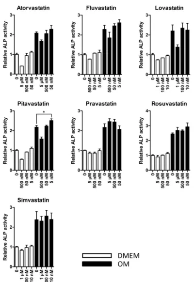
Fig. 2. Effects of various statins on ALP activities during osteogenic differentiation of POMSCs. POMSCs were cultured in DMEM (white bars) or induced to osteogenic differentiation with OM (black bars) for 14 days. Treatments of pitavastatin (at 5 nM) increased ALP activities in 14-day POMSC cul- tures with OM. One asterisk (*) indicates p<0.05.
tatin, 그리고 rosuvastatin은 5 μM, 500 nM 그리고 50 nM;
fluvastatin 및 pitavastatin은 500 nM, 50 nM 그리고 5 nM;
lovastatin 및 simvastatin은 1 μM, 100 nM 그리고 10 nM).
POMSCs의 골세포 분화유도 초기에 따른 ALP활성에 대 한 statin의 영향 분석
POMSCs의 초기 골세포 분화유도에 대한 각각의 statin의 영향을 알아보기 위해, POMSCs를 세포증식과 생존능력에 영 향을 미치지 않는 농도의 statin이 첨가된 골세포분화 OM배양 액으로 세포배양하였다. 14일간의 골세포분화 유도 후에 초기 조골세포 분화 표지자로 잘 알려진 ALP의 활성을 측정하였 다. Fig. 2에서 보여지는 바와 같이, 골세포분화 유도효과가 없는 통상적인 DMEM배양액을 사용한 대조군에 비해, 골세 포분화를 유도하는 OM배양액 그룹에서는 ALP 활성이 14일 간의 배양에서는 약 2배 정도 증가하였다. 이러한 결과는 본 실험에 사용되는 POMSCs가 OM배양액에 반응하여 골세포 분화의 초기단계인 조골세포로 분화하는 능력이 있음을 분명 하게 제시해 준다. OM 배양액과 POMSCs의 생존능력에 영향
을 주지 않는 농도 범위에서 각각의 statin약물처리는 OM 배 양액 단독 조건에서와 비슷한 ALP 활성을 나타내었다. 그러 나, atorvastatin, fluvastatin, pitavastatin, pravastatin, 그리고 rosuvastatin 처리는 ALP 활성을 작지만 다소 증가시키는 경 향을 보였으며, 이 중에서 pitavastatin 5 nM처리에서는 통계 적으로 유의하게 ALP 활성이 증가됨이 관찰되었다. 이러한 실험결과는 일부 statin 계열 약물 처리가 POMSCs의 골세포 분화 초기과정을 다소 촉진할 수 있음을 제시한다.
POMSCs의 골세포 분화유도 후기에 따른 칼슘 침착에 대 한 statin의 영향 분석
POMSCs의 후기 골세포 분화유도에 대한 각각의 statin의 영향을 알아보기 위해, POMSCs를 생존능력에 영향을 미치지 않는 농도 범위의 statin약물이 처리된 OM배양액으로 세포배 양하였다. 4주간 골세포분화 유도 후에 후기 골세포분화 표지 자로 잘 알려진 칼슘 침착을 측정하였다. Fig. 3에서와 같이, 각각의 statin약물 처리는 OM 배양액 단독 조건에서와 비슷한
Fig. 3. Effects of various statins on calcium deposits during osteogenic differentiation of POMSCs.
POMSCs were cultured and induced to osteo- genic differentiation with OM for 4 weeks.
Treatments of pravastatin (at 5 μM and 500 nM) increased calcium deposits in 4-week POMSC cultures with OM. One asterisk (*) indicates p<0.05.
정도의 칼슘 침착 양상을 나타내었다. 그러나, pravastatin 처 리의 경우에는 5 μM과 500 nM 농도에서 처리하지 않는 대조 군에 비해 통계적으로 유의하게 소폭 증가시키는 효과를 보였 다. 이러한 실험결과는 pravastatin 약물 처리가 골바탕질의 무기질 축적에 긍정적인 효과가 있음을 제시한다.
POMSCs 골세포 분화 촉진에서 statin의 역할
최근 대사 증후군이 골다공증의 발병과 밀접한 연관이 있다 는 보고가 있다[18, 41, 47]. 대사 증후군의 한 구성 요소인 지질 대사이상(dyslipidemia) 또한 골다공증과 밀접한 관련이 있음 이 밝혀졌다[31, 32, 45]. 지질대사이상은 PPAR-γ를 활성화하 고 이는 RANKL/RANK/OPG pathway를 통해 파골세포를 활성화하여 골흡수를 촉진하는 것으로 알려져 있다[5, 21]. 또 한 Wnt-β-catenin pathway를 억제하여 조골세포를 억제하고 이는 골형성을 억제한다[43]. 이에 더하여, 실험동물 수준에서 statin을 이용한 지질대사이상 치료는 골다공증의 개선 효과가 있음이 확인되었고, 임상적으로도 statin은 고지혈증을 감소시 켜 골밀도를 개선하는 효과가 있었다[1, 12, 23, 48]. 본 연구는
다양한 statin계열 약물을 사용하여 POMSCs의 골세포 분화과 정에 미치는 효과를 체계적으로 분석하였고, 일부 statin약물 이 골세포 분화과정의 표지자인 ALP활성 및 칼슘 침작 증가 를 유도하여 POMSC의 골세포 분화를 촉진하는 효과를 확인 하였다. 이러한 조골세포의 분화 및 활성을 상향조절하는 효 과는 임상적으로 중요한 의미를 가질 수 있다. 그 이유는 현재 광범위하게 사용중인 골다공증 치료제는 대부분 골흡수를 억 제하는 약물이며, 그 중 가장 광범위하게 사용되는 비스포스 포네이트(bisphosphonate)는 파골세포의 대사장애를 초래하 여 골흡수를 강력하게 억제한다[8]. 하지만 이러한 약제는 위 장관계 부작용, 턱뼈의 괴사, 비전형 골절과 같은 여러 부작용 이 문제가 된다[35, 39]. 또한 골흡수 억제로 인하여 정상 생리 적인 낡은 골의 제거 과정이 지연되며 새로운 골형성 저해도 야기한다[13]. 따라서 최근 골다공증 치료 연구는 골형성을 촉 진하는 조골세포의 활성을 증가시키는 물질로 많은 노력이 이 루어지는 추세이다. 본 연구에서 일부 statin의 경우 POMSCs 의 골세포 분화와 조골세포 활성을 촉진하는 효과를 보였는 데, 향후 추가적인 관련 연구를 통하여 골형성을 촉진하는 물
질로서 statin의 영향을 더욱 면밀히 분석한다면 골다공증의 치료제 개발에 도움이 될 것으로 사료된다.
감사의 글
본 연구는 교육부와 미래창조과학부의 재원으로 한국연구 재단의 지원(과제번호: NRF-2016R1D1A1B03931722, NRF-2019 R1F1A1060013, 및 NRF-2017R1D1A1B03035996)을 받아 이루 어졌으며 이에 대해 감사드립니다.
References
1. An, T., Hao, J., Sun, S., Li, R., Yang, M., Cheng, G. and Zou, M. 2017. Efficacy of statins for osteoporosis: a system- atic review and meta-analysis. Osteoporos. Int. 28, 47-57.
2. Armas, L. A. and Recker, R. R. 2012. Pathophysiology of osteoporosis: new mechanistic insights. Endocrinol. Metab.
Clin. North Am. 41, 475-486.
3. Berebichez-Fridman, R., Gomez-Garcia, R., Granados-Mon- tiel, J., Berebichez-Fastlicht, E., Olivos-Meza, A., Granados, J., Velasquillo, C. and Ibarra, C. 2017. The holy grail of or- thopedic surgery: mesenchymal stem cells-their current uses and potential applications. Stem Cells Int. 2017, 2638305.
4. Bu, D. X., Griffin, G. and Lichtman, A. H. 2011. Mechanisms for the anti-inflammatory effects of statins. Curr. Opin.
Lipidol. 22, 165-170.
5. Campos, R. M., de Piano, A., da Silva, P. L., Carnier, J., Sanches, P. L., Corgosinho, F. C., Masquio, D. C., Lazaretti- Castro, M., Oyama, L. M., Nascimento, C. M., Tock, L., de Mello, M. T., Tufik, S. and Damaso, A. R. 2012. The role of pro/anti-inflammatory adipokines on bone metabolism in NAFLD obese adolescents: effects of long-term interdiscipli- nary therapy. Endocrine 42, 146-156.
6. Choudhery, M. S., Badowski, M., Muise, A., Pierce, J. and Harris, D. T. 2014. Donor age negatively impacts adipose tissue-derived mesenchymal stem cell expansion and differ- entiation. J. Transl. Med. 12, 8.
7. Chung, J. E., Park, J. H., Yun, J. W., Kang, Y. H., Park, B.
W., Hwang, S. C., Cho, Y. C., Sung, I. Y., Woo, D. K. and Byun, J. H. 2016. Cultured human periosteum-derived cells can differentiate into osteoblasts in a perioxisome prolifer- ator-activated receptor gamma-mediated fashion via bone morphogenetic protein signaling. Int. J. Med. Sci. 13, 806-818.
8. Coxon, F. P., Thompson, K., Roelofs, A. J., Ebetino, F. H.
and Rogers, M. J. 2008. Visualizing mineral binding and up- take of bisphosphonate by osteoclasts and non-resorbing cells. Bone 42, 848-860.
9. De Bari, C., Dell'Accio, F. and Luyten, F. P. 2001. Human periosteum-derived cells maintain phenotypic stability and chondrogenic potential throughout expansion regardless of donor age. Arthritis Rheum. 44, 85-95.
10. De Bari, C., Dell'Accio, F., Vanlauwe, J., Eyckmans, J., Khan, I. M., Archer, C. W., Jones, E. A., McGonagle, D., Mitsiadis, T. A., Pitzalis, C. and Luyten, F. P. 2006. Mesenchymal mul-
tipotency of adult human periosteal cells demonstrated by single-cell lineage analysis. Arthritis Rheum. 54, 1209-1221.
11. Dominici, M., Le Blanc, K., Mueller, I., Slaper-Cortenbach, I., Marini, F., Krause, D., Deans, R., Keating, A., Prockop, D. and Horwitz, E. 2006. Minimal criteria for defining multi- potent mesenchymal stromal cells. The international society for cellular therapy position statement. Cytotherapy 8, 315- 317.
12. El-Nabarawi, N., El-Wakd, M. and Salem, M. 2017. Atorvas- tatin, a double weapon in osteoporosis treatment: an experi- mental and clinical study. Drug Des. Devel. Ther. 11, 1383- 1391.
13. Ettinger, B., Burr, D. B. and Ritchie, R. O. 2013. Proposed pathogenesis for atypical femoral fractures: lessons from materials research. Bone 55, 495-500.
14. Ferreira Junior, D. B., Pizziolo, V. R., Oliveira, T. T., Matta, S., Piccolo, M. S. and Queiroz, J. H. 2018. Biometric, histo- morphometric, and biochemical profile in atorvastatin cal- cium treatment of female rats with dexamethasone-induced osteoporosis. Rev. Bras. Ortop. 53, 607-613.
15. Ferretti, C. and Mattioli-Belmonte, M. 2014. Periosteum de- rived stem cells for regenerative medicine proposals: Boost- ing current knowledge. World J. Stem Cells 6, 266-277.
16. Hanayama, R., Shimizu, H., Nakagami, H., Osako, M. K., Makino, H., Kunugiza, Y., Tomita, T., Tsukamoto, I., Yoshi- kawa, H., Rakugi, H. and Morishita, R. 2009. Fluvastatin improves osteoporosis in fructose-fed insulin resistant mod- el rats through blockade of the classical mevalonate path- way and antioxidant action. Int. J. Mol. Med. 23, 581-588.
17. Hernandez-Vallejo, S. J., Beaupere, C., Larghero, J., Capeau, J. and Lagathu, C. 2013. HIV protease inhibitors induce sen- escence and alter osteoblastic potential of human bone mar- row mesenchymal stem cells: beneficial effect of pravastatin.
Aging Cell 12, 955-965.
18. Hwang, D. K. and Choi, H. J. 2010. The relationship between low bone mass and metabolic syndrome in Korean women.
Osteoporos. Int. 21, 425-431.
19. Jadhav, S. B. and Jain, G. K. 2006. Statins and osteoporosis:
new role for old drugs. J. Pharm. Pharmacol. 58, 3-18.
20. Jain, M. K. and Ridker, P. M. 2005. Anti-inflammatory ef- fects of statins: clinical evidence and basic mechanisms. Nat.
Rev. Drug Discov. 4, 977-987.
21. Khosla, S. 2001. Minireview: the OPG/RANKL/RANK system. Endocrinology 142, 5050-5055.
22. Mendoza, S., Noa, M., Mas, R. and Mendoza, N. 2005.
Comparison of the effects of D-003, a mixture of high-molec- ular-weight aliphatic acids from sugarcane wax, and pravas- tatin on bones and osteoclast apoptosis of ovariectomized rats. Drugs Exp. Clin. Res. 31, 181-191.
23. Mohamed, M. T., Abuelezz, S. A., Atalla, S. S., El Aziz, L.
F. A. and Gorge, S. S. 2017. The anti-osteoporotic and an- ti-atherogenic effects of alendronate and simvastatin in ovariectomized rats fed high fat diet: A comparative study of combination therapy versus monotherapy. Biomed. Phar- macother. 89, 1115-1124.
24. Monjo, M., Rubert, M., Ellingsen, J. E. and Lyngstadaas, S.
P. 2010. Rosuvastatin promotes osteoblast differentiation and regulates SLCO1A1 transporter gene expression in MC3T3-E1 cells. Cell Physiol. Biochem. 26, 647-656.
25. Mundy, G., Garrett, R., Harris, S., Chan, J., Chen, D., Rossini, G., Boyce, B., Zhao, M. and Gutierrez, G. 1999. Stimulation of bone formation in vitro and in rodents by statins. Science 286, 1946-1949.
26. Nakashima, Y. and Haneji, T. 2013. Stimulation of osteoclast formation by RANKL requires interferon regulatory factor-4 and is inhibited by simvastatin in a mouse model of bone loss. PLoS One 8, e72033.
27. Nissen, S. E., Tuzcu, E. M., Schoenhagen, P., Brown, B. G., Ganz, P., Vogel, R. A., Crowe, T., Howard, G., Cooper, C.
J., Brodie, B., Grines, C. L. and DeMaria, A. N. 2004. Effect of intensive compared with moderate lipid-lowering ther- apy on progression of coronary atherosclerosis: a random- ized controlled trial. JAMA. 291, 1071-1080.
28. Oda, Y., Sasaki, H., Miura, T., Takanashi, T., Furuya, Y., Yoshinari, M. and Yajima, Y. 2018. Bone marrow stromal cells from low-turnover osteoporotic mouse model are less sensitive to the osteogenic effects of fluvastatin. PLoS One 13, e0202857.
29. Ohnaka, K., Shimoda, S., Nawata, H., Shimokawa, H., Kaibuchi, K., Iwamoto, Y. and Takayanagi, R. 2001. Pitavas- tatin enhanced BMP-2 and osteocalcin expression by in- hibition of Rho-associated kinase in human osteoblasts. Bio- chem. Biophys. Res. Commun. 287, 337-342.
30. Pagkalos, J., Cha, J. M., Kang, Y., Heliotis, M., Tsiridis, E.
and Mantalaris, A. 2010. Simvastatin induces osteogenic dif- ferentiation of murine embryonic stem cells. J. Bone Miner.
Res. 25, 2470-2478.
31. Pelton, K., Krieder, J., Joiner, D., Freeman, M. R., Goldstein, S. A. and Solomon, K. R. 2012. Hypercholesterolemia pro- motes an osteoporotic phenotype. Am. J. Pathol. 181, 928-936.
32. Pirih, F., Lu, J., Ye, F., Bezouglaia, O., Atti, E., Ascenzi, M.
G., Tetradis, S., Demer, L., Aghaloo, T. and Tintut, Y. 2012.
Adverse effects of hyperlipidemia on bone regeneration and strength. J. Bone Miner. Res. 27, 309-318.
33. Qadir, F., Alam, S. M., Zehra, T., Mehmood, A. and Siddiqi, A. Q. 2016. Role of pitavastatin in prevention of osteopenic changes in ovariectomized rats. J. Coll. Physicians Surg. Pak.
26, 41-45.
34. Reginster, J. Y. and Burlet, N. 2006. Osteoporosis: a still in- creasing prevalence. Bone 38, S4-9.
35. Rizzoli, R., Akesson, K., Bouxsein, M., Kanis, J. A., Napoli, N., Papapoulos, S., Reginster, J. Y. and Cooper, C. 2011.
Subtrochanteric fractures after long-term treatment with bi- sphosphonates: a european society on clinical and economic aspects of osteoporosis and osteoarthritis, and international osteoporosis foundation working group report. Osteoporos.
Int. 22, 373-390.
36. Ruan, F., Zheng, Q. and Wang, J. 2012. Mechanisms of bone anabolism regulated by statins. Biosci. Rep. 32, 511-519.
37. Shahrezaee, M., Oryan, A., Bastami, F., Hosseinpour, S., Shahrezaee, M. H. and Kamali, A. 2018. Comparative impact of systemic delivery of atorvastatin, simvastatin, and lova- statin on bone mineral density of the ovariectomized rats.
Endocrine 60, 138-150.
38. Sobolev, M. S., Faitelson, A. V., Gudyrev, O. S., Rajkumar, D. S. R., Dubrovin, G. M., Anikanov, A. V., Koklina, N. U.
and Chernomortseva, E. S. 2018. Study of endothelio- and osteoprotective effects of combination of rosuvastatin with L-norvaline in experiment. J. Osteoporos. 2018, 1585749.
39. Tadrous, M., Wong, L., Mamdani, M. M., Juurlink, D. N., Krahn, M. D., Levesque, L. E. and Cadarette, S. M. 2014.
Comparative gastrointestinal safety of bisphosphonates in primary osteoporosis: a network meta-analysis. Osteoporos.
Int. 25, 1225-1235.
40. Takayama, T., Hiro, T., Yamagishi, M., Daida, H., Hirayama, A., Saito, S., Yamaguchi, T. and Matsuzaki, M. 2009. Effect of rosuvastatin on coronary atheroma in stable coronary ar- tery disease: multicenter coronary atherosclerosis study measuring effects of rosuvastatin using intravascular ultra- sound in Japanese subjects (COSMOS). Circ. J. 73, 2110-2117.
41. Wang, D., Liu, N., Gao, Y., Li, P. and Tian, M. 2014.
Association between metabolic syndrome and osteoporotic fracture in middle-aged and elderly Chinese peoples. Cell Biochem. Biophys. 70, 1297-1303.
42. Wang, Y. L., Hong, A., Yen, T. H. and Hong, H. H. 2018.
Isolation of mesenchymal stem cells from human alveolar periosteum and effects of vitamin D on osteogenic activity of periosteum-derived cells. J. Vis. Exp. 135, E57166.
43. Wong, S. K., Chin, K. Y., Suhaimi, F. H., Ahmad, F. and Ima-Nirwana, S. 2016. The relationship between metabolic syndrome and osteoporosis: A review. Nutrients 8, 347 44. Xie, Y., Liu, C., Huang, H., Huang, J., Deng, A., Zou, P. and
Tan, X. 2018. Bone-targeted delivery of simvastatin-loaded PEG-PLGA micelles conjugated with tetracycline for osteo- porosis treatment. Drug Deliv. Transl. Res. 8, 1090-1102.
45. Yamaguchi, T., Sugimoto, T., Yano, S., Yamauchi, M., Sowa, H., Chen, Q. and Chihara, K. 2002. Plasma lipids and osteo- porosis in postmenopausal women. Endocr. J. 49, 211-217.
46. Yang, P. M., Liu, Y. L., Lin, Y. C., Shun, C. T., Wu, M. S.
and Chen, C. C. 2010. Inhibition of autophagy enhances an- ticancer effects of atorvastatin in digestive malignancies.
Cancer Res. 70, 7699-7709.
47. Yaturu, S., Humphrey, S., Landry, C. and Jain, S. K. 2009.
Decreased bone mineral density in men with metabolic syn- drome alone and with type 2 diabetes. Med. Sci. Monit. 15, CR5-9.
48. Zhou, H., Xie, Y., Baloch, Z., Shi, Q., Huo, Q. and Ma, T.
2017. The effect of atorvastatin, 3-hydroxy-3-methylglutaryl coenzyme A reductase inhibitor (HMG-CoA), on the pre- vention of osteoporosis in ovariectomized rabbits. J. Bone Miner. Metab. 35, 245-254.
초록:스타틴(statin) 약물이 성체줄기세포의 골분화에 미치는 영향
문동규1․윤정원2․김보규3․이아람3․문선영3․변준호2․황선철1*․우동균3*
(1경상대학교병원 정형외과, 2경상대학교병원 구강악안면외과, 3경상대학교 약학과)
골다공증의 진행은 뼈질량 감소와 골절위험 증가를 야기한다. 골다공증은 노인 인구에서 흔하며, 최근 들어 급 속한 고령화 사회로 인해 그 환자수도 동반하여 크게 증가하고 있다. 현재 처방되는 골다공증 치료제의 대부분은 파골세포 억제 효과에 기반하여 골흡수를 방지한다. 그러나 이러한 골다공증 치료제는 새로운 뼈형성을 증가시키 지는 못하며 수반되는 여러 부작용도 보고되고 있다. 따라서 골다공증의 새로운 제어와 치료법 개발을 위해 성체 줄기세포의 골세포 분화유도와 조골세포 활성을 도모하는 재생의학적 접근이 활발히 연구되고 있다. 스타틴 (statin) 계열 약물은 혈중 콜레스테롤 강하제로 심혈관 질환에 흔히 처방되는 치료제이다. 흥미롭게도 최근 일련 의 연구에서 이러한 스타틴이 조골세포 활성에 긍정적인 영향을 주어 뼈형성을 촉진한다는 보고가 발표되고 있 다. 따라서, 본 연구에서는 이러한 스타틴 약물이 인체 골막유래 성체줄기세포의 골세포 분화과정이나 조골세포 활성에 영향이 있는 지를 분석하였다. 현재 임상적으로 처방되는 총 7 종류의 스타틴 약물에 대해, 골막유래 성체 줄기세포의 골세포 분화과정에서 조골세포 활성과 관련된 초기와 후기 표지자인 alkaline phosphatase의 활성과 칼슘 침착을 각각 분석하였다. 본 연구에서 일부 스타틴(pitavastatin과 pravastatin)은 약하지만 뼈형성을 증가시 키는 효과가 있음을 알 수 있었다. 이러한 연구결과는 스타틴이 골막유래 줄기세포로부터 골세포로의 분화나 조 골세포 활성을 조절할 수 있는 물질이 될 수 있으며, 이러한 약물이 골세포분화나 재생의학의 새로운 조절 물질로 서 골다공증 치료에 응용될 수 있음을 제시한다.