Thoracic Hyperkyphosis affects Scapular Orientation and Trunk Motion During Unconstrained Arm Elevation
Jae-man Park1, MSc, PT, Jong-duk Choi2, PhD, PT, Song-i Han3, MSc, PT
1Dept. of Physical Therapy, College of Graduate School, Daejeon University
2Dept. of Physical Therapy, College of Health & Medical Science, Daejeon University
3Dept, of Physical Therapy, Daejeon Barun mind hospital
Abstract
1)Background: Shoulder function is achieved by the coordinated movements of the scapula, humerus, and thoracic spine, and shoulder disorders can be associated with altered scapular kinematics. The trunk plays an important role as the kinematic chain during arm elevation.
Objects: The purpose of this study was to determine the effects of thoracic hyperkyphosis on scapular orientation and trunk motion.
Methods: Thirty-one subjects (15 in the ideal thorax group and 16 in the thoracic hyperkyphosis group) performed right-arm abduction and adduction movements in an unconstrained plane. The scapular orientation and trunk motion were recorded using a motion analysis system.
Results: Those subjects with thoracic hyperkyphosis displayed greater scapular posterior tilting at a 120˚ shoulder elevation, greater scapular internal rotation throughout the arm raising phase, and greater trunk axial rotation at the upper ranges of the shoulder elevation, compared to those subjects with an ideal thorax (p<.05).
Conclusion: Thoracic hyperkyphosis can cause scapular instability, greater trunk rotation and greater scapular posterior tilting, and may contribute to preventing the achievement of a full range of humeral abductions in an unconstrained plane.
Key Words: Scapular orientation; Shoulder abduction; Thoracic hyperkyphosis; Trunk.
Introduction
Due to scapula placement on the posterior-lateral plane of thorax, the glenoid fossa is towards the di- rection of about 35˚ anterior to the coronal plane.
This plane has been defined as the scapular plane, and motion of the scapula and humerus tend to be performed across this plane during arm elevation over head (Meumann, 2010). But scapular position is altered in poor upper body posture; this position can bring about reduced shoulder abduction strength and altered activity of the scapular stabilizer, because the orientation of the scapula affects the length-tension relationship of the muscles associated with the
shoulder complex (Kebaetese et al. 1999; Mottram, 1997; Thigpen et al. 2010; Wegner et al. 2010). It has been suggested that altered shoulder bio- mechanics could be an effect of shoulder disorders and pain. Recent work has found that patients with shoulder disorders have the decreased upward scap- ular rotation and posterior tilting, and researchers have suggested that this scapula position leads to a decrease in the subacromial space (Borstad and Ludewig, 2002; Ludewig and Cook, 2000; Mell et al.
2005). The alignment of the cervical and thoracic spine is associated with neck pain (Lau et al. 2010).
Also, a slouched posture with greater thoracic ky- phosis creates a decrease in shoulder range of mo-
Corresponding author: Jong-duk Choi, [email protected]
tion (ROM) (Bullock et al. 2005; Kebaetse et al.
1999). Some studies have suggested that restricted ROM and dysfunction of the shoulder results from altered scapular kinematics and muscle imbalance and weakness (Finley and Lee 2003; Kebaetse et al.
1999; Thigpen et al. 2010; Yang et al. 2009).
The thoracic spine plays an important role in the kinematic sequence of arm elevation (Crosbie et al.
2008, Theodoridis and Ruston, 2002). Lateral flexion and axial rotation of the thoracic spine are strongly coupled. The ranges and patterns of coupled-motion lateral flexion and axial rotation in the thoracic spine vary according to thoracic posture and initial motion, as well as thoracic spine segment (Edmondston et al.
2007; Sizer et al. 2007). The trunk contributes to the kinematic chain for arm movement. It has been sug- gested that the increased trunk movement in patients with shoulder tightness may compensate for impaired arm elevation (Fayad et al. 2008).
Despite this close relationship among thoracic ky- phosis, scapular kinematics, and trunk movement during arm elevation, studies of the effects of in- creased thoracic kyphosis posture on scapular and trunk motion have so far been lacking. The purpose of this study was to determine the effects of in- creased thoracic kyphosis posture on scapular and trunk motion during arm elevation. We hypothesized that individuals with increased thoracic kyphosis would display increased trunk motion as well as al- tered scapular motion compared to individuals with ideal thorax posture.
Methods
Thoracic angle measurement
Thoracic kyphosis angle was assessed in 35 vol- unteers divided into two groups (ideal thorax group and thoracic hyperkyphosis group) using a motion analysis system (LUKOtronic AS202, Lutz-Kovasc- Electronics, Innsbruck, Austria). Active infrared skin markers were placed over the spinous processes of
first, fifth and tenth thoracic vertebrae. Next, partic- ipants were instructed to look straight ahead in a natural standing posture with their arms resting at their sides. Thoracic kyphosis angle was recorded while they maintained this posture for three seconds.
The thoracic kyphosis angle was derived from the line segments between T1-T5 and T5-T10. In this study, the ideal thorax group was defined as having a thoracic kyphosis angle ≤ 25˚, while the thoracic hyperkyphosis group was defined as having a thora- cic kyphosis angle ≥ 35˚ (Claus et al, 2009).
Subjects
Thirty-one healthy right-hand dominant university students who met the thoracic angle parameters as described above in the 35 volunteers were recruited for the study. Mean (standard deviation) height, weight, and age were 163.63 (5.18) ㎝, 56.47 (9.12)
㎏, and 21.8 (1.9) years in the ideal thorax group (men=3, women=12), and 165.94 (8.71) ㎝, 56.69 (8.84)
㎏, and 22.13 (1.75) years in the thoracic hyper- kyphosis group (men=6, women=10). Thoracic ky- phosis angle was 21.03 (5.85)˚ in the ideal thorax group and 37.09 (4.19)˚ in the thoracic hyperkyphosis group, and there was a significant difference between groups (p=.000). Subjects did not have a history of shoulder girdle surgery, fracture and/or injury, cur- rent shoulder and/or neck limited ROM and pain, or scoliosis. Prior to data collection all subjects read and signed a consent form.
Data acquisition
The scapular and trunk kinematics were recorded using a motion analysis system (LUKOtronic AS202, Lutz-Kovasc-Electronics, Innsbruck, Austria). This system consists of three cameras and active infrared skin markers. All kinematics data were recorded at a rate of 100 ㎐. Active infrared skin markers were attached on the following anatomical landmarks (Wu et al. 2005): spinous processe of the first and tenth thoracic vertebrae, right and left tenth posterior rib 10 ㎝ from midline, inferior angle and spinal root of
A B
C
D
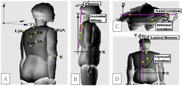
Figure 1. Angle setting (a) location of the infra-red markers used for analysis of scapular orientation and trunk motion, (b) scapular orientation and trunk motion in the sagittal plane, (c) scapular orientation and trunk motion in the transverse plane, (d) scapular orientation and trunk motion in the frontal plane.
the scapula, posterior-lateral acromion, and lateral epicondyle of humerus on the right side. Reliability of scapular orientation measurement using three-di- mensional motion analysis ranged between .99 and 1.0 in upward rotation, between .97 and .99 in poste- rior tilting, and between .78 and .98 in internal rota- tion (Yano et al. 2010).
In this study, scapular internal border, scapular spine, humerus, and thoracic spine were defined as the line from the spinal root to the inferior angle of the scapula, as the line from the spinal root of the scapula to the acromion, as the line from the acro- mion to the lateral epicondyle of humerus, and as the line from T1 to T10, respectively.
Data analysis
Humeral angle, trunk motions, and scapular ori- entations were collected while subjects performed dynamically to raise and lower the arm. Scapular orientations included upward rotation, anterior tilting, and internal rotation of the scapula. Trunk motions included flexion, lateral flexion, and axial rotation.
The humeral angle was calculated from the angle between the humerus and the thoracic spine. The
angle of upward rotation, anterior tilting, and internal rotation were calculated from the angle between the scapular internal border and the thoracic spine in the frontal plane, from the angle between the scapula and the thoracic spine in the sagittal plane, and from the angle between the scapular spine and a horizon- tal line in the transverse plane, respectively (Emery et al. 2010; Kebaetse et al. 1999; Yano et al. 2010).
The angle between the thoracic spine and a pure vertical line was projected in the sagittal and frontal plane for collection of flexion and lateral flexion of the trunk, and the angle between the line from right to left of the tenth posterior rib and a horizontal line was projected in the transverse plane for collection of trunk axial rotation (Emery et al, 2010; Fayad et al. 2008) (Figure 1).
Experimental procedure
The motion analysis system was set up in a laboratory. The participants was standing and kept looking straight ahead in a natural static posture with arms resting at their sides. After calibration was completed, each subject performed five abduction and adduction trials in an unconstrained plane while
Parameters ANOVA factor df F-ratio p-value Scapular orientation
Upward rotation
Group 1 .000 .986
Group x humeral angle 3 .960 .962
Group x phase 1 3.560 .677
Group x humeral angle x phase 3 .192 .902
Anterior tilting
Group 1 2.459 .128
Group x humeral angle 3 4.973 .003*
Group x phase 1 .066 .799
Group x humeral angle x phase 3 .518 .671
Internal rotation
Group 1 1.632 .212
Group x humeral angle 3 .355 .786
Group x phase 1 4.730 .038*
Group x humeral angle x phase 3 .571 .635
Trunk motion
Flexion
Group 1 2.699 .111
Group x humeral angle 3 2.279 .085
Group x phase 1 3.851 .059
Group x humeral angle x phase 3 1.941 .129
Lateral flexion
Group 1 .000 .985
Group x humeral angle 3 .070 .976
Group x phase 1 .000 .999
Group x humeral angle x phase 3 .131 .941
Axial rotation
Group 1 8.615 .006*
Group x humeral angle 3 5.442 .002*
Group x phase 1 1.124 .298
Group x humeral angle x phase 3 1.385 .253
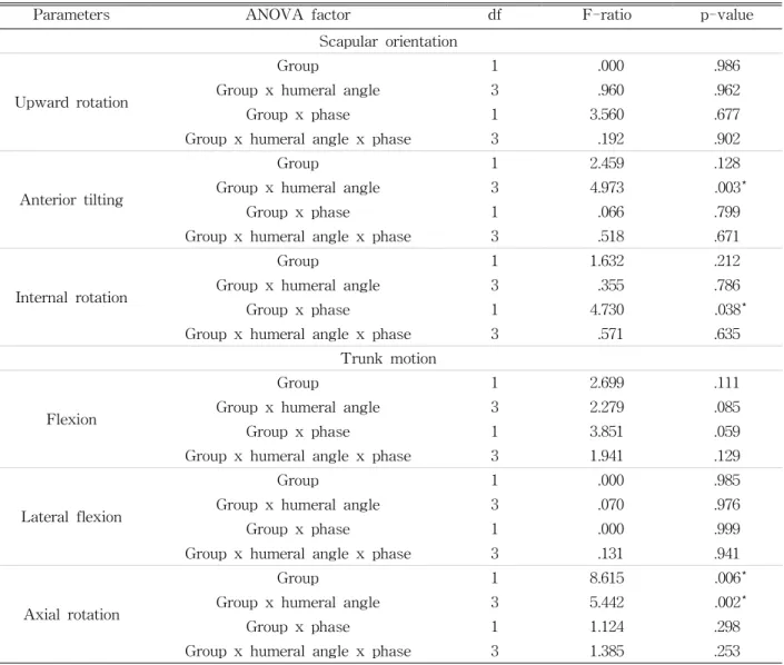
*Indicates statistical significance where p<.05.
Table 1. Mixed model analysis of variance for the effects of group (ideal thorax, hyperkyphosis thracic), phase (raising, lowering) and shoulder angle (30°, 60°, 90° ,120°) on scapular orientation and trunk motion
we recorded their scapular and trunk kinematics.
They attempted to raise the arm in four seconds (raising phase), maintain maximum elevation for one second, and then lower the arm in four seconds (lowering phase). Subjects were allowed a thirty seconds rest between trials. This task did not control the plane of elevation or elbow and hand position, and only required the subject to reach maximal ab- duction range comfortably and to lower to the start- ing arm position for adduction. The scapular and trunk kinematics were collected continuously at a
rate of 100 ㎐ for the five trials, and scapular ori- entations and trunk motions at 30˚, 60˚, 90˚, and 120˚
of the humeral angle in the middle three of the five trials were averaged for data analysis.
6. Statistical analysis
Mixed model ANOVAs (groups-humeral angle x phase) were used to compare scapular orientation (upward rotation, anterior tilting and internal rotation) and trunk motion (flexion, lateral flexion and axial rotation) (dependent variables) between ideal thorax
Ideal thorax group Hyperkyphosis thoracic group
Parameters 30° 60° 90° 120° Phase
total 30° 60° 90° 120° Phase
total Upward rotation
Raising (˚) 1.34 (7.89)a
11.04 (9.46)
21.68 (8.73)
31.10 (9.04)
16.29 (2.01)
2.39 (6.86)
11.42 (8.18)
21.55 (9.58)
30.07 (9.68)
16.57 (1.95) Lowering (˚) 3.10
(7.46)
11.45 (7.92)
21.71 (8.83)
30.73 (9.97)
16.97 (1.85)
3.92 (7.06)
11.92 (6.44)
21.15 (7.67)
30.12 (9.08)
16.78 (1.79) HAb total (˚) 2.66
(1.85)
11.25 (1.96)
21.70 (2.16)
30.91 (2.42)
3.14 (1.79)
11.67 (1.9)
21.35 (2.09)
30.54 (2.34) Group total
(˚)
16.63 (1.89)
16.68 (1.82) Anterior tilting
Raising (˚) -1.12 (2.61)
-5.06 (1.82)
-6.85 (1.74)
-9.95 (3.64)
-5.74 (.67)
-1.56 (2.68)
-5.44 (2.96)
-9.19 (3.56)
-13.78 (4.59)
-7.49 (.65) Lowering (˚) -1.65
(5.21)
-6.11 (4.53)
-8.37 (4.63)
-10.59 (4.75)
-6.68 (.94)
-2.66 (2.54)
-6.39 (2.70)
-9.96 (3.88)
-13.77 (4.28)
-8.20 (.91) HA total (˚) -1.38
(.74)
-5.58 (.69)
-7.61 (.83)
-10.27*
(1.07)
-2.11 (7.1)
-5.92 (.67)
-9.58 (.81)
-13.77*
(1.04) Group total
(˚)
-6.21 (.75)
-7.84 (.72) Internal rotation
Raising (˚) 21.93 (6.20)
20.35 (6.07)
19.44 (6.00)
17.67 (6.08)
19.85† (1.71)
26.55 (7.51)
25.44 (7.02)
23.86 (7.20)
23.47 (8.72)
24.83† (1.66) Lowering (˚) 22.53
(7.52)
20.52 (7.54)
18.60 (10.37)
18.71 (11.9)
20.09 (2.01)
24.45 (7.56)
20.78 (7.29)
20.32 (7.42)
20.71 (8.54)
21.56 (2.01) HA total (˚) 22.23
(1.76) 2043
(1.70) 19.02
(1.87)
18.19 (2.20)
25.50 (1.71)
23.11 (1.65)
22.09 (1.81)
22.09 (2.13) Group total
(˚)
19.97 (1.82)
23.20 (1.76)
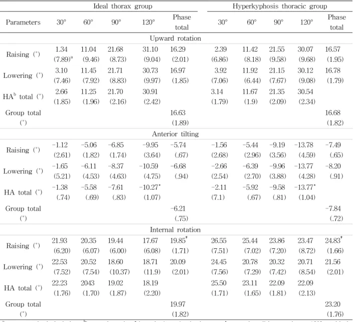
amean±standard deviation, bhumeral angle, *thoracic hyperkyphosis group’s anterior tilting angle at 120° < ideal thorax group, †thoracic hyperkyphosis group’s internal rotation angle at raising phase > ideal thorax group.
Table 2. Mean and standard deviation of scapular orientation across groups, phases or shoulder angles
and thoracic hyperkyphosis groups (independent vari- able). Each analysis included humeral angles (30˚, 60˚, 90˚ and 120˚) and phase (raising phase and lowering phase) as within subject factors. Statistical sig- nificance was set at α<.05 for all analyses and effect sizes were calculated. Post-hoc Tukey tests with Bonferroni adjustments were run where statistically significant interactions were noted. All statistical analyses were performed using SPSS for Windows
software ver. 25.0, (IBM corp., Armonk, NY, USA).
Results
Scapular orientation
For scapular upward rotation, there were no sig- nificant main effects for the group, and interactions for the group by humeral angle, group by phase, or
Ideal thorax group Hyperkyphosis thoracic group
Parameters (˚) 30° 60° 90° 120° Phase
total 30° 60° 90° 120° Phase
total Flexion
Raising 4.96
(4.77)a 5.36 (4.39)
3.93 (4.61)
3.63 (4.22)
4.47 (1.64)
7.97 (7.49)
7.72 (7.77)
7.40 (8.11)
7.59 (9.10)
7.67 (1.59) Lowering 4.10
(4.42) 3.03 (5.54)
2.50 (5.38)
1.80 (6.06)
2.86 (1.77)
8.23 (7.60)
7.20 (8.17)
7.27 (8.78)
6.85 (8.32)
7.39 (1.72) HAb total 4.53
(1.6) 4.20 (1.68)
3.22 (1.79)
2.72 (1.74)
8.10 (1.55)
7.46 (1.63)
7.33 (1.73)
7.22 (1.97)
Group total 3.66
(1.69)
7.53 (1.64) Lateral flexion
Raising .50
(2.73)
2.44 (2.93)
3.42 (3.37)
5.44 (4.26)
2.95 (.73)
.39 (2.05)
2.43 (2.21)
3.59 (2.81)
5.31 (3.73)
2.93 (.70) Lowering 1.18
(2.89)
2.15 (3.52)
3.24 (3.60)
4.95 (4.48)
2.88 (.81)
1.00 (2.28)
2.27 (2.62)
3.27 (3.06)
4.88 (3.68)
2.86 (.79)
HA total .84
(.62)
2.29 (.71)
3.33 (.81)
5.19 (1.03)
.70 (.60)
2.35 (.69)
3.43 (.78)
5.10 (1.00)
Group total 2.91
(.76)
2.89 (.73) Axial rotation
Raising .48
(.30)
-1.29 (2.97)
-2.46 (4.01)
-5.34 (5.63)
-2.15 (.73)
.44 (.28)
-3.65 (2.83)
-6.95 (4.14)
-10.82 (4.77)
-5.24 (.71)
Lowering .64
(4.80)
-3.87 (4.37)
-5.55 (6.69)
-7.28 (5.64)
-4.01 (1.13)
-2.24 (4.76)
-7.53 (4.42)
-10.54 (4.65)
-12.21 (5.07)
-8.13 (1.09)
HA total .56
(.61)
-2.58*
(.81)
-4.00*
(1.10)
-6.31*
(1.30)
-.91 (.59)
-5.59*
(.79)
-8.74*
(1.06)
-11.51*
(1.26)
Group total -3.70†
(.51)
-6.07† (.78)
amean±standard deviation, bhumeral angle, *thoracic hyperkyphosis group’s axial rotation angle at 60°, 90° and 120°
>ideal thorax group, †thoracic hyperkyphosis group’s axial rotation angle >ideal thorax group Table 3. Mean and standard deviation of trunk motion across groups, phases or shoulder angle
group by humeral angle by phase (Table 1).
For scapular anterior tilting, there was a sig- nificant group by humeral angle interaction (p=.003;
effect size=.146) (Table 1). Scapular tiling angles in the raising and lowering phase at each humeral an- gle were averaged for further analysis of each hum- eral angle; there was a significant main effect of the group at 120° of humeral angle (p=.026; effect size=.16) (Table 2). There were no other significant main effects of group or interactions for group by phase, or group by humeral angle by phase.
For scapular internal rotation, there was a sig- nificant group by phase interaction (p=.038; effect
size=.14) (Table 1). Scapular internal rotation angles at 30°, 60°, 90° and 120° of the humeral angle at each phase were averaged for further analysis of each phase, there was a significant main effect of the group at the raising phase (p=.46; effect size=.131) (Table 2). There were no other significant main effects of the group or interactions for the group by humeral angle, or group by humeral angle by phase.
Trunk motion
For trunk flexion, there were no significant main effects of the group or interactions for the group by
humeral angle, group by phase, or group by humeral angle by phase (Table 1).
For trunk lateral flexion, there were no significant main effects of the group or interactions for the group by humeral angle, group by phase or group by humeral angle by phase (Table 1).
For trunk axial rotation, there was a significant main effect of the group (p=.006; effect size=.229) (Table 1). Average trunk axial angles in the thoracic hyperkyphosis group displayed greater trunk axial rotation angles than the ideal thorax group (Table 3).
There was a significant group by humeral angle in- teraction (p=.002; effect size=.158) (Table 1). Trunk axial rotation angles in the raising and lowering phase at each humeral angle were averaged for fur- ther analysis of each humeral angle; there was a significant main effect of the group at 60°, 90°, and 120° of the humeral angle (p=.013; effect size=.194, p=.004; effect size=.249, p=.008; effect size=.221, re- spectively) (Table 3). There were no other significant interactions for group by phase, or group by humeral angle by phase.
Discussion
Scapular orientation
We had assumed that the scapular plane in in- dividuals with thoracic hyperkyphosis posture would naturally be altered due to altered scapular position.
Therefore, subjects were instructed to abduct and adduct across their natural plane. In the current study, individuals with thoracic hyperkyphosis pos- ture displayed significant greater posterior tilting at 120° of the humeral angle compared to those with ideal thorax posture, though scapular upward rotation was not significantly different between groups. In past studies, no significant relationship was revealed between thoracic posture and impingement (Lewis et al. 2005; McClure et al. 2006). Previous studies had reported that less scapular upward rotation, less pos- terior tilting, and greater internal rotation were dis-
played in shoulders with impingement or rotator cuff disease, and less scapular upward rotation and pos- terior tilting of these would result in a lack of ele- vation of the anterior acromion, contributing to im- pingement (Ludewig and Braman, 2011; Ludewig and Cook, 2000; Ludewig and Reynodls, 2009). Our re- sults can explain that thoracic hyperkyphosis posture may not produce an alteration of scapular orientation that could cause impingement or rotator cuff disease.
A previous study reported that when the same subjects adopted a slouched posture compared to an erect posture during abduction in the scapular plane, the posterior tilting relative to the thoracic spine was increased at rest and horizontal arm position and de- creased at maximal arm position, and shoulder max- imal abduction angle was decreased (Kebaetse et al.
1999). The scapular posterior tilting as well as hum- eral external rotation occurred remarkably after 90°
of abduction in the scapular plane and were essential secondary motion for greater humeral elevation, whereas scapular upward rotation occurred approx- imately linearly through humeral elevation (Yano et al. 2009). Scapular posterior tilting and upward rota- tion were accompanied by humeral external rotation (McClure et al, 2001). It has been suggested that in- creases in scapular orientation appeared in order to assist reduced motion of the glenohumeral joint and for protection of the subacromial space (Chopp et al.
2011, McClure et al. 2006). Therefore, an increase of scapular posterior tilting at 120° in subjects with thoracic hyperkyphosis posture may contribute to achievement of full range of humeral elevation.
In our study, subjects in the thoracic hyper- kyphosis group displayed greater internal rotation at the raising phase; this result is consistent with re- ports examining the effects of forward head position and rounded shoulder posture on scapular rotation angle during reaching tasks (Thigpen et al. 2010).
The greater scapular internal rotation produced a protraction such of the scapula that scapular medial border moves away from the thorax (Bourne et al.
2007). Previous studies have found that this position
leads to altered activity of the upper and lower tra- pezius and serratus anterior, reduced shoulder abduc- tion, and external/internal rotation strength due to instability of the scapulothoracic and glenohumeral joints (Ludewig and Reynolds, 2009; Picco et al. 2010;
Smith et al. 2003; Smith et al. 2006). A previous study found that greater upper thoracic angle as well as a lesser craniovertebral angle are displayed in a person with neck pain, and the upper thoracic angle has stronger correlation with neck pain severity than forward head angle (Lau et al. 2010). We suggest that it is possible to cause neck pain if the increased thoracic kyphosis angle related to a greater incline of the cervical spine and protraction of the scapula was sustained for a prolonged period.
We found that subjects with thoracic hyper- kyphosis posture have greater axial rotation of the trunk during arm elevation. In a previous report comparing trunk motion between subjects without and with frozen shoulder, increased axial rotation of the trunk was displayed during abduction in subjects with frozen shoulder compared to healthy subjects, suggesting that increased trunk motion may compen- sate for impaired arm elevation (Fayad et al. 2008).
We may state that the trunk has an important role as kinematic chain during arm elevation, and in- creased trunk torsion in subjects with thoracic hy- perkyphosis posture may represent compensatory motion for greater range of humeral elevation.
Limitations
Subjects in this study were healthy individuals in their early 20 s without pain in the neck or shoulder.
It was difficult to represent the general population or, the relationship between pain and degree of thoracic kyphosis. Altered actions of shoulder musculature can change the motion of scapulothoracic and gleno- humeral joints. There is some lack of explanation as to why there seems to be a difference between our study and previous study results, because we did not measure muscle activity or other spine alignment.
We measured global trunk motion, whereas the pre-
vious study measured rotation within the thoracic spine. This difference between measurements should be considered in the interpretation of these results.
Conclusion
The results of this study show that subjects with thoracic hyperkyphosis posture free from neck/should- er pain displayed greater scapular posterior tilting at 120° of shoulder elevation, greater scapular internal rotation throughout arm raising, as well as greater trunk axial rotation at the upper ranges of shoulder elevation. Our finding provides evidence that thoracic hyperkyphosis posture leads to scapular instability by placing the scapula in a protraction position. Greater trunk rotation and greater scapular posterior tilting was compensatory for full range of abduction instead of scapular instability.
References
Broeks JG, Lankhorst GJ, Rumping K, et al. The long-term outcome of arm function after stroke:
Results of a follow-up study. Disabil Rehabil.
1999;21(8):357-364. https://doi.org/10.1080/0963828 99297459
Borstad JD, Ludewig PM. Comparison of scapular kinematics between elevation and lowering of the arm in the scapular plane. Clin Biomech.
2002;17(9):650–659. https://doi.org/10.1016/s0268- 0033(02)00136-5
Bourne DA, Choo AM, Regan WD, et al. Three-di- mensional rotation of the scapula during func- tional movements: An in vivo study in healthy volunteers. J Shoulder Elbow Surg. 2007;16(2):
150-162. https://doi.org/10.1016/j.jse.2006.06.011 Braman JP, Engel SC, Laprade RF, et al. In vivo
assessment of scapulohumeral rhythm during unconstrained over head reaching in asympto- matic subjects. J Shoulder Elbow Surg. 2009;
18(6):960-967.
Bullock MP, Foster NE, Wright CC. Shoulder im- pingement: the effect of sitting posture on shoulder pain and range of motion. Manual Ther. 2005;10(1):28-37. https://doi.org/10.1016/
j.math.2004.07.002
Chopp JN, Fischer SL, Dickerson CR. The specificity of fatiguing protocols affects scapular orientation:
Implications for subacromial impingement. Clin Biomech. 2011;26(1):40-45. https://doi.org/10.1016 /j.clinbiomech.2010.09.001
Claus AP, Hides JA, Moseley GL, et al. Is ‘ideal’
sitting posture real?: Measurement of spinal curves in four sitting postures. Manual Ther.
2009;14(4):404–408. https://doi.org/10.1016/j.math.
2008.06.001
Crosbie J, Kilbreath SL, Hollmann L, et al.
Scapulohumeral rhythm and associated spinal motion. Clin Biomech. 2008;23(2):184–192.
https://doi.org/10.1016/j.clinbiomech.2007.09.012 Edmondston SJ, Aggerholm M, Elfving S, et al.
Influence of posture in the range of axial rota- tion and coupled lateral flexion of the thoracic spine. J. Manipulative Physiol Ther. 2007;30(3):
193-199. https://doi.org/10.1016/j.jmpt.2007.01.010 Emery K, De Serres SJ, McMillan A, et al. The ef-
fects of a Pilates training program on arm-trunk posture and movement. Clin Biomech. 2010;25(2):
124-130. https://doi.org/10.1016/j.clinbiomech.2009.
10.003
Fayad F, Hanneton S, Lefevre-Colau MM, et al. The trunk as a part of the kinematic chain for arm elevation in healthy subjects and in patients with frozen shoulder. Brain Res. 2008;1191(29):107-115.
https://doi.org/10.1016/j.brainres.2007.11.046 Finley MA, Lee RY, Effect of sitting posture on
3-dimensional scapular kinematics measured by skin mounted electromagnetic tracking sensors.
Arch Phys Med Rehabil. 2003;84(4):563-368.
https://doi.org/10.1053/apmr.2003.50087
Kebaetse M, McClure P, Pratt NA. Thoracic position effect on shoulder range of motion, strengthand
three-dimensional scapular kinematics. Arch Phys Med Rehabil. 1999;80(8):945-950. https://doi.org/
10.1016/s0003-9993(99)90088-6
Kuo YL, Tully EA, Galea MP. Video analysis of sagittal spinal posture in healthy young and older adults. J Manipulative Physiol Ther. 2009;
32(3):210-215. https://doi.org/10.1016/j.jmpt.2009.
02.002
Lau KT, Cheung KY, Chan KB, et al. Relationships between sagittal postures of thoracic and cer- vical spine, presence of neck pain, neck pain se- verity and disability. Manual Ther. 2010;15(5):
457-462. https://doi.org/10.1016/j.math.2010.03.009 Lewis JS, Green A, Wright C. Subacromial impinge-
ment syndrome: The role of posture and muscle imbalance. J Shoulder Elbow Surg. 2005;14(4):
385-392. https://doi.org/10.1016/j.jse.2004.08.007 Ludewig PM, Braman JP. Shoulder impingement: bi-
omechanical considerations in rehabilitation.
Manual Ther. 2011;16(1):33-39.
Ludewig PM, Cook TM. Alterations in shoulder kin- ematics and associated muscle activity in people with symptoms of shoulder impingement. Phys Ther. 2000;80(3):276-291. https://doi.org/10.1016/
j.math.2010.08.004
Ludewig PM, Reynolds JF. The association of scapular kinematics and glenohumeral joint pathologies. J Orthop Sports Phys Ther. 2009;39(2):90-104.
https://doi.org/10.2519/jospt.2009.2808
McClure PW, Michener LA, Karduna AR. Shoulder function and 3-dimensional scapular kinematics in people with and without shoulder impingement syndrome. Phys Ther. 2006;86(8):1075-1090.
McClure PW, Michener LA, Sennett BJ, et al. Direct 3-dimensional measurement of scapular kine- matics during dynamic movements in vivo. J Shoulder Elbow Surg. 2001;10(3):269-277.
Mell AG, LaScalza S, Guffey P, et al. Effect of rota- tor cuff pathology on shoulder rhythm. J Shoulder Elbow Surg. 2005;14(1):58-64.
https://doi.org/10.1016/j.jse.2004.09.018
Meumann DA. Kinesiology of the Musculoskeletal
This article was received October 18, 2019, was re- viewed October 18, 2019, and was accepted November 15, 2019.
System: foundations for rehabilitation, second ed.
Mosby, NY, 2010
Mottram SL. Dynamic stability of the scapula.
Manual Ther. 1997;2(3):123-131. https://doi.org/
10.1054/math.1997.0292
Picco BR, Fischer SL, Dickerson CR. Quantifying scapula orientation and its influence on maximal hand force capability and shoulder muscle activity.
Clin Biomech. 2010;25(1):29-36. https://doi.org/10.
1016/j.clinbiomech.2009.09.008
Pribicevic M, Pollard H, Bonello R. An epidemiologic survey of shoulder pain in chiropractic practice in Australia. J Manipulative Physiol Ther. 2009;
32(2):107-117. https://doi.org/10.1016/j.jmpt.2008.
12.005
Sheikhzadeh A, Yoon J, Pinto VJ, Kwon YW.
Three-dimensional motion of the scapula and shoulder during activities of daily living. J Shoulder Elbow Surg. 2008;17(6):936-942.
https://doi.org/10.1016/j.jse.2008.04.008
Sizer PS, Brisme´e JM, Cook C. Coupling behavior of the thoracic spine: a systematic review of the literature. J Manipulative Physiol Ther. 2007;
30(5):390-399. https://doi.org/10.1016/j.jmpt.2007.
04.009
Smith J, Dietrich CT, Kotajarvi BR, et al. The effect of scapular protraction on isometric shoulder ro- tation strength in normal subjects. J Shoulder Elbow Surg. 2006;15(3):339-343. https://doi.org/
10.1016/j.jse.2005.08.023
Smith J, Kotajarvi BR, Padgett DJ, et al. Effect of scapular protraction and retraction on isometric shoulder elevation strength. Arch Phys Med Rehabil. 2002;83(3):367-370. https://doi.org/10.1053/
apmr.2002.29666
Tempelhof S, Rupp S, Seil R. Age-related prevalence of rotator cuff tears in asymptomatic shoulders.
J Shoulder Elbow Surg 1999;8(4):296-299.
https://doi.org/10.1016/s1058-2746(99)90148-9 Theodoridis D, Ruston S. The effect of shoulder
movements on thoracic spine 3D motion. Clin Biomech. 2002;17(5):418-421. https://doi.org/10.
1016/s0268-0033(02)00026-8
Thigpen CA, Padua DA, Michener LA, et al. Head and shoulder posture affect scapular mechanics and muscle activity in overhead tasks. J Electromyogr Kinesiol. 2010;20(4):701-709. https://doi.org/10.1016/
j.jelekin.2009.12.003
Van der Windt DA, Koes BW, Boeke AJ, et al.
Shoulder disorders in general practice: prog- nostic indicators of outcome. Br J Gen Pract.
1996;46(410):519-523.
Wegner S, Jull G, O’Leary S, et al. The effect of a scapular postural correction strategy on tra- pezius activity in patients with neck pain. Man Ther. 2010;15(6):562-566. https://doi.org/10.1016/
j.math.2010.06.006
Wu G, van der Helm FC, Veeger HE, et al. ISB recommendation on definitions of joint coordinate systems of various joints for the reporting of human joint motion—Part II: shoulder, elbow, wrist and hand. J Biomech. 2005;38(5):981-992.
https://doi.org/10.1016/j.jbiomech.2004.05.042 Yang JL, Lu TW, Chou FC, et al. Secondary mo-
tions of the shoulder during arm elevation in patients with shoulder tightness. J Electromyogr Kinesiol. 2009;19(6):1035-1042. https://doi.org/10.
1016/j.jelekin.2008.10.011
Yano Y, Hamada J, Tamai K, et al. Different scap- ular kinematics in healthy subjects during arm elevation and lowering: glenohumeral and scap- ulothoracic patterns. J Shoulder Elbow Surg.
2010;19(2):209-215. https://doi.org/10.1016/j.jse.
2009.09.007