Introduction
The patella is located between the femur and tibia, and the knee joint acts as a pulley [1]. In general, the area of contact between the patella and the femur is larger and thicker than the medial articular surface, resulting in a more significant load [2]. Abnormal alignment of the patella and an imbalance in muscle strength are pathological causes of patellofemoral pain syndrome (PFPS) [3]. The imbalance of muscle strength causes chronic pain by repeating functionally incorrect movements and decreasing motor performance [4]. For
the correct alignment of the patella, strength exercises are essential for ensuring the balance and stability of the medial and lateral masses [5].
Strength training can be divided into a closed-kinetic chain (CKC) exercise that is performed while the distal limb is fixed and an open-kinetic chain (OKC) exercise that is performed in the free state without a distal limb [6]. Among these exercises, CKC exercise mobilizes many muscles of the body segment. Moreover, as the distal limb is fixed, it provides stability through a joint compression force and promotes a proprioceptive sensation[7]. CKC exercise is essential for rehabilitation https://doi.org/10.14474/ptrs.2021.10.2.98
eISSN 2287-7584 pISSN 2287-7576
Phys TherRehabil Sci 2021, 10(2), 98-105
www.jptrs.org
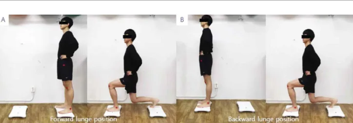
Comparative Study of the Biomechanical Factors in Range of Motion, Muscle Activity, and Vertical Ground Reaction Force between a Forward Lunge and Backward Lunge
Samho Park
a, TianZong Huang
a, Junyoung Song
a, Myungmo Lee
ba
Department of Physical Therapy, Graduate School, Daejeon University, Republic of Korea
b
Department of Physical Therapy, Daejeon University, Republic of Korea
Objective: The purpose of this study was to examined the kinematic relationship and differences through the range of motion (ROM), muscle activity, and vertical ground reaction force (VGRF) during forward and backward lunge movements, which are effective in improving muscle strength and balance ability of the lower extremities, and to provide clinical information on more efficient lunge movements.
Design: Cross-sectional study
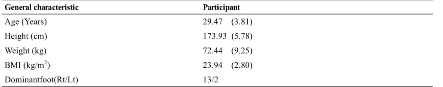
Methods: Fifteen adult males who met the selection criteria were tested for their dominant feet.Forward and backward lunges were then performed, and the ROM, muscle activity, and VGRF were measured for kinematic analysis during the lunge movement.The differences betweenthe forward lunge and backward lunge intervention were examined using a paired t-test.
Results: A significant increase in the ROM of the knee and ankle was observed during the forward and backward lunges (p<0.05).
In addition, in terms of the muscle activity, the peak values of the vastus medialis oblique (VMO) and VGRF also showed a significant increase in the forward lunge compared to the backward lunge (p<0.05).
Conclusions: This study showed an increase in VGRF peak value, knee and ankle ROM, and VMO muscle activity during forward lunge. Based on these results, it is considered necessary to apply differently depending on the direction of progress in consideration of the musculoskeletal situation and physical ability during the lunge movement.
Key Words: Electromyography, Exercise, Kinematics, Quadriceps Muscle, Weight-Bearing
Received: Apr 8, 2021 Revised: May 12, 2021 Accepted: May 14, 2021
Corresponding author: Myungmo Lee(ORCID https://orcid.org/0000-0002-2192-1701) Department of Physical Therapy, Daejeon University
62, Daehak-ro, Dong-gu, Daejeon city, Republic of Korea [34520]
Tel: Fax: 82-42-280-4295, E-mail: [email protected]
This is an Open-Access article distributed under the terms of the Creative Commons Attribution Non-Commercial License (http://creativecommons.org/licenses/
by-nc/4.0) which permits unrestricted non-commercial use, distribution, and reproduction in any medium, provided the original work is properly cited.
Copyright © 2021 Korean Academy of Physical Therapy Rehabilitation Science