• 검색 결과가 없습니다.

Role of the PLA2-Activated Neutrophilic Oxidative Stress in Oleic Acid-Induced Acute Lung Injury

N/A
N/A
Protected

Academic year: 2021

Share "Role of the PLA2-Activated Neutrophilic Oxidative Stress in Oleic Acid-Induced Acute Lung Injury"

Copied!
7
0
0

로드 중.... (전체 텍스트 보기)

전체 글

(1)

Tuberc Respir Dis 2010;68:55-61

CopyrightⒸ2010. The Korean Academy of Tuberculosis and Respiratory Diseases. All rights reserved.

Role of the PLA2-Activated Neutrophilic Oxidative Stress in Oleic Acid-Induced Acute Lung Injury

Young Man Lee, M.D., Byung Yong Kim, M.D., Yoon Yub Park, M.D.

Department of Physiology, Catholic University of Daegu School of Medicine, Daegu, Korea

Background: The underlying pathogenesis of fat embolism-induced acute lung injury (ALI) has not been elucidated.

In the present study, the pathogenesis of fat embolism-induced ALI was probed in association with neutrophilic oxidative stress in oleic acid (OA)-induced ALI of S-D rats.

Methods: OA was injected intravenously to provoke ALI in experimental rats. Five hours later, indices of ALI were measured to confirm the role of the neutrophilic respiratory burst. The effect of an inhibition of phospholipase A2 (PLA2) was also evaluated.

Results: The accumulation of neutrophils in the lung due to OA caused increased neutrophilic oxidative stress in lung, which was ameliorated by mepacrine. What were the results from inhibition of PLA2.

Conclusion: Excess neutrophilic oxidative stress contributes to OA-induced ALI, which is lessened by the inhibition of PLA2.

Key Words: Acute Lung Injury; Embolism, Fat; Neutrophils; Phospholipase A2

Address for correspondence: Young Man Lee, M.D.

Department of Physiology, Catholic University of Daegu School of Medicine, 3056-6, Daemyung 4-dong, Nam-gu, Daegu 705-718, Korea

Phone: 82-53-650-4472, Fax: 82-53-850-3293 E-mail: [email protected]

Received: Dec. 10, 2009 Accepted: Feb. 9, 2010

Introduction

Acute respiratory distress syndrome (ARDS) is charac- terized by an acute inflammatory pulmonary edema re- sulting in severe hypoxemia and respiratory failure

1

. One of the etiologies of this morbid malady is the pul- monary fat embolism encountered in multiple traumas including multiple fracture

2

. Multiple fractures can be corrected by suitable surgical intervention but the leak- age of minute fat droplets into microvasculatures is un- avoidable during operation or injury. Even during major operations including abdominal surgery, minute fat droplets are leaked into the vasculatures and cause fat embolism.

Among many types of lipids, oleic acid (OA) is well known to provoke acute lung injury during pulmonary

fat embolism

3

. Up to now, however, the underlying pathogenesis of the fat embolism-induced acute lung in- jury (ALI) has not been well known. Some insist that neutrophils play a key role in the genesis of acute al- veolar flooding by the release of elastase and free radi- cals during pulmonary fat embolism

4

. Whereas Hill et al

5

suggested that neutrophils were not involved in the ALI caused by infusion of OA in rats.

In addition, although free radicals are one of the can-

didates altering the function of alveolar capillary barrier

in OA-induced (ALI), the main source of free radicals

has not been elucidated. Since fat embolisminduced ALI

or ARDS are currently serious clinical problem, espe-

cially closely related to sepsis or severe form of in-

fection, some authors reported that the phospholipase

A2 (PLA2), a rate limiting enzyme of inflammation,

regulated the inflammatory reaction in the lung during

ARDS

6

. The activation of PLA2 prior to that of NADPH

oxidase is indispensable for the initiation of neutrophilic

respiratory burst

7

. Many evidences have been issued

that the activation of the PLA2 evokes neutrophilic res-

piratory burst in several organs

8

. Several proinflam-

(2)

matory lipid molecules released by the PLA2 activation are thought to be responsible for the respiratory burst

9

. Needless to say, neutrophils constitute a very important mobile defensive system of the body by releasing pro- tease and free radicals to the vicinity of the inflamed area. But the altering or imbalance of the production of the free radicals and deterring of the antioxidant sys- tem by the neutrophils in the body ensue hazardous tis- sue injury

10

.

On the bases of these reports and hypotheses, we at- tempted to elucidate whether the neutrophilic respira- tory burst is involved in the pathogenesis of OA-induced ALI. By carrying out these experiments we would like to confirm the role of neutrophils in OA-induced ALI, from which could be followed by the development of new modalities for the treatment or prevention of multi- ple trauma or fracture-induced ARDS.

Mepacrine was used for the inhibition of PLA2 of which activation is crucial for the neutrophilic respira- tory burst.

Materials and Methods 1. Materials

Neutrophils were isolated from fresh human blood.

Male Sprague-Dawley rats (BW 250 g) were pathogen free and purchased from Korean Scientific Animal Supplier. All chemicals were purchased from Sigma- Aldrich Chemical Co, if not commented otherwise.

2. Methods

1) Evaluation of the generation of free radicals from neutrophils

(1) Isolation of human neutrophils: To know the effect of OA on the generation of free radicals, neutrophils were isolated according to the methods of Haslett et al

11

. Briefly, 30 mL of blood containing heparin (100 IU) mixed with equal volume of Pentaspan in 50 mL syringe and stand for 45 minutes in two syringes.

After separation of plasma which contained neu- trophils, it was overlayed on the gradient-made percoll solution (74, 55%) and centrifuged 1,500 rpm for 20 mi-

nutes at room temperature. Layer of neutrophils was collected and washed twice and resuspended with Hank's balance salt solution (HBSS with 1 mM CaCl

2

, pH 7.4).

(2) Cytochrome-c reduction assay: According to the method of Botha

12

, the generation of superoxide anion was assessed by cytochrome-c reduction assay. In tubes containing neutrophils (1×10

7

cell/mL), HBSS, sodium oleate (100 μM), mepacrine were added and incubated for 30 minutes at 37

o

C in water bath. The amount of produced superoxide anion was evaluated as the a- mount of reduced cytochrome-c (nmol/1×10

7

cells).

(3) Confirmation of the neutrophilic respiratory burst by nitro blue tetrazolium (NBT) test: Nitro blue tetrazo- lium is reduced by superoxide anion and to form for- mazan granules. If neutrophils are in the state of respi- ratory burst, neutrophils react with NBT and engulf for- mazan granules which can be shown in the cytoplasm.

For NBT test, isolated human neutrophils (2×10

7

cells/mL) were reacted in tubes containing 0.02% NBT, 100 μM sodium oleate and 100 μM mepacrine at 37

o

C for 20 minutes. After incubation, the suspension of cells was centrifuged at 800 rpm for 10 minutes, the pellet was put on the slides, then the slides were wright stained. NBT positive neutrophils were counted per 200 neutrophils and the percentage of NBT positive cells was calculated.

(4) Test of neutrophilic chemotaxis by OA and mepa- crine; neutrophil aggregation test: To confirm the che- motactic effect of OA to neutrophils, neutrophil ag- gregation test was performed. In tube containing iso- lated neutrophils (1×10

7

cell/mL), NBT, sodium oleate and mepacrine (0.02%, 100 μM, 100 μM, respectively) were added. Then tubes were incubated at 37

o

C for 20 minutes and the formation of clump consisted of neu- trophils, OA and formazan was identified. Mepacrine was tried to know the effect of the inhibition of PLA2 which is known to be essential for the neutrophilic res- piratory burst.

2) Induction of ALI in Sprague-Dawley rats

For the induction of ALI simulating OA-induced

ARDS, OA (30 μg) was mixed with 0.1% bovine serum

(3)

Table 1. Cytochrome-c reduction assay

Control (n=9) OA (n=9) OA+Mepa (n=9)

Cytochrome-creduced (nmol/1.0×10

7

pMNs) 46.1±1.5 103.4±6.4* 38.9±0.3

Values are given as mean±SD, n indicates number of experiments.

*p<0.001, control vs OA,

p<0.001, OA vs OA+Mepa.

Mepa: mepacrine; OA: oleic acid.

albumin (270 μL) and was injected into the saphenous vein of the rats

13

. Twenty four hours after the injection, rats were sacrificed by overdose injection of the mixture of ketamine hydrochloride and xylazine. The rats were thoracotomized and lungs were resected. Then the lungs were weighed, dipped into the liquid nitrogen for being frozen immediately and stored at -70

o

C till analyzed. To know the effect of the inhibition of PLA2, mepacrine (20 mg/kg) was given intraperitoneally 30 minutes after OA injection.

(1) Calculation of L/B ratio: At the time of sacrifice, body weights and resected lung weights were measured before the calculation of the lung weight/body weight ratio (L/B ratio). The ratio denotes the accumulation of lung water after treatment of OA and mepacrine.

(2) Measurement of lung MPO: The lung MPO activity (U/g of wet lung) was measured according to the Gold- blum's method

14

. Briefly, left lungs were thawed and homogenized in 4.0 mL of phosphate buffer (20 mM, pH 7.4) before centrifugation at 30,000×g for 30 mi- nutes at 4

o

C. Then the pellet was resuspended in 4.0 mL of phosphate buffer (50 mM, pH 6.0) with 0.5%

hexadecyl tetramethyl ammonium bromide. Samples were sonicated (Polytron, Swizland) for 90 seconds on ice at maximal power and then incubated for 120 mi- nutes at 60

o

C. MPO activity was measured using o-dia- nisidine as the chromogenic substrate and hydrogen peroxide to initiate the reaction. Absorbance at 460 nm was proportional to MPO activity.

(3) Measurement of BALF protein content: To obtain BALF, bronchoalveolar lavage was carried out with Tris- buffered saline (pH 7.4). With 8.0 mL of buffer solution, 3 times of pushing and withdrawing through the trachea was done and collected 6.0 mL of BALF. The BALF was centrifuged 1,500 rpm for 20 minutes at room temper-

ature to sediment cellular components. The supernat- ants were used for the determination of protein accord- ing to the method of Brown et al

15

.

(4) Enumeration of WBC in BALF: To assess the mi- gration of white blood cells into the alveolar lumen, we counted the number of WBC in the BALF. After BAL, BALFs were centrifuged and supernatants were col- lected for the measurement of protein. Then the sedi- ments were resuspended with 0.1 mL of Tris-buffered saline and aliquots were used to count WBC with hemocytometer.

3) Statistical analyses

Data were analyzes using a one-way analysis of var- iance with a Tukey-Kramer multiple comparison test. A p-value of <0.05 was considered statistically signifi- cant.

Results

Following experimental results were obtained by the study to elucidate the role of neutrophilic oxidative stress in OA-induced ALI.

In cytochrome-c reduction assay, OA stimulated iso- lated human neutrophils to increase the generation of superoxide anion which resulted in reduction of 103.4

±6.42 nmol of cytochrome-c/1.0×10

7

PMNs (n=9).

Comparing with 46.1±1.51 nmol of cytochromec/1.0×

10

7

PMNs (n=9) in control neutrophils, OA increased the production of free radicals from neutrophils significantly (p<0.001). And mepacrine (100 μM) suppressed the OA-provoked superoxide generation effectively (p<

0.001) which resulted in the reduction of 38.9±0.3 nmol of cytochrome-c/1×10

7

PMNs (n=9) (Table 1).

In NBT test for the confirmation of neutrophilic respi-

ratory burst, OA intensified neutrophilic respiratory

(4)

Figure 1. Microscopic finding of OA-neutrophil aggregate after NBT reaction in vitro. Massive accumulation of for- mazan-positive neutrophils (asterisk) was noted at the vi- cinities of OA droplets (arrows). Original magnification

×100.

Figure 2. Morphological evidence of chemotactic activity of OA on neutrophils. Around OA droplets (asterisk), dense accumulation of formazan positive neutrophils was noted. Almost all the neutrophils were formazan positive.

The accumulation of neutrophils at the vicinities of OA droplets was much more dense compared with that of the remote area from OA droplets (arrow head). Original magnification ×40.

Table 2. NBT test for the evaluation of PMN respiratory burst

Control OA OA+Mepa

(n=10) (n=10) (n=10)

Formazan positive 4.6±2.9 96.1±2.4* 9.1±5.7

cells, %

Values are given as mean±SD, n indicates number of experiments.

*p<0.001, control vs OA,

p<0.001, OA vs OA+Mepa.

Mepa: mepacrine; OA: oleic acid.

Table 3. Neutrophil aggregation test in vitro

Control OA OA+Mepa

(n=10) (n=10) (n=10)

Aggregation of PMN 0 100 0

(1×10

7

cell/mL), %

n indicates number of experiments.

burst which resulted in the increase of formazan pos- itive cells to 96.1±2.42% (n=10) compared with 4.6±

2.9% (n=10) of non-stimulated neutrophils. Mepacrine (100 μM) suppressed the neutrophilic respiratory burst which was resulted in the decrease of NBT positive cells to 9.1±5.72% (n=10, p<0.001) compared with 96.1±

2.42% (n=10) of OA-stimulated neutrophils (Table 2).

Neutrophilic aggregation test for the confirmation of the neutrophilic chemotaxis of OA showed dramatic ag- gregation and chemotaxis of neutrophils (Figures 1, 2).

The light microscopic examination revealed that the formed clumps were composed of neutrophils contain- ing formazan granules and lipid droplets. Without OA, neutrophils did not form the aggregate and mepacrine (100 μM) inhibited the formation of clumps or ag-

gregate completely. That is, the formation of the ag- gregate in OA stimulated neutrophils was 100% (n=10).

However, in control and mepacrine treated neutrophils, the formation of aggregates was nil (n=10, respectively) (Table 3).

As one of the indices of the accumulation of the fluid in the lung, L/B ratio (lung weight×10

3

/body weight) was calculated. Comparing with 5.45±0.45 of control rats (n=11), the L/B ratios of OA given rats were 8.07±

0.54 (n=6) and higher (p<0.001) than that of control rats. Mepacrine (100 μM) decreased (p<0.001) L/B ra- tios to 5.36±0.60 (n=15) in OA-given rats (Table 4).

The protein content (O.D at 562 nm) in the BALF of

OA-given rats were 0.45±0.15 (n=12) and was higher

(p<0.001) than 0.27±0.05 of control rats (n=17). In

(5)

Table 4. Comparison of lung weight/body weight ratio (L×10

3

/B)

Control OA OA+Mepa

(n=11) (n=6) (n=15)

L×10

3

/B 5.45±0.45 8.07±0.54* 5.36±0.60

Values are given as mean±SD, n indicates number of experiments.

*p<0.001, control vs OA,

p<0.001, OA vs OA+Mepa.

Mepa: mepacrine; OA: oleic acid.

Table 5. Comparison of protein content (OD at 562 nm)

Control OA OA+Mepa

(n=17) (n=12) (n=10)

OD at 562 nm 0.27±0.05 0.45±0.15* 0.33±0.09

Values are given as mean±SD, n indicates number of experiments.

*p<0.001, control vs OA,

p<0.001, OA vs OA+Mepa.

Mepa: mepacrine; OA: oleic acid.

Table 6. Comparison of lung MPO (μ/g of wet lung) and number of WBC (millions/two lungs) in BALF

Control OA OA+Mepa

Nr. WBC in BALF 5.82±1.50 (n=10) 20.15±0.39 (n=11)* 6.76±2.47 (n=10)

MPO 9.96±2.64 (n=8) 33.38±10.44 (n=9) 35.98±10.53 (n=7)

Values are given as mean±SD, n indicates number of experiments.

*p<0.001, control vs OA,

p<0.001, OA vs OA+Mepa.

Mepa: mepacrine; OA: oleic acid.

comparison, mepacrine (100 μM) decreased (p<0.001) protein content to 0.33±0.09 (n=10) in OA given rats (Table 5).

The number of WBC (millions/two lungs) in BALF af- ter OA treatment increased (p<0.001) to 20.15±0.39 (n=11) compared with 5.82±1.50 (n=10) of contol rats.

Mepacrine (100μM) decreased the number of WBC ef- fectively to 6.76±2.47 (n=16, p<0.001) in OA given rats (Table 6).

The lung MPO activities (U/g of wet lung) of OA giv- en rats were increased (p<0.001) to 33.38±10.44 (n=

19) by OA compared with 9.96±2.64 (n=8) of control rats. The treatment of mepacrine (100μM) resulted in the MPO activity of 35.98±10.53 (n=7) and did not change the increased MPO activities by OA (Table 6).

Discussion

The present study was performed to know whether neutrophilic oxidative stress played a role in the patho- genesis of acute inflammatory edema in OA-induced ALI.

As is known, the exact pathogenesis of ARDS has not been explained, and accordingly the appropriate way

of management of ARDS has not been established. Even if the pathological findings of full-blown ARDS shows very similar traits regardless of the various etiologies, still there is a possibility of diverse pathogeneses of ARDS according to their etiologies. In this regards, ARDS caused by pulmonary fat embolism is entitled to be studied meticulously to get a clue to understand the pathogenesis and a way of novel treatment of this noto- rious malady. In other words, we do not think ARDS is a single disease entity in spite of its identical patho- logical and physiological outcomes regardless of multi- ple etiologies.

In this point of view, we tried to figure out the role of neutrophils in OA-induced ARDS. Despite some au- thors

16

insisted that neutrophilic oxidative stress had no functional, pathophysiological relationship with patho- genetic pathway of OA-induced ARDS, we have found opposite evidences to confirm the neutrophilic roles in OA-induced ALI.

Firstly, OA stimulated the generation of neutrophilic superoxide anion conspicuously and it was suppressed by the inhibition of PLA2 with mepacrine (100 μM).

The effect of OA on the neutrophilic oxidative stress

was confirmed again by the NBT test. OA increased

(6)

NBT positive cells, which was inhibited by mepacrine (100 μM). As Dana et al

17

demonstrated, the activation of PLA2 was prerequisite of neutrophilic superoxide generation. Therefore OA appeared to activate PLA2 of neutrophils causing the release of superoxide anion from neutrophils. This assumption was confirmed by cytochrome-c reduction assay and NBT test in this study. Very interestingly, in our neutrophil aggregation test, OA-induced neutrophilic aggregation and respira- tory burst. In microscopic examination of the ag- gregates, it was revealed that the formazan positive neu- trophils were intermingled with minute OA-droplets.

These findings are thought to be very important to un- derstand the pathogenesis of OA-induced ALI, and one step forward, fat embolism-induced ARDS. If fat drop- lets in the capillaries, induces chemotaxis of neutrophils into the microvasculatures and is followed by respira- tory burst, these processes definitely ensues the destruc- tion of alveolar and endothelial barriers very rapidly. As was confirmed in our study, OA was chemotactic and provoked the generation of superoxide anion, which could promote the oxidative stress in pulmonary capillaries. Thus OA is the instigator of neutrophilic oxi- dative stress in the pulmonary capillaries and the ini- tiator of pulmonary edema, at least in OA-induced ARDS.

As Repine

18

pointed out, localized, abrupt and ex- plosive generation of free radicals from neutrophils can't be opposed or neutralized by antioxidant system in the body. In OA-induced ALI, the OA-droplets block the pulmonary capillaries, attract neutrophils and make ag- gregates which are the massive source of the generation of superoxide anion.

Secondly, the role of PLA2 is also crucial for the evoking of OA-induced ARDS. Long before, Lee et al

19

stressed that PLA2 is the pivot of the inflammatory cas- cade during ARDS. Similarly, Furue et al

20

demonstrated that PLA2 was crucial in OA-induced ARDS in rabbits.

In our present study, mepacrine suppressed the gen- eration of superoxide anion from neutrophils, resulted in the suppression of oxidative stress and decreased the number of NBT positive neutrophils. Even if mepacrine

(100 μM) suppressed the respiratory burst, it could not prevent the accumulation of neutrophils in the micro- vessels because it did not have the resolving activity on chemotactic OA droplets. As was pointed out, OA is a chemotactic, especially to neutrophils, which was the reason why OA increased lung MPO activity. The in- creased lung MPO activity was not decreased by mepa- crine because of the chemotactic property of OA. Only the production of superoxide anion was suppressed by mepacrine by way of the inhibition of PLA2.

In association with OA-induced neutrophilic aggrega- tion, another possible assumption is that the neutrophi- lic plug induced pulmonary hypertension. Young and his colleagues

21

demonstrated that the strong antihyper- tensive nitroprusside ameliorated pulmonary edema and pulmonary hypertension in OA-induced ALI. In con- junction with our experimental evidences to Young's re- port, it is highly probable that OA induced neutrophilic aggregation in microvasculature which blocks the capil- lary lumen, then increases hydrostatic pressure causing the pulmonary edema. In the present study, the in- hibition of PLA2 with mepacrine suppressed the for- mation of the aggregates.

From these results, we would like to propose that ox- idants and OA were the initiator of the formation of neutrophilic clump in the microvessels. When we take into account of Vadasz's report

22

that OA inhibition of alveolar fluid absorption, the OA's role to cause pulmo- nary edema is much more evident in association with neutrophilic oxidative stress. It's analagous to increase pressure into the punctured tube increases the output of the fluid through the holes but not through the im- permeable walls.

Thirdly, in our in vivo study, OA increased lung MPO

activity. Since OA is regarded as a chemotactic material

for neutrophils, the increased accumulation of neu-

trophils in the lung is natural. The accumulated neu-

trophils are thought to be the source of free radicals

causing oxidative stress, thus the increased amount of

protein in BALF after OA is not unexpected. By the in-

hibition of PLA2 with mepacrine, the generation of su-

peroxide anion was diminished and followed by the de-

(7)

crease of protein content in BALF. Although mepacrine did not decrease the MPO activity in OA-induced ALI, it decreased the migration of the neutrophils into the alveolar lumen. Thus the inhibition of PLA2 appears to decrease the oxidative stress in OA-induced ALI.

Taken together, on the basis of our present study, we concluded that neutrophilic oxidative stress played a pivotal role in OA-induced ALI in Sprague-Dawley rats.

And some appropriate interventions to decrease the ag- gregation and oxidative stress of neutrophils could lead to the novel modalities for prevention and cure of the OA-induced ALI.

References

1. Ware LB, Matthay MA. The acute respiratory distress syndrome. N Engl J Med 2000;342:1334-49.

2. Bernard GR. Acute respiratory syndrome: a historical perspective. Am J Respir Crit Care Med 2005;172:

798-806.

3. Zhou Z, Kozlowski J, Schuster DP. Physiologic, bio- chemical, and imaging characterization of acute lung injury in mice. Am J Respir Crit Care Med 2005;

172:344-51.

4. Liu H, Zhang D, Zhao B, Zhao J. Superoxide anion, the main species of ROS in the development of ARDS induced by oleic acid. Free Radical Res 2004;38:1281-7.

5. Hill LL, Chen DL, Kozlowski J, Schuster DP. Neutro- phils and neutrophil products do not mediate pulmo- nary hemodynamic effects of endotoxin on oleic acid- induced lung injury. Aneth Analg 2004;98:452-7.

6. Tighe D, Moss R, Boghossian S, Heath MF, Chessum B, Bennett ED. Multi-organ damage resulting from ex- perimental faecal peritonitis. Clin Sci (Lond) 1989;76:

269-76.

7. Dana R, Malech HL, Levy R. The requirement for phos- pholipase A2 for activation of the assembled NADPH oxidase in human neutrophils. Biochem J 1994;297:

217-23.

8. Jaeschke H, Smith CW. Mechanisms of neutrophil-in- duced parenchymal cell injury. J Leukoc Biol 1997;

61:647-53.

9. Bulger EM, Maier RV. Lipid mediators in the patho- physiology of critical illness. Crit Care Med 2000;28:

N27-36.

10. Babior BM. Phagocytes and oxidative stress. Am J Med 2000;109:33-44.

11. Haslett C, Guthrie LA, Kopaniak MM, Johnston RB Jr, Henson PM. Modulation of multiple neutrophil func- tions by preparative methods or trace concentrations of bacterial lipopolysaccharide. Am J Pathol 1985;119:

101-10.

12. Botha AJ, Moore FA, Moore EE, Fontes B, Banerjee A, Peterson VM. Postinjury neutrophil priming and activa- tion states: therapeutic challenges. Shock 1995;3:

157-66.

13. Shiue S, Thrall RS. Effect of corticosteroid therapy on the acute lung injury and recovery stage of oleic acid induced lung injury in the rat. Exp Lung Res 1991;

17:629-38.

14. Goldblum SE, Wu KM, Jay M. Lung myeloperoxidase as a measure of pulmonary leukostasis in rabbits. J Appl Physiol 1985;59:1978-85.

15. Brown RE, Jarvis KL, Hyland KJ. Protein measurement using bicinchoninic acid: elimination of interfering sub- stances. Anal Biochem 1989;180:136-9.

16. McGuigan RM, Mullenix P, Norlund LL, Ward D, Walts M, Azarow K. Acute lung injury using oleic acid in the laboratory rat: establishment of a working model and evidence against free radicals in the acute phase. Curr Surg 2003;60:412-7.

17. Dana R, Leto TL, Malech HL, Levy R. Essential require- ment of cytosolic phospholipase A2 for activation of the phagocyte NADPH oxidase. J Biol Chem 1998;273:441- 5.

18. Repine JE. Scientific perspectives on acute respiratory distress syndrome. Lancet 1992;339:466-9.

19. Lee YM, Hybertson BM, Terada LS, Repine AJ, Cho HG, Repine JE. Mepacrine decreases lung leak in rats given interleukin-1 intratracheally. Am J Repir Crit Care Med 1997;155:1624-8.

20. Furue S, Kuwabara K, Mikawa K, Nishina K, Shiga M, Maekawa N, et al. Crucial role of group IIA phospholi- pase A2 in oleic acidinduced acute lung injury in rabbits. Am J Repir Crit Care Med 1999;160:1292-302.

21. Young JS, Rayhrer CS, Edmisten TD, Cephas GA, Tribble CG, Kron IL. Sodium nitroprusside mitigates oleic acid-induced acute lung injury. Ann Thorac Surg 2000;69:224-7.

22. Vadász I, Morty RE, Kohstall MG, Olschewski A, Grim-

minger F, Seeger W, et al. Oleic acid inhibits alveolar

fluid reabsorption: a role in acute respiratory distress

syndrome? Am J Respir Crit Care Med 2005;171:469-79.

수치

Table  1.  Cytochrome-c  reduction  assay
Table  2.  NBT  test  for  the  evaluation  of  PMN  respiratory burst
Table  6.  Comparison  of  lung  MPO  (μ/g  of  wet  lung)  and  number  of  WBC  (millions/two  lungs)  in  BALF

참조

관련 문서

Although the inhibition of NADPH oxidase in isolated neutrophils by apocynin decreased the generation of free radicals significantly, the oxidative stress in the lung

Moxifloxacin Alleviates Oleic Acid-provoked Neutrophilic Respira- tory Burst in the Rat Lung through the Inhibition of Cytosolic Phospholipase A 2.. Young Man

Given that Mac-1 is upregulated in neutrophils that are sequestered in the lung, we investigated the effects of a Mac-1 inhibitor on the pulmonary microcirculation. To extend

In addition, arginase inhibition attenuated the oxidative stress and inflammation and decreased the severity of lung injury caused by pneumoperitoneum.. Conclusions: By increasing

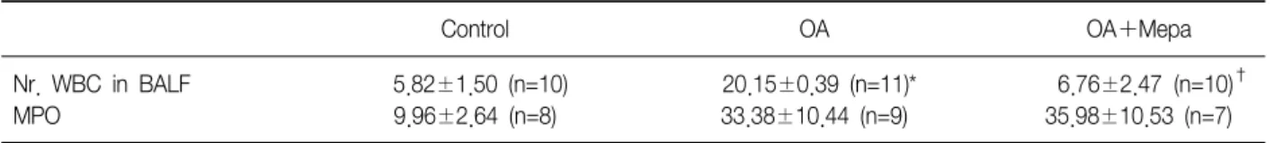
The following parameters caused by LPS treatment were observed ; body weights, lung weights, pulmonary transcapillary albumin transit, arterial gas parameters

RESULTS: Treatment with EAF resulted in significant suppression of oxidative stress in RAW264.7 macrophages as demonstrated by increased endogenous superoxide dismutase (SOD)

Moxifloxacin Alleviates Oleic Acid-provoked Neutrophilic Respira- tory Burst in the Rat Lung through the Inhibition of Cytosolic Phospholipase A 2.. Young Man Lee,

Abstract - Nonalcoholic fatty liver disease (NAFLD) is a kind of liver inflammation caused by an accumulation of fat in the liver. Patients with NAFLD have an increased risk to