Tuberc Respir Dis 2010;68:334-344
CopyrightⒸ2010. The Korean Academy of Tuberculosis and Respiratory Diseases. All rights reserved.
Moxifloxacin의 Secretory PLA 2 억제가 올레인 산으로 유도된 호중구성 급성 폐손상에 미치는 영향
대구가톨릭대학교 의과대학 생리학교실
김병용, 이영만Moxifloxacin Ameliorates Oleic Acid-induced Acute Lung Injury by Modulation of Neutrophilic Oxidative Stress in Rats
Byung Yong Kim, M.D., Young Man Lee, M.D.
Department of Physiology, Daegu Catholic University School of Medicine, Daegu, Korea
Background: Based on the known immunoregulatory functions of moxifloxacin on phagocytes, the therapeutic effect of moxifloxacin on oleic acid (OA)-induced acute lung injury (ALI) was investigated.
Methods: Moxifloxacin (10 mg/kg) was given to male Sprague-Dawley rats that had been given oleic acid (OA, 30 μL) intravenously. Five hours after OA injection, parameters demonstrating ALI were assessed to measure the effects of moxifloxacin on acute lung injury.
Results: The pathological findings of OA-induced ALI's was diminished by moxifloxacin. Through ultrastructural and CeCl3 EM histochemistry, moxifloxacin was confirmed to be effective in decreasing oxidative stress in the lung as well. Indices of ALI, such as lung weight/body weight ratio, protein content in bronchoalveolar lavage fluid, and lung myeloperoxidase were decreased by moxifloxacin. In diaminobenzidine immunohistochemistry, fluorescent immunohistochemistry, and Western blotting of the lung, moxifloxacin had decreased the enhanced expression of secretory phospholipase A2 (sPLA2) by OA.
Conclusion: We concluded that moxifloxacin was effective in lessening acute inflammatory pulmonary edema caused by OA, by inhibiting the neutrophilic respiratory burst, which was initiated by the activation of sPLA2. Key Words: Acute Lung Injury; Neutrophils; Oleic Acid; Free Radicals; Phospholipases A2, Secretory
Address for correspondence: Young Man Lee, M.D.
Department of Physiology, Daegu Catholic University School of Medicine, 3056-6, Daemyung 4-dong, Nam-gu, Daegu 705-718, Korea
Phone: 82-53-650-4473, Fax: 82-53-850-3292 E-mail: leeym.cu.ac.kr
Received: Mar. 26, 2010 Accepted: May. 20, 2010
서 론
올레인 산(oleic acid)을 비롯한 다양한 원인에 의해 발 생하는 급성 폐손상, 특히 급성 호흡곤란증후군의 병리학 적 소견은 염증세포의 심한 침윤에 따른 급성 폐부종이며 이러한 급성 염증성 변화는 면역학적인 기전이 작용한다1. 염증성 사이토카인인 tumor necrosis factor (TNF), inter-
leukin-1 (IL-1), transforming growth factor-β (TGF-β) 등은 급성 폐손상의 발병에 관여하며 특히 TNF나 IL-1은 폐장의 phospholipase A2 (PLA2)의 활성화를 통해 염증성 지질분자를 생성하여 호증구에서 산소기를 생성하게 한 다2. 실제로 급성 호흡곤란증후군 환자의 폐세척액 내에 는 TNF, IL-1 등의 사이토카인의 농도가 증가해 있으며 PLA2의 활성도 또한 증가해 있음이 알려져 있다3. 호중구의 조직 내 침윤에 따른 조직의 손상은 호중구에 서 유리되는 단백분해효소나 산소기의 유리가 그 원인이 라고 알려져 있으며4, 특히 산소기의 유리에는 호중구 막 의 NADPH oxidase의 활성화가 선행된다. 호중구에서의 이러한 산소기 유리에는 염증조절효소로 알려진 PLA2가 관여하며5 최근에는 여러 종류의 PLA2 아형(subtype)이 발견되면서 각각의 아형이 염증반응에 관여하는 기전들
이 연구되고 있다6.
급성 호흡곤란증후군의 병리학적 소견은 폐장의 간질 및 폐포강 내의 호중구의 출현인데 침윤된 호중구 주변의 혈관 내피세포 및 제1, 2형 폐포세포의 손상이며7 또한 제 2형 폐포세포의 미세구조적 변화도 일어난다8. 올레인 산 에 의한 급성 호흡곤란증후군에서는 위에서 말한 PLA2 활 성도의 증가, 호중구의 침윤, 산소기 생성에 따른 산화성 스트레스에 의해 급성 폐부종이 일어난다9.
한편 fluoroquinolone계의 항생제의 일종인 moxifloxa- cin (1-cyclopropyl-7-[(S,S)-2,8-diazabi-cyclo[4.3.0]-non-8- yl]-6-fluoro-8-methoxy-1,4-dihydro-4-oxo-3-quinoline carboxylic acid)은 항균작용 외에도 면역변환작용이 있는 것으로 알려져 있다10,11. 특히 조직에서의 호중구의 이동 을 억제하며 탐식구에서의 산소기의 생성을 억제함이 최 근 연구에서 밝혀졌다12.
또한 moxifloxacin은 폐장에서 염증성 사이토카인인 TNF 및 IL-1의 생성을 감소시켜 감염 시의 보호작용 및 면역변환작용이 있다고 한다13. 이러한 점에 착안하여 본 연구에서는 moxifloxacin의 면역변환효과가 흰 쥐에서 올 레인 산에 의해 유도된 급성 폐손상의 감소 내지는 치료 효과가 있는지, 만약 그러한 효과가 있다면 어떤 기전에 의한 것인지를 밝히는 것을 그 목적으로 하였다.
대상 및 방법
1. 실험동물 및 시약
실험동물은 체중 300 g 내외의 Sprague-Dawley종 수컷 흰 쥐를 사용하였다. 실험동물에게는 물과 먹이를 자유로 이 먹게 허용하였고(
ad libitum
) 실험 전 12시간 전부터는 절식시켰다. 조직 및 세포화학면역 검사(immunohisto- chemistry)를 위해서는 Santa Cruz사제(Santa Cruz Bio- technology, Santa Cruz, CA, USA)의 goat anti-human sPLA2 polyclonal antibody 및 goat anti-human sPLA2polyclonal antibody와 2차 항체인 rabbit anti-goat anti- body와 biotinylated anti-goat IG를 사용하였다. Moxi- floxacin hydrochloride는 Bayer (Leverkusen, Germany) 사로부터 기증받아 사용하였다. 그 외의 시약들은 특별한 언급이 없는 한 Sigma-Aldrich (St. Louis, Mo, USA)사 제 품을 사용하였다.
2. 흰쥐에서의 급성 폐손상의 유발 및 moxifloxacin의 투여
급성 폐손상을 유발하기 위하여 흰쥐의 대퇴정맥으로 300 μL의 올레인 산 혼합액(30 μL 올레인 산+0.1% bo- vine serum albumin 270 μL)을 주사하였다. 올레인 산 주사 후 5시간 후에는 대부분의 흰 쥐에서 급성 폐손상이 유발되었다14. Moxifloxacin은 올레인 산 주사 직후에 10 mg/kg를 복강 내로 주사하였다.
3. 실험군
실험군은 0.1% BSA를 투여한 대조군(sham group), 올 레인 산을 투여한 군(OA-group) 및 올레인 산과 moxi- floxacin을 투여한 군(Moxi-group)으로 구분하였다.
4. 폐장의 무게/체중의 비의 계산
폐장의 무게/체중의 비(lung weight/body weight ra- tio, L/B ratio)는 급성 폐부종의 유용한 지표로 사용될 수 있다. 즉, 대조군의 L/B ratio와 실험군의 L/B ratio를 비교 시 L/B ratio의 증가는 급성 폐부종이 일어났음을 시사한 다. L/B ratio를 계산하기 위하여 흰쥐를 xylazine (6 mg/kg) 및 엔프루란으로 마취한 뒤 기도를 절개하여 Harvard Rodent Ventilator에 연결한 뒤 개흉술을 시행하 였다. 개흉술 후에 좌측 및 우측 폐장을 절제하여 무게를 측정하고 이를 이용 L/B ratio를 계산하였다.
5. 폐 세척액 내의 단백함량의 측정
올레인 산에 의한 폐손상으로 혈관 내 단백질이 폐포강 내로 이동(alveolar flooding)을 평가하기 위하여 기관지 폐포세척(bronchoalveolar lavage, BAL)을 시행 후 폐포 세척액(bronchoalveolar lavage fluid, BALF) 내의 단백함 량을 Brown 등의 방법15에 따라 측정하였다.
6. Lung myeloperoxidase의 측정
폐장 내 호중구침윤의 정도를 측정하기 위하여 Gold- blum 등의 방법16에 따라 폐장 내 myeloperoxidase (MPO) 의 활성도를 측정하였다. MPO는 호중구 세포질 내에 존 재하는 효소로서 조직 내의 호중구의 침윤이 증가하면 그 활동도가 증가하므로 호중구 침윤의 정도를 정량적으로 평가할 때 그 활동도를 측정한다. 실험동물의 기관지를 절개하여 Harvard Rodent Ventilator에 연결하고 개흉술 을 시행하였다. 그 후 우심실에 1,000 IU의 헤파린을 주입
한 뒤 폐동맥에 삽관하여 Masterflex perfusion pump를 이용하여 생리적 식염수를 관류시켜 폐장 내 혈액을 제거 하였다. 좌, 우측 폐장을 절제 후 무게를 측정한 뒤 액체 질소에 담궈 급속 냉동시킨 후 분석 시까지 -70oC에서 냉동 보관하였다. MPO의 측정은 폐장을 조직 분쇄기 (Polytron, Switzerland)를 이용하여 4.0 mL, pH 7.4의 20 mM 인산칼륨완충용액 내에서 간 뒤, 18,000 rpm, 4oC에 서 30분간 원심분리 후 상등액은 버렸다. 그 후 침전층 (pellet)을 다시 4.0 mL 50 mM 인산완충용액(pH 6.0)에 재부유시키고 4oC에서 Vibracell (Sonics & Materials Inc., Danbury, CT, USA)를 이용하여 초음파 처리하여 세포막 을 파열시켰다. 그 후 균질액을 60oC 항온 수조에서 120분 간 처리하여 단백분해효소를 불활성시키고 12,000 rpm으 로 원심분리 후 상등액은
o
-dianisidine이 함유된 500 μM 과산화수소용액과 반응시켜 파장 460 nm에서 분광비색 계를 이용하여 MPO의 활성도를 측정하였다.7. 형태학적 관찰
1) 광학 현미경적인 관찰
실험동물의 기관을 절개하여 4% paraformaldehyde 용 액(PFA, pH 7.4)을 기도 내로 주입한 뒤 기도를 결찰하였 다. 그 후 폐를 적출하여 무작위로 폐조직의 절편을 절취 한 뒤 4% PFA에 24시간 후고정하였다. 고정이 끝난 조직 은 알코올의 농도를 순차적으로 증가시켜 탈수시키고 xy- lene으로 남은 알코올을 제거한 후 파라핀(Paraplast; Ox- ford, St Louis, MO, USA)에 포매하였으며, RM2155 Micro- tome (Leica, Wetzlar, Germany)으로 박절하여 4 μm 두께 의 연속 절편을 만든 후 hematoxylin & eosin (H&E)으로 염색하여 광학현미경으로 관찰하였다.
2) 전자현미경을 이용한 제2형 폐포세포의 관찰(폐장에 서의 산화성 스트레스의 관찰)
올레인 산에 의한 급성 폐손상 시의 제2형 폐포세포의 미세구조를 관찰하기 위하여 절제된 폐조직을 2.5% glu- taraldehyde로 고정하고 밀폐된 용기 내에서 공기를 주입 하여 조직 내의 공기를 제거하였다. pH 7.4의 인산용액으 로 수세 후에 1% osmium teroxide로 후고정 하였다. 그 뒤 alcohol-propylene으로 탈수한 다음 epoxy-resin에 포 매하고 열중합한 뒤에 초박절편을 제작한 뒤 uranyl ace- tate와 lead citrate로 염색한 뒤 Hitachi 9H-600 (Hitachi, Tokyo, Japan) 투과전자현미경으로 관찰하였다.
3) 전자현미경을 이용한 조직의 과산화수소의 생성 검 사(Electron microscopic CeCl3 cytochemistry) 폐장 내의 산소기의 형성을 검사하기 위하여 Hobson 등의 방법17에 따라 CeCl3 cytochemical electon micro- scopy를 시행하였다. 폐장조직을 적출 즉시 2.0 mM ce- rium chloride, 10 mM 3-amino-1,2,4-triazol, 0.1 M tris- maleate buffer (pH 7.5), 7% sucrose, 0.002% triton X- 100로 조제된 기질에 담그고 30분간 반응시켰다. 반응이 끝난 조직을 수세와 탈수의 과정을 거친 후 epoxy-resin에 포매한 뒤 열중합 시키고 초박절 후 대조염색을 하지 않고 Hitachi (9H-600) 투과전자현미경으로 관찰하였다.
4) DAB를 이용한 폐장의 sPLA2 조직면역화학법 대조군과 실험군의 조직절편을 xylene에서 탈파라핀 하고 알코올을 거쳐 물로 수세한 후 PBS에 세척하였다.
조직 내의 내인성과산화효소의 활성을 저해하기 위하여 0.3% H2O2를 함유한 PBS로 30분간 처리하였고, 비특이적 인 반응을 억제하기 위하여 10% 정상토끼혈청으로 30분 동안 처리하였다. 1차 항체로는 goat anti-human sPLA2
polyclonal antibody (N-16; Santa Cruz Biotechnology)를 1:50으로 희석하여 실온에서 3시간 적용하였다. 그 후 PBS로 10분간씩 3회 세척한 후 2차 항체인 biotinylated anti-goat IgG (DAKO, Carpinteria, CA, USA)를 1:100으 로 희석하여 실온에서 1시간 반응하였다. PBS로 세척한 후 ABC kit (avidin-biotin-peroxidase complex; Vector Laboratories Inc., Burlingame, CA, USA)로 1시간 동안 반응시켰고, PBS로 3차례 세척한 후 0.01% H2O2와 0.05%
3, 3'-diaminobenzidine (DAB; Sigma, St Louis, MO, USA)가 포함된 0.05 M tris-HCl 완충 용액(pH 7.4)으로 발색시켰다. PBS로 세척하고 hematoxylin으로 대조 염색 한 후 Axiophot Photomicroscope (Carl Zeiss, Oberko- chen, Germany)으로 관찰하고, AxioCam MRc5 (Carl Zeiss)로 촬영하였다.
5) 형광면역화학법을 이용한 조직에서의 sPLA2의 발현 검사
대조군과 실험군의 조직절편을 DAB를 이용한 조직면 역화학법에서와 같이 처리한 후 비특이적인 반응을 억제 하기 위하여 10% 정상토끼혈청으로 30분 동안 처리하였 다. 1차 항체로는 goat anti-human sPLA2 polyclonal anti- body (N-16)를 1:50으로 희석하여 실온에서 3시간 적용 하였다. 그 후 PBS로 10분간씩 3회 세척한 후 2차 항체인 biotinylated anti-goat IgG를 1:100으로 희석하여 실온 에서 1시간 반응하였다. PBS로 세척한 후 propidium io-
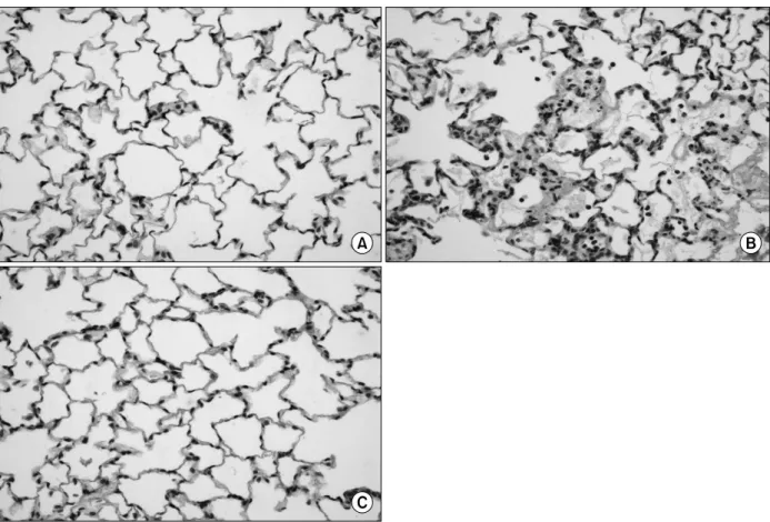
Figure 1. (A) The effect of oleic acid on the morphological changes in the lung. Intraalveolar phagocytes, especially neutrophils were found abundantly. Hyaline membrane and wide spread hemorrhage were found also (H&E stain, ×40).
(B) The effect of moxifloxacin on the histological changes in the lung of rat given oleic acid intravenously. Alveoli are patent and the accumulation and migration of neutrophils into the alveolar lumen were significantly lessened by moxifloxacin. Generalized inflammatory changes were diminished by moxifloxacin (H&E stain, ×100).
dide로 대조염색한 후 PBS로 수세하고 Axiophot Photo- microscope로 관찰하고, AxioCam MRc5로 촬영하였다.
8. Western blot analysis
적출한 조직에 IPH lysis buffer (50 mM Tris [pH 8.0], 150 mM NaCl, 5 mM EDTA, 0.5% NP40, 100 mM PMSF, leupeptin 1 mg/mL, aprotinin 1 mg/mL and 1 M DTT)를 첨가하여 조직분쇄기를 이용 조직을 파쇄하였다. 파쇄한 조직은 4oC에서 30분 동안 융해시켜 12,000 rpm에서 10 분 동안 원심분리 하여 조직으로부터 상등액을 분리하였 다. 분리한 상등액은 단백질 정량 kit인 Bio-Rad protein assay system을 이용하여 정량하였고, 정량한 단백질을 일정한 농도로(30 μg) 만들어 DTT가 첨가된 2×SDS- loading (Trin-Cl [pH 6.8] 100 mM, glycerol 20%, bromo- phenol blue 0.2%, SDS 4%, dithiothreitol 200 mM) buf- fer와 섞어 98oC에서 5분간 가열하였다. 그리고 SDS- PAGE gel로 전기영동하여 Immobilion-P-membrane (Mili- pore, Bedford, MA, USA)에 transfer시켰다. Membrane은 일차 항체 anti-sPLA2 (Santa Cruz Biotechnology)와 반응 시킨 후 이차항체와 반응시켰다. 항체들에 대한 발현분석 은 Horseradish Peroxidase-Linked 이차항체에 의해 발현 되는 ECL Western Blot Analysis system (Amersham Pharmacia Biotech, Amersham, UK)을 이용하여 확인하 였다. 확인된 단백질의 발현 차이는 densitometry를 이용 하여 분석하였다(Quantity One; Bio-Rad, Hercules, CA,
USA).
9. 통계처리
모든 성적은 평균±표준오차로 나타내었다. 성적을 변 수별로 정규분포화한 후 로그변환 된 변수를 이용하여 ANOVA 검정 후, 오류를 보정하기 위하여 Student-Neu- mann-Keul test를 이용하였고 p<0.05를 유의하다고 인 정하였다.
결 과
1. H&E 염색을 통한 폐장 조직의 검경
H&E 염색 후 광학 현미경을 이용하여 폐장의 조직을 검경한 결과 sham group에서는 폐포강 내에 염증세포가 관찰되지 않았으나 OA-group에서는 폐포강 내의 염증세 포 특히 호중구의 출현이 현저하였다. 또한 폐포강 내의 단백질의 유출, 혈관의 부종(perivascular cuffing) 등이 현 저하고 혈관에서 간질 및 폐포강 내로의 호중구 및 적혈구 의 이동이 관찰되었다. Moxi-group에서는 이러한 병리학 적인 소견이 현저히 감소하여 moxifloxacin의 급성 폐손 상의 감소 효과를 확인할 수 있었다(Figure 1).
2. L/B Ratio
급성 폐부종의 지표인 L/B ratio는 sham group에서 5.3±0.4였고 OA-group에서는 8.8±1.0으로서 sham
Table 1. Parameters of lung injury in rats given oleic acid and moxifloxacin
Sham OA OA+Moxi
(L/B)×10
35.3±0.4 (n=9) 8.6±1.0* (n=8) 5.8±1.0
†(n=8)
BAL protein (mg/two lungs) 1.8±2.3 (n=13) 8.4±2.3* (n=7) 4.8±1.6
†(n=7) Values are given as mean±SE.
n indicates number of experiments.
OA: oleic acid; Moxi: moxifloxacin; BAL: bronchoalveolar lavage; L/B: lung weight/body weight ratio.
*p<0.001 Sham vs. OA,
†p<0.001 OA vs. OA+Moxi.
Table 2. Effect of OA and moxifloxacin on the infiltration of neutrophils in the lung
Sham OA OA+Moxi
Lung MPO (U/g of lung) 8.1±2.5 (n=9) 38.9±8.2* (n=10) 20.1±4.8
†(n=9)
Values are given as mean±SE.
n indicates number of experiments.
OA: oleic acid; Moxi: moxifloxacin; MPO: myeloperoxidase.
*p<0.001, Sham vs. OA,
†p<0.001, OA vs. OA+Moxi.
Figure 2. The ultrastructural changes of alveolar type II cells of the lung in oleic acid (OA), OA with moxifloxacin treated rats. Well preserved la- mellar bodies were found in alveolar type II cells of the control lung (A).
In OA treated rat, a vacuolization of lamellar bodies of alveolar type II cells was prominent, a direct evidence of oxidative stress (B). In contrast, OA with moxifloxacin treated rats, lamellar bodies of alveolar type II cells were relatively well preserved (C) (Original magnification ×3,000).
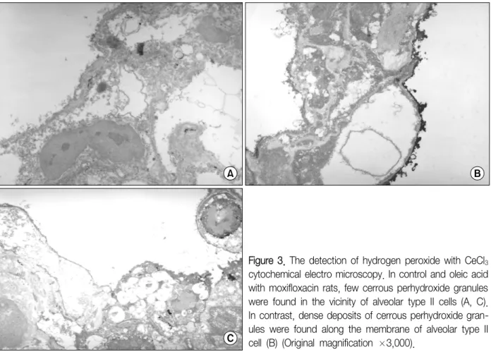
Figure 3. The detection of hydrogen peroxide with CeCl3
cytochemical electro microscopy. In control and oleic acid with moxifloxacin rats, few cerrous perhydroxide granules were found in the vicinity of alveolar type II cells (A, C).
In contrast, dense deposits of cerrous perhydroxide gran- ules were found along the membrane of alveolar type II cell (B) (Original magnification ×3,000).
group에 비해 증가(p<0.001)하였고 Moxi-group에서는 5.8±1.0으로서 OA-grouop에 비해(p<0.001) 감소하였 다(Table 1).
3. BALF 내의 단백함량
폐장 내 모세혈관의 손상에 따른 alveolar flooding을 나 타내는 지표인 BALF 내의 단백질의 함량(mg/two lungs) 은 sham group에서 1.8±0.4, OA-group에서 8.4±2.3으 로서 현저히(p<0.001) 증가하였으나 Moxi-group에서는 4.8±1.6으로서 OA-group에 비해 감소(p<0.001)하였다 (Table 1).
4. Lung MPO
폐장 내 호중구의 침윤의 정도를 나타내는 Lung MPO (U/g of lung)는 sham group에서 8.1±2.5였고 OA- group에서는 38.9±8.2로서 sham group보다 높았다(p<
0.001). 이에 비하여 Moxi-group에서는 20.1±4.8로서 OA- group에 비하여 낮았다(p<0.001)(Table 2).
5. 제2형 폐포의 미세구조적 변화
OA를 주사한 흰쥐 폐장의 제2형 폐포세포는 층상체의 공포화 및 계면활성물질의 변성이 대조군과 비교 시 현저 하였다(Figure 2A, B). OA와 moxifloxacin을 동시에 준 경우에는 제2형 폐포 내의 층상체 내의 층상구조가 비교 적 온전하게 보존되어 있고 계면활성제의 변성도 거의 관 찰 되지 않았다(Figure 2C).
6. 폐장에서의 과산화수소의 생성관찰
전자현미경을 이용한 CeCl3 세포화학법에서는 대조군 의 경우 제2형 폐포세포의 변연에는 cerrous perhydr- oxide의 과립이 거의 발견되지 않았으나(Figure 3A) OA 를 투여한 쥐에서는 다량의 cerrous perhyroxide 과립이 제2형 폐포세포의 막을 따라 관찰되었다(Figure 3B). OA 와 moxifloxacin을 동시에 투여한 군에서는 제2형 폐포세 포의 변연에 cerrous perhydroxide의 과립이 거의 관찰되 지 않았다(Figure 3C).
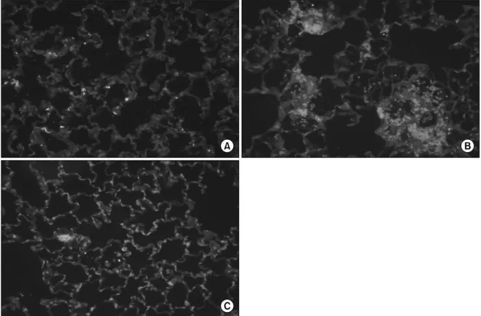
Figure 4. (A) An immunohistochemistry for the detection of secretory phospholipase A2 (sPLA2) in the normal rat's lung.
Staining sPLA2 with diaminobenzidine revealed few reactive cells against sPLA2 antibody. Primary antibody against sPLA2
was goat anti-human sPLA2 polyclonal antibody (dilutional factor ×50) and secondary antibody was biotinylated anti-goat IgG (dilutional factor ×100) (Original magnification ×200). (B) A representative of immunohistochemistry for detection of sPLA2 in the lung of oleic acid (OA) given rat. In the alveoli, abundant migrated neutrophils of which cytoplasm and cell membrane were intensely reacted with diaminobenzidine signifying strong activation of sPLA2 in these cells. Hyaline membranes were found also in the alveolar lumen. Primary antibody against sPLA2 was goat anti-human sPLA2 polyclonal antibody (dilutional factor ×50) and secondary antibody was biotinylated anti-goat IgG (dilutional factor ×100) (Original magnification ×200). (C) An immunohistochemical photograph of detecting sPLA2 in the lung of rat given OA and moxifloxacin. Few phagocytes were found in the lung and diaminobenzidine positive cell was not found. Primary antibody against sPLA2 was goat anti-human sPLA2 polyclonal antibody (dilutional factor ×50) and secondary antibody was bio- tinylated anti-goat IgG (dilutional factor ×100) (Original magnification ×200).
7. DAB를 이용한 조직 면역법의 결과
폐장조직 내 sPLA2의 발현 부위를 알아보기 위해 시행 한 DAB 조직 면역법의 결과는 Figure 4와 같다. 올레인 산은 호중구에서 sPLA2의 발현을 현저히 증가시키고 moxifloxacin은 이러한 sPLA2 발현의 증가를 현저히 억제 하였다.
8. 폐장에서의 형광 세포면역법의 결과
폐장에서 형광 면역법을 시행하여 sPLA2에 대한 발현을
검사한 결과는 Figure 5와 같다. 대조군에 비해서 OA group에서는 조직에서의 sPLA2의 발현이 현저하고 이러 한 변화는 moxifloxacin에 의하여 강력히 억제되었다.
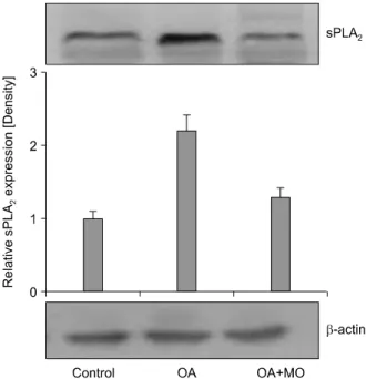
9. Western blot analysis
폐장조직의 면역침전법에 의한 sPLA2의 발현 검사는 OA 에 의하여 현저한 증가를 보였다. Moxifloxacin은 OA에 의 한 이러한 sPLA2의 발현 증가를 억제하였다(Figure 6).
Figure 5. (A) A fluorescent immunohistochemistry for detection of secretory phospholipase A2 (sPLA2) in the normal rat's lung. Counter-staining with propidium iodide of sPLA2 revealed few cells of sPLA2 positive against sPLA2 antibody.
Primary antibody against sPLA2 was goat anti-human sPLA2 polyclonal antibody (dilutional factor ×50) and secondary antibody was biotinylated anti-goat IgG (dilutional factor ×100) (Original magnification ×200). (B) A representative of fluorescent immunohistochemistry for detection of sPLA2 in the lung of oleic acid (OA) given rat. Dense, highly fluorescent area were found in the lung depicting the strong expression of PLA2 in the lung. Primary antibody against sPLA2 was goat anti-human sPLA2 polyclonal antibody (dilutional factor ×50) and secondary antibody was biotinylated anti-goat IgG (dilutional factor ×100) (Original magnification ×200). (C) A fluorescent immunohistochemical photograph of detect- ing sPLA2 in the lung of rat given OA and moxifloxacin. Fluorescent area of lung by OA was strikingly diminished by moxifloxacin signifying the inhibition of PLA2. Primary antibody against sPLA2 was goat anti-human sPLA2 polyclonal antibody (dilutional factor ×50) and secondary antibody was biotinylated anti-goat IgG (dilutional factor ×100) (Original magnification ×200).
고 찰
올레인 산은 실험동물에서 급성 호흡곤란증후군과 유 사한 폐부종을 유발하며 인체에도 소량 존재하는 지질의 일종이다18. 올레인 산이 혈중으로 유입되면 일단 지방입 자에 의한 혈전증(fat embolism)이 유발될 것으로 보이며 올레인 산이 호중구에 대한 화학주성을 가짐과 동시에 PLA2를 활성화시키는 성질이 있으므로19 폐장의 모세혈관 내로 호중구가 밀집하고 이에 따라 모세혈관의 폐쇄가 일
어날 것으로 생각된다. 결과적으로 모세혈관 내 정수압의 증가하고, 호증구에서의 산소기 및 단백 분해 효소의 유리 에 따라 혈관 내피세포의 손상이 올 수 있다. 이러한 일련 의 과정은 본 연구의 결과를 통해서도 유추할 수 있다.
즉, 폐장의 형태학적인 검사에서도 보이듯이 올레인 산은 폐장의 심한 염증반응을 유발하고 있다. 폐포강 내의 호 중구의 출현은 정상적인 폐장에서는 볼 수 없는 현상이며 동시에 폐장에서는 혈관의 부종 및 폐포강 내의 유리간질 막(hyaline membrane)의 형성이 두드러져 급성 호흡곤란
Figure 6. Western blot analysis of secretory phospholi- pase A2 (sPLA2) in the lung. Oleic acid (OA) treatment in- creased the expression of sPLA2 but this increased ex- pression was down-regulated by moxifloxacin (MO).
증후군과 유사한 형태학적인 변화를 보인다. 급성 폐부종 의 지표라 할 수 있는 L/B ratio와 혈관내피세포의 손상지 표로 볼 수 있는 BALF 내의 단백함량도 올레인 산에 의해 증가하며 이러한 변화와 연관하여 폐장의 MPO의 활동도 도 증가해 있다. 이러한 올레인 산에 의한 변화들은 moxi- floxacin에 의해 현저히 감소하고 있다.
Moxifloxacin에 의한 이러한 변화들은 호중구의 작용과 연관시켜 볼 때 일단은 호중구의 폐장 내 침윤의 감소가 그 원인이며 호중구 침윤의 감소는 moxifloxacin의 면역 변환작용 중 탐식구의 이동 억제작용20에 의한 듯하다. 최 근의 보고들에 의하면 moxifloxacin이 내독소에 의해 탐 식구에서 생성되는 염증성사이토카인의 생성, 특히 호중 구의 화학주성을 유발하는 interleukin-8을 억제하거나21 염증성 사이토카인에 의해 유발되는 호중구에서의 산소 기의 생성을 억제한다22고 한다. 올레인 산에 의한 폐장조 직의 손상 기전에는 PLA2가 관여하는데, 잘 알려진 대로 PLA2는 염증반응을 조절하는 효소이고 이 효소의 활성화 는 단 시간 내에 대량의 염증성 지질분자를 조직으로 유리 하게 한다. 올레인 산에 의한 sPLA2 활동도의 증가는 본 연구에서도 관찰되며 moxifloxacin은 올레인 산에 의한 sPLA2 활성도의 증가를 억제하고 있다. 올레인 산에 의한 염증성 사이토카인의 분비가 moxifloxacin에 의해 억제됨
으로서 PLA2의 활성화가 감소될 수도 있을 것이다. Moxi- floxacin이 올레인 산에 의한 폐장 내 호중구의 침윤 및 이동을 현저히 감소시킬 뿐만 아니라 sPLA2를 억제하는 것은 산화성 스트레스에 의한 조직의 손상이라는 관점에 서 보면 moxifloxacin이 산화성 스트레스를 효과적으로 차단함으로써 폐장의 손상을 감소시킨다고 볼 수 있다.
본 연구에서 전자현미경을 이용한 미세구조 및 CeCl3 세 포화학법을 이용하여 관찰한 결과는 올레인 산에 의한 제 2형 폐포세포의 층상체의 공포화 및 과산화수소와 cerium chloride의 반응물질인 cerrous peroxide의 생성이다. 이 러한 결과는 올레인 산에 의한 폐장의 산화성 스트레스에 의해 조직의 손상이 일어났음을 의미하고, moxifloxacin 에 의해 이러한 손상이 줄고 cerrous peroxide 과립이 거 의 생성되지 않았다는 사실은 moxifloxacin이 산화성 스 트레스를 억제하였음을 의미한다.
올레인 산에 의한 급성 폐손상에 PLA2가 관여하는 기전 은 본 연구의 조직면역법의 결과에서도 분명히 나타나고 있다. 폐장 조직의 DAB 조직면역화학법은 올레인 산에 의한 조직 내의 호중구의 sPLA2의 발현 증가를 보여주고 있고 moxifloxacin은 이러한 호중구에서의 sPLA2의 발현 을 분명히 억제하고 있다. 이것은 올레인 산에 의한 조직 의 손상이 부분적으로는 호중구의 PLA2의 활성화에 따른 것이며 이 중에서도 특히 sPLA2의 활성화가 중요한 역할 을 한다는 것을 의미한다. 또 한 가지 주목할 것은 올레인 산과 호중구의 상호작용에 의한 모세혈관 내 혈전의 생성 이다. 올레인 산은 모세혈관 내에서 지방색전을 형성한 뒤 화학주성에 의해 호중구를 끌어들여 올레인 산과 호중 구의 덩어리를 형성하며19 이 결과 호중구에서의 산소기 및 단백 분해 호소의 유리가 혈관 내피 세포의 손상으로 이어질 것으로 보인다. 본 연구에서는 형광면역법에 의한 조직에서의 sPLA2의 발현 검사에서도 이 사실을 확인할 수 있다. 올레인 산은 조직에서의 sPLA2의 발현을 증가시 키고 moxifloxacin은 이를 효과적으로 차단하고 있다.
PLA2의 억제는 올레인 산-호중구의 응집도 거의 완전히 차단함으로써 혈관 내피 세포의 손상을 줄여 혈중 단백질 이 폐포강 내로 이동하는 것을 억제한다.
올레인 산이 어떠한 경로를 통해 폐장의 PLA2의 활성도 를 증가시키고 이에 따른 산소기의 형성이 증가하는지는 명확하지 않다. TNFα를 비롯한 여러 종류의 염증성사이 토카인이 PLA2 활성도에 관여하고 특히 TNF-α가 급성 폐 손상에 관여함23, 그리고 TNF의 염증유발효과는 PLA2의 활성화에 의한다는 것이 잘 알려져 있어24,25 올레인 산에
의한 급성 폐손상 시 moxifloxacin에 의한 염증성 사이토 카인의 분비에 대한 연구도 이뤄져야 한다고 생각된다.
요약하면 올레인 산에 의해 유발된 급성 폐손상을 호중 구의 작용이라는 측면에서 올레인 산이 호중구에 대한 화 학주성, sPLA2의 활성화를 통해 산화성 스트레스를 일으 키고 moxifloxacin은 PLA2 작용의 억제 및 호중구의 res- piratory burst의 억제를 통해 손상을 감소시킨다. Moxi- floxacin이 항균작용 외에도 면역변환 효과를 가지고 있다 는 사실에 착안하여 시행된 본 연구의 결과는 moxifloxacin 이 호중구의 산소기 생성을 억제하여 급성 폐손상을 감소 시킨다는 사실을 확인시켜 주었고, 적어도 다발성 외상, 특 히 골절에 따른 지방색전증에 따르는 급성 폐손상 시에는 moxifloxacin의 유용성이 검토되어야 함을 시사한다.
참 고 문 헌
1. Bernard GR, Artigas A, Brigham KL, Carlet J, Falke K, Hudson L, et al. The American-European Consensus Conference on ARDS: definitions, mechanisms, relevant outcomes, and clinical trial coordination. Am J Respir Crit Care Med 1994;149:818-24.
2. Pruzanski W, Vadas P. Phospholipase A2: a mediator between proximal and distal effectors of inflammation.
Immunol Today 1991;12:143-6.
3. Pittet JF, Mackersie RC, Martin TR, Matthay MA.
Biological markers of acute lung injury: prognostic and pathogenetic significance. Am J Respir Crit Care Med 1997;155:1187-205.
4. Chenevier-Gobeaux C, Simonneau C, Therond P, Bon- nefont-Rousselot D, Poiraudeau S, Ekindjian OG, et al.
Implication of cytosolic phospholipase A2 (cPLA2) in the regulation of human synoviocyte NADPH oxidase (Nox2) activity. Life Sci 2007;81:1050-8.
5. Dana R, Malech HL, Levy R. The requirement for phos- pholipase A2 for activation of the assembled NADPH oxidase in human neutrophils. Biochem J 1994;297(Pt 1):217-23.
6. Schaloske RH, Dennis EA. The phospholipase A2 su- perfamily and its group numbering system. Biochim Biophys Acta 2006;1761:1246-59.
7. Lee YM, Hybertson BM, Cho HG, Terada LS, Cho O, Repine AJ, et al. Platelet-activating factor contributes to acute lung leak in rats given interleukin-1 intratrach- eally. Am J Physiol Lung Cell Mol Physiol 2000;279:
L75-80.
8. Martensson J, Jain A, Stole E, Frayer W, Auld PA,
Meister A. Inhibition of glutathione synthesis in the newborn rat: a model for endogenously produced oxi- dative stress. Proc Natl Acad Sci U S A 1991;88:9360-4.
9. Furue S, Kuwabara K, Mikawa K, Nishina K, Shiga M, Maekawa N, et al. Crucial role of group IIA phospholi- pase A(2) in oleic acid-induced acute lung injury in rabbits. Am J Respir Crit Care Med 1999;160:1292-302.
10. Werber S, Shalit I, Fabian I, Steuer G, Weiss T, Blau H. Moxifloxacin inhibits cytokine-induced MAP kinase and NF-kappaB activation as well as nitric oxide syn- thesis in a human respiratory epithelial cell line. J Antimicrob Chemother 2005;55:293-300.
11. Weiss T, Shalit I, Blau H, Werber S, Halperin D, Levitov A, et al. Anti-inflammatory effects of moxifloxacin on activated human monocytic cells: inhibition of NF- kappaB and mitogen-activated protein kinase activation and of synthesis of proinflammatory cytokines. Antimi- crob Agents Chemother 2004;48:1974-82.
12. Uriarte SM, Molestina RE, Miller RD, Bernabo J, Farinati A, Eiguchi K, et al. Effects of fluoroquinolones on the migration of human phagocytes through Chlamydia pneumoniae-infected and tumor necrosis factor alpha- stimulated endothelial cells. Antimicrob Agents Chem- other 2004;48:2538-43.
13. Shalit I, Horev-Azaria L, Fabian I, Blau H, Kariv N, Shechtman I, et al. Immunomodulatory and protective effects of moxifloxacin against Candida albicans-in- duced bronchopneumonia in mice injected with cyclo- phosphamide. Antimicrob Agents Chemother 2002;46:
2442-9.
14. Shiue ST, Thrall RS. Effect of corticosteroid therapy on the acute injury and recovery stage of oleic acid in- duced lung injury in the rat. Exp Lung Res 1991;17:
629-38.
15. Brown RE, Jarvis KL, Hyland KJ. Protein measurement using bicinchoninic acid: elimination of interfering substances. Anal Biochem 1989;180:136-9.
16. Goldblum SE, Wu KM, Jay M. Lung myeloperoxidase as a measure of pulmonary leukostasis in rabbits. J Appl Physiol 1985;59:1978-85.
17. Hobson J, Wright J, Churg A. Histochemical evidence for generation of active oxygen species on the apical surface of cigarette-smoke-exposed tracheal explants.
Am J Pathol 1991;139:573-80.
18. Matalon S, Ji HL. Oleic acid damages ion transport and promotes alveolar edema: the dark side of healthy living. Am J Respir Crit Care Med 2005;171:424-5.
19. Lee YM, Kim BY, Park YY. Role of the PLA2-activated neutrophilic oxidative stress in oleic acid-induced acute
lung injury. Tuberc Respir Dis 2010;68:55-61.
20. Williams AC, Galley HF, Webster NR. The effect of moxifloxacin on release of interleukin-8 from human neutrophils. Br J Anaesth 2001;87:671-2.
21. Zimmermann GS, Neurohr C, Villena-Hermoza H, Hatz R, Behr J. Anti-inflammatory effects of antibacterials on human Bronchial epithelial cells. Respir Res 2009;10:89.
22. Hand WL, Hand DL, Vasquez Y. Increased poly- morphonuclear leukocyte respiratory burst function in type 2 diabetes. Diabetes Res Clin Pract 2007;76:44-50.
23. Ljungman AG, Tagesson C, Lindahl M. Endotoxin stim- ulates the expression of group II PLA2 in rat lung in vivo and in isolated perfused lungs. Am J Physiol
1996;270:L752-60.
24. Arbibe L, Vial D, Rosinski-Chupin I, Havet N, Huerre M, Vargaftig BB, et al. Endotoxin induces expression of type II phospholipase A2 in macrophages during acute lung injury in guinea pigs: involvement of TNF-al- pha in lipopolysaccharide-induced type II phospholi- pase A2 synthesis. J Immunol 1997;159:391-400.
25. Lindbom J, Ljungman AG, Lindahl M, Tagesson C.
Increased gene expression of novel cytosolic and secre- tory phospholipase A(2) types in human airway epi- thelial cells induced by tumor necrosis factor-alpha and IFN-gamma. J Interferon Cytokine Res 2002;22:947-55.