Characterization of the Growth, Total Lipid and Fatty Acid Profiles in Microalga, Nannochloropsis oceanica under Different Nitrogen Sources
Majid Mahdieh*, Salimeh Shabani, and Mohammad Reza Amirjani Department of Biology, Faculty of Science, Arak University, Arak 38156-8-8349, Iran
Received: January 10, 2018 / Revised: March 7, 2018 / Accepted: May 31, 2018
Introduction
It seems that continual trust on energy of fossil fuel resources is labile, due to both reduction of world reserves and the emission of greenhouse gases con- nected with their application. Hence, there are strong studies aimed at replacing a renewable resource, con- taining potential biofuels, as energy sources. Recently, biodiesel fuel has received significant concern, because biodiesel is a biodegradable, reproducible and also non- toxic fuel. This fuel emits neither net carbon dioxide nor sulfur to the atmosphere and exhale less gaseous impu-
rity than common diesel [1]. Microalgae are single-celled photosynthetic aquatic organisms. They use sunlight as energy and make various components with carbon diox- ide and other elements. These organisms have a higher photosynthetic efficiency than plants for the production of biomass [2]. Various studies showed that biofuels spe- cifically derived from microalgae are considered to be a technically viable alternative resource of energy [3−7].
Microalgae with high lipid contents are new reproduc- ible resources for biofuel production, and pilot produc- tion of biofuel from microalgae has been documented [8].
However, the commercial production of biofuel from these organisms has not begun due to its high cost. The main factors contributing to the lipid production of microalgae are: the growth rate, cell density and lipid content. Currently, the selection of microalgae with The properties of microalgae as bioresources for biodiesel production can be improved by adding nitrogen sources into the culture medium. Thus, Nannochloropsis oceanica CCAP 849/10 was cultured in f/2 media supplemented with five different forms of nitrogen at 0.88 mmol-N l-1 each: ammonium bicarbonate (NH4HCO3), ammonium sulfate ((NH4)2SO4), sodium nitrate (NaNO3), ammonium nitrate (NH4NO3), and urea. The cell density, lipid content, and fatty acid profile of the microalga were determined after 15 days of cultivation. The growth of N. oceanica based on cell number was lowest in the medium with NH4NO3, and increased significantly in the medium with NH4HCO3. Cells treated with (NH4)2SO4, and NH4NO3 produced the highest total lipid contents (i.e., 65% and 62% by dry weight, respectively). The fatty acid profiles of the microalga were significantly different in the various nitrogen sources. The major fatty acids detected in cultures supplemented with NH4HCO3, (NH4)2SO4, NH4NO3, or urea were C14:0, C16:0, C16:1, C18:0, C18:1, C18:2, C20:5, and C22:6. However, the C16:1 content in the NaNO3-supplementedculture was very low. This study highlights that the nitrogen source can strongly influence lipid production in N. oceanica and its fatty acid composition.
Keywords: Biodiesel, Chlorophyll a, nutrient, microalga, urea
*Corresponding author
Tel: +98-86-34173317, Fax: +98-86-34173406 E-mail: [email protected]
© 2019, The Korean Society for Microbiology and Biotechnology
higher lipid contents and improvements to the quantity and quality of the lipids produced by microalgae are con- sidered for the technology of biodiesel production from microalgae. Previous studies [9−11] have revealed that it is possible to manipulate the lipid yield and lipid proper- ties of algal cells by the optimizing microalgae culture conditions (e.g. temperature and also light intensity) or features of nutrient medium (nitrogen, phosphates as well as iron concentrations). Microalgae biomass and biofuels production are changed by various physico- chemical factors such as nutrients, light intensity, tem- perature, pH and salinity [12, 13]. Especially between diverse nutritional agents, nitrogen is one of the most critical nutrients for algae growth, because this nutrient is a precipitant in all structural and also functional pro- teins such as enzymes, peptides, chlorophylls, energy transfer molecules, and genetic materials in algal cells [14, 15]. Wang et al. [16] reported that the nitrogen con- centration in culture medium strongly affects both cell growth rate and cellular biochemical compositions in microalgae. In addition, numerous investigations have demonstrated that when the nitrogen is restricted in cul- ture medium, microalgae decrease cell growth rate and raise their lipid or carbohydrate content, reducing pro- tein synthesis [17].
Many microalgae species prefer ammonium, science less energy is needed for its assimilating into amino acids. In contrary, some microalgae such as Botryococcus braunii and Dunaliella tertiolecta prefer nitrate over ammonium for growth [18, 19]. Recent studies con- firmed that some species of Chlorella also prefer nitrate rather than ammonium for growth, and these species also effectively use a variety of organic nitrogen sources such as urea, glycine, yeast extract (YE) and peptone [20, 21]. The results of Norici et al. [22] investigations proved that relevancy on the nitrogen source, biochemi- cal composition can also be changed. For instance, pro- tein content of Dunaliella salina was 2-times higher with ammonium than nitrate supplementation. In con- trary, the lipid amount of Chlorella sorokiniana was over 2-folds more with ammonium than urea or nitrate supplementation [23]. Since the desirable source of nitrogen for growth varies from species to species, and the biochemical composition also can be differed by the supplemented nitrogen sources, it is essential to mea- sure different nitrogen sources and take the most suit-
able source for each taxon in order to increase the efficiency of the goal product, like lipid and carbohydrate for biodiesel and bioethanol, respectively. Therefore, comprehension of the nitrogen sources effect on growth and also lipid value will ameliorate lipid yield and help large-scale commercial producers in selecting an appro- priate fertilizer [24].
In this study, a oily microalga, Nannocholoropsis oce- anica, was selected for lipid production. The effects of various nitrogen forms such as nitrate, ammonium, and organic nitrogen (urea) on the cell growth, and the bio- chemical composition of N. oceanica were analyzed. Also, the microalgal lipid was converted to fatty acid methyl esters (FAME), and the fatty acid composition was assessed for measuring the effect on the resulted bio- diesel properties.
Materials and Methods
Organism and culture treatments
Nannochloropsis oceanic CCAP 849/10 was purchased from the Culture Collection of Algae and Protozoa at Scotland. The microalga was grown in 500 ml flasks with f/2 medium [25] and continues aeration with air. N.
oceanic was grown at 23± 1℃ and 100 µmol photons m-2s-1 light intensity (L/D = 14:10). In order to study the effect of different nitrogen forms on the cell growth and biochemical composition of N. oceanica, sodium nitrate (NaNO3, 0.88 mmol N L-1) in F/2 medium was replaced by different nitrogen sources, including ammonium nitrate (NH4NO3), ammonium bicarbonate (NH4HCO3), ammonium sulfate (NH4)2SO4) and urea (CO(NH2)2).
Initial nitrogen concentration was the same, at 0.88 mmol N L-1 and pH was adjusted to 8 after addition of each nitrogen source.
The cultures were grown for 15 days. The biomass, based on cell number and lipid quantity and quality of microalga were then assessed. All of the experiments were carried out in triplicate.
Cell density
The cell densities of the cultures were measured by hemacytometer cell counter.
The chlorophyll a content
The chlorophyll a (Chla) was determined spectropho-
tometry according to Mackinney [26]. Briefly, a volume of 2 ml culture sample was withdrawn. Cells were cen- trifuged at 3000 rpm for 10 min. The supernatant was removed and cells were then resuspended in 2 ml of dis- tilled water to remove any salts and centrifuged. This washing process was repeated twice. Then, cells were resuspended in 2 ml of methanol (99.8%) with strong vortex mixing for 15 s. After 20 min, the cells were har- vested via centrifugation at 4000 rpm for 5 min and the supernatant absorbance was read at the wavelength of 665 nm.
Fluorescence microscopy
Nile Red (NR) staining is a rapid diagnostic method to measure the amount of biodiesel-convertible lipid that the cells accumulate. Its fluorescence features made NR a natural candidate for lipid staining and quantification.
For lipids fluorescence microscopy analysis, the cells were stained with 0.5 mg/ml NR (Sigma, USA) stock solution after fixing cells with 5% paraformaldehyde.
Next the stained cells viewed under a fluorescent micro- scope (Olympus, IX70) with 100 × objective lens was used to visualize the fluorescent yellow-gold lipid in microalgal cells. Images were taken with a cooled CCD camera at the same exposure time. NR emission was observed with 460± 10 nm excitation and 560−640 nm band pass emission filters.
Lipid content
For determining lipid content, the algal cells were col- lected after 15 days by centrifugation at 4,000 rpm for 15 min, and pellets freeze-dried at -46℃. The modified method of Bligh and Dyer [27] was used for lipid extraction. In the method, chloroform-methanol (2:1, v/v) solution was applied for extracting lipid of the freeze- dried microalga cell. For this, 0.2 g of freeze-dried microalgae was added in 50 ml of chloroform-methanol solution over 24 h, furthermore at this time, sonicated (70-Hz) twice per each 30 min. The obtained suspension was filtered and washed twice with a KCl solution.
Then, the lower phase was transferred into a pre- weighed glass vial. The chloroform-methanol solution was vaporized to dryness at 40℃ under vacuum. The content of lipid was determined gravimetrically and esti- mated by the follow equation:
Y(%) = WL/WDA
Where, WL is the weight of the extracted lipid and WDA is the dry algae biomass.
Transesterification and FAME analysis
The extracted total lipid was used for testing its fatty acid profile. Fatty acid methyl esters (FAMEs) were trans-sterificated with 0.4 M KOH-methanol. FAMEs were analytically verified by gas chromatography analy- ses. Fatty acid methyl esters were declared using flame ionization detection after injecting the sample into an Agilent 6890N gas chromatograph equipped with a col- umn of Omega wax 320, 30 m × 0.32 mm I.D., 0.25µm.
Both temperatures of the injector and detector were 260℃. The temperature of the column was gained from its initial value of 60 to 170℃ at a rate of 50℃ min-1, pur- sued by an increment of 180℃ at 2℃ min-1. The tempera- ture retained stable for 2 min, then, it was increased to 230℃ at 2℃ min-1 and preserved fixed for 1 min; eventu- ally, the temperature was augmented to 240℃ at 1℃
min-1, wherein the temperature was stable until all FAMEs had been washed. The washing gas was helium, and the flow speed was 30 ml min-1. The obtained Peaks were characterized by comparing retention times with known standards (Sigma Chemical Co., USA). The per- cent of fatty acids was defined using the normalization approach.
Data analysis
Two-way analysis of variance analyses (2-way ANOVA) were employed to assess the significance of lipid content variation between groups. When ANOVA confirmed significant variation, manifold comparisons among means value were done with Duncan’s test. SPSS v16 was used for statistical analyses.
Results and Discussion
Cell density in various nitrogen sources
Cell density with ammonium bicarbonate reached the highest value of 25.5 × 10⁶ after 15 days. Sodium nitrate showed the second highest cell concentration of 23.3 × 10⁶, followed by ammonium sulfate (16.7 × 10⁶), urea (14.3 × 10⁶), and ammonium nitrate (8 × 10⁶). How-
ever, in culture supplemented with ammonium nitrate, cell growth was slowed down, resulting in a lower cell density (Fig. 1).
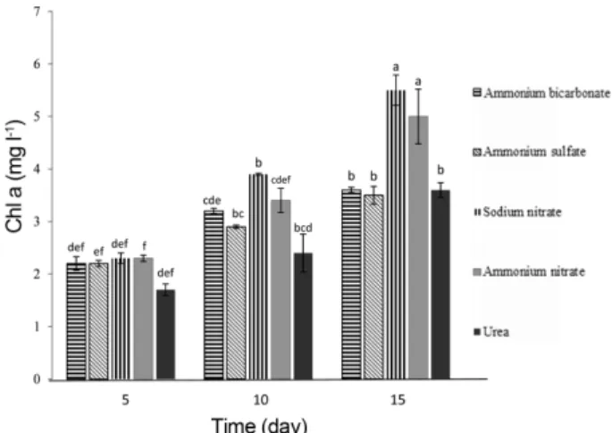
Chlorophyll a is often used as an estimate of algal bio- mass. However, Chla content was lower in media sup- plemented with ammonium bicarbonate (3.5± 0.30 mg l-1) than sodium nitrate (5.53± 0.32 mg l-1) (Fig. 2). It seems algal biomass estimated by Chla content was not matched with cell density in microalga. Many species of microalgae are able to use different forms of nitrogen, containing nitrate, nitrite, ammonium and other organic nitrogen sources like urea [28]. Each nitrogen source is primary reduced to the ammonium form, then assimi- lated into amino acids via a diversity of pathways.
Although ammonium bicarbonate was the fastest con- sumed nitrogen source by Nannocholoropsis cells, it seems that the resulted highest cell density is not merely due to the nitrogen, as the ammonium bicarbon- ate also contains bicarbonate as carbon source. Nanno- choloropsis seemingly utilizes both nitrogen and bicarbonate compounds, and as a result, the cell growth was more triggered. While the initial nitrogen concen- tration was similar in all cultures, the initial concentra- tion of total carbon in the medium with ammonium bicarbonate was higher than that in medium with other N sources.
Urea, as organic nitrogen source, is relatively energet-
ically cost effective than other nitrogen sources and also be easily utilized after being converted to ammonium and bicarbonate by urease in most microalgae species [29]. Nannocholoropsis oceanica grew fine in culture supplemented with urea, and the final cell concentration as well as biomass productivity was higher than observed with ammonium nitrate supplementation.
While, Campos et al. [30] stated that Nannochloropsis salina grows better in the presence of urea rather than nitrate or ammonium. Isochrysis galbana was also found to achieve to the highest cell concentration in urea rather than nitrate or nitrite [31]. These results indicate that the preference for nitrogen source and the ability of nitrogen utilization were changed from species to species.
Experiments confirmed that many microalgae usually prefer ammonium rather than other sources of nitrogen such as nitrate or nitrite. Because, ammonium is the reduced form of nitrogen and can be directly assimilated into amino acids, while other forms (e.g. nitrate or nitrite) must first be reduced to ammonium within the cells before its consumption [32]. In the present study, it was found that ammonium bicarbonate but not ammo- nium nitrate is more favorable for the growth of Nanno- choloropsis cells (Figs. 1 and 2). But, many species of microalgae, for example, D. tertiolecta, I. galbana, Neo- chloris oleoabundans, C. sorokiniana, and Botryococcus braunii, prefer nitrate over ammonium for their growth Fig. 1. Effects of different forms nitrogen supplementation
of the culture medium on the cell density of Nannochloro- psis oceanica. Data represent the mean values of triplicates
± standard deviation (SD). Values with the same lower case let- ter are not significantly different at p = 0.05 significance level based on Duncan’s multiple range test.
Fig. 2. Effects of different forms nitrogen supplementation of the culture medium on the chlorophyll a contents Nan- nochloropsis oceanica. Data represent the mean values of triplicates± standard deviation (SD). Values with the same lower case letter are not significantly different at p = 0.05 sig- nificance level based on Duncan’s multiple range test.
and development [18, 20, 28, 33, 34].
The growth of Nannocholoropsis cells was slowed by ammonium nitrate supplementation, thus it seem had a toxic effect on the cell growth, at the certain concentra- tion of 0.88 mM. Ramanna et al. [35] suggested that the negative effect on cell growth is due to the fact that the excessive transport of ammonium to the cells can pre- vent the ATP formation in the chloroplast, leading to photosynthesis inhibition. According to Norici et al. [22]
findings, the transport of nitrate is more regulated in algal cells, while the influx of ammonium is not easily controlled, especially when the extracellular ammonium concentration is high. Some investigators [33, 36]
reported that ammonium oversaturation in the medium can strongly decreased the pH by releasing H+ ions, resulting in preventing growth of the cell and even caus- ing cell lysis.
Changes in lipid content
The lipid content of Nannocholoropsis oceanica obtained after 15 days of incubation with different nitro- gen forms is shown in Fig. 3. The level of increase in lipid content was very different, depending on the sup- plied N sources. In the cultures supplemented with ammonium sulfate and ammonium nitrate, the cells mostly produced higher lipid contents than those with other nitrogen sources (The highest total lipid contents
were 65 and 62 %, respectively).
In cultures supplemented with organic-N (Urea) and nitrate, the lipid content moderately increased up to 51% and 42% respectively on the 15th day, while the supplementation with ammonium bicarbonate, result- ing in the lowest lipid content (26%) on the final day (Fig. 3). As there is a reverse relation between microal- gal growth and cellular lipid content, thus in culture supplemented with ammonium bicarbonate, the lowest lipid content was resulted. Although more lipid content was obtained from the cultures with ammonium sulfate, the lipid productivity was lower than that of nitrate- or organic-N supplementation due to a lower biomass pro- duction, resulting from the cell growth inhibition. Simi-
Fig. 3. Effects of different nitrogen source supplementation of the culture medium on the total lipid contents of Nan- nochloropsis oceanica. Data represent the mean values of triplicates± standard deviation (SD). Values with the same lower case letter are not significantly different at p = 0.05 sig- nificance level based on Duncan’s multiple range test.
Fig. 4. Fluorescence micrographs of Nannochloropsis oceanica stained with Nile Red fluorescence dye and screened under 1000X magnification with different N sources: (A) Ammo- nium bicarbonate, (B) Ammonium sulfate, (C) Sodium Nitrate (D) Ammonium nitrate and (E) Urea. The lipid drop- lets appear as yellow color.
larly, some algae species such as I. galbana and C.
sorokiniana accumulate higher lipids when supple- mented with ammonium as a nitrogen source rather than nitrate or urea, however, it is important to know that the final biomass concentrations were very low, because ammonium inhibits the cell growth [23, 34].
Fluorescence microscopic analysis of Nile Red-stained cells
To visually observe the effect of nitrogen sources on lipid accumulation in cells, we collected and stained cells with Nile Red and observed by fluorescence microscopy.
Microscopy results were in strong agreement with our previous results, as lipid accumulation (increased size and number of cytoplasmic lipid droplets) in ammonium
sulfate and ammonium nitrate was higher than the con- trol (sodium nitrate) and ammonium bicarbonate (Fig.
4A−E).
Fatty acid profiles
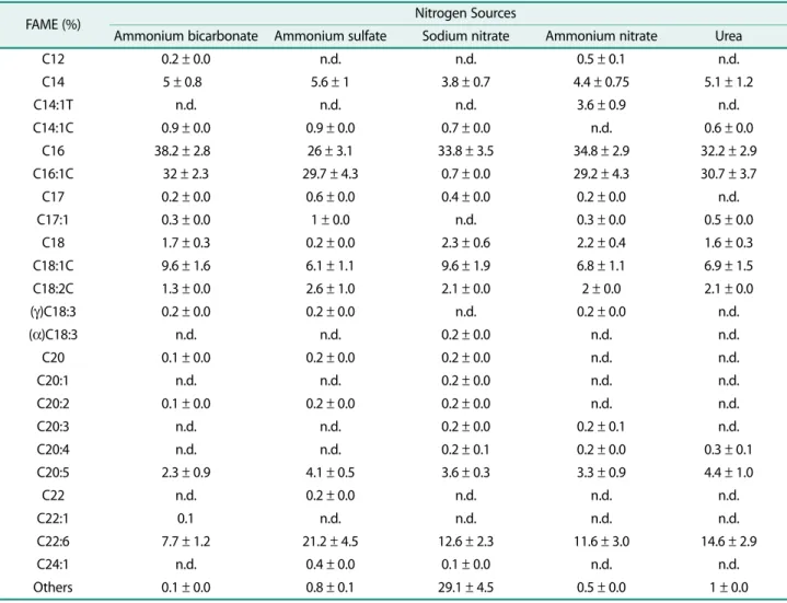
The fatty acid profiles of Nannochloropsis oceanica grown under different nitrogen sources are shown in Table 1. In cultures supplemented with ammonium bicarbonate, ammonium sulfate, ammonium nitrate and urea, C14:0, C16:0, C16:1, C18:0, C18:1, C18:2, C20:5 and C22:6 were detected. However, in culture supple- mented with sodium nitrate, C16:1 had very small peak.
Other small peaks were detected using all nitrogen sources. From the area of the main peak, the fatty acid component ratio was calculated (Table 1). The relative
Table 1. Fatty acid methyl ester (FAME) profile of Nannochloropsis oceanica cells cultivated under different nitrogen sources.
Values are presented as mean ± SD (n = 3).
FAME (%) Nitrogen Sources
Ammonium bicarbonate Ammonium sulfate Sodium nitrate Ammonium nitrate Urea
C12 0.2± 0.0 n.d. n.d. 0.5± 0.1 n.d.
C14 5± 0.80 5.6± 1 3.8± 0.7 4.4± 0.75 5.1± 1.2
C14:1T n.d. n.d. n.d. 3.6± 0.9 n.d.
C14:1C 0.9± 0.0 0.9± 0.0 0.7± 0.0 n.d. 0.6± 0.0
C16 38.2± 2.80 26± 3.1 33.8± 3.5 34.8± 2.9 32.2± 2.9
C16:1C 32± 2.3 29.7± 4.3 0.7± 0.0 29.2± 4.3 30.7± 3.7
C17 0.2± 0.0 0.6± 0.0 0.4± 0.0 0.2± 0.0 n.d.
C17:1 0.3± 0.0 1± 0.0 n.d. 0.3± 0.0 0.5± 0.0
C18 1.7± 0.3 0.2± 0.0 2.3± 0.6 2.2± 0.4 1.6± 0.3
C18:1C 9.6± 1.6 6.1± 1.1 9.6± 1.9 6.8± 1.1 6.9± 1.5
C18:2C 1.3± 0.0 2.6± 1.0 2.1± 0.0 2± 0.0 2.1± 0.0
(γ)C18:3 0.2± 0.0 0.2± 0.0 n.d. 0.2± 0.0 n.d.
(α)C18:3 n.d. n.d. 0.2± 0.0 n.d. n.d.
C20 0.1± 0.0 0.2± 0.0 0.2± 0.0 n.d. n.d.
C20:1 n.d. n.d. 0.2± 0.0 n.d. n.d.
C20:2 0.1± 0.0 0.2± 0.0 0.2± 0.0 n.d. n.d.
C20:3 n.d. n.d. 0.2± 0.0 0.2± 0.1 n.d.
C20:4 n.d. n.d. 0.2± 0.1 0.2± 0.0 0.3± 0.1
C20:5 2.3± 0.9 4.1± 0.5 3.6± 0.3 3.3± 0.9 4.4± 1.0
C22 n.d. 0.2± 0.0 n.d. n.d. n.d.
C22:1 0.1 n.d. n.d. n.d. n.d.
C22:6 7.7± 1.2 21.2± 4.5 12.6± 2.3 11.6± 3.0 14.6± 2.9
C24:1 n.d. 0.4± 0.0 0.1± 0.0 n.d. n.d.
Others 0.1± 0.0 0.8± 0.1 29.1± 4.5 0.5± 0.0 1± 0.0
n.d.= not detected
abundance of main fatty acid products is different between microalga grown under different nitrogen sources. The main fatty acid composition of nitrate- grown alga comprised 3.8%, 33.8%, 2.3%, 9.6%, 2.1%, 3.6% and 12.6% of C14:0, C16:0, C18:0, C18:1, C18:2, C20:5 and C22:6. In contrast, main fatty acid composi- tion of urea-grown alga comprised 5.1%, 32.2%, 30.7%, 1.6%, 6.9%, 2.1%, 4.4% and 14.9% of C14:0, C16:0, C16:1, C18:0, C18:1, C18:2, C20:5 and C22:6. Also, the main fatty composition of ammonium-grown algae con- sisted C14:0, C16:0, C16:1, C18:0, C18:1, C18:2, C20:5 and C22:6. In addition, a clear difference can be seen in C16:1 and C22:6 for cells grown in different nitrogen sources. Thus, it is obvious that different nitrogen sources can affect the composition of fatty acid in microalgae. Campos et al. [30] reported the same FAME fraction of lipid in N. salina irrespective of nitrogen source. Furthermore, the main fatty acids of N. oceanica IMET1 when grew in the modified f/2 medium were C16:0 and C16:1 [37].
One of the most prominent factors that affects the properties of biodiesel is profile of fatty acid, because the molecular features of FAMEs, including length of carbon chain and the double bond number, directly influence some characteristics of biodiesel such as: the viscosity, ignition quality, oxidative constancy, and property of cold flow [38, 39].
Different factors, such as various nutritional condi- tions, physicochemical conditions as well as growth phases can change the composition of fatty acid [40, 41].
Serrano et al. [42] found that oxidation stability and cold flow performance have reverse relationships to varia- tions in composition of fatty acids.
For example, the raise in unsaturated fatty acids (UFAs) would improve the cold flow performance, while decreasing the oxidative stability. Inversely, the increase of saturated fatty acids (SFAs) could result in better oxi- dative stability but poor cold flow property.
Lapuerta et al. [43] stated that the great fraction attendance of unsaturated fatty acids outcome in a small cetane number of biodiesel, fathering a poor ignition state. Because the UFA fraction is lower in all forms of nitrogen, quality of ignition would be better. However, higher fraction of SFA can result in an inferior cold-flow property; it is possible to achieve the fuel quality by using some additives [38].
In this research, the effects of different nitrogen sources were examined on the different physiological parameters such as the growth, lipid production and composition of N. oceanica CCAP 849/10. The obtained data confirmed that N. oceanica produces a much higher lipid content when cultivated with ammonium sulfate than with other nitrogen sources. In the presence of ammonium sulfate, higher amount of C22:6 fatty acid yield in comparison with other nitrogen sources. Ammo- nium bicarbonate-grown cells have higher cell number than other nitrogen source-grown cells. Therefore, we concluded that replacement of nitrate in f/2 medium with ammonium bicarbonate will have a negative effect on lipid content but boost cell growth. Thus we suggest that ammonium sulfate is a better nitrogen source with respect to lipid productivity as biodiesel in Nannochloro- psis oceanica.
Acknowledgments
This research was financially supported (No. 94/11138) by Research and Technology office of Arak University and authors thank Arak Uni- versity.
Conflict of Interest
The authors have no financial conflicts of interest to declare.
References
1. Antolín G, Tinaut FV, Briceño Y, Castaño V, Pérez C, Ramírez AI.
2002. Optimization of biodiesel production by sunflower oil transesterification. Bioresour. Technol. 4: 111-114.
2. Miao X, Wu Q. 2006. Biodiesel production from heterotrophic microalgal oil. Bioresour. Technol. 97: 841-846.
3. McGinnis KM, Dempster TA, Sommerfeld MR. 1997. Characteriza- tion of the growth and lipid content of the diatom Chaetoceros muelleri. J. Appl. Phycol. 9: 19-24.
4. Scragg AH, Morrison J, Shales SW. 2003. The use of a fuel contain- ing Chlorella vulgaris in a diesel engine. Enzyme Microb. Technol.
33: 884-889.
5. Spolaore P, Joannis-Cassan C, Duran E, Isambert A. 2006. Com- mercial applications of microalgae. J. Biosci. Bioeng. 101: 87-96.
6. Takagi M, Karseno Yoshida T. 2006. Effect of salt concentration on intracellular accumulation of lipids and triacylglyceride in marine microalgae Dunaliella cells. J. Biosci. Bioeng. 101: 223-226.
7. Schenk PM, Thomas-Hall, Skye R, Stephens E, Marx UC, Muss- gnug JH, et al. 2008. Second generation biofuels: High-efficiency microalgae for biodiesel production. Bioenergy Res. 1: 20-43.
8. Arenas EG, Rodriguez Palacio MC, Juantorena AU, Fernando SEL,
Sebastian PJ. 2017. Microalgae as a potential source for biodiesel production: techniques, methods, and other challenges. Int. J.
Energy Res. 41: 761-789.
9. Emdad ID, Berland B. 1989. Variation in lipid class composition during batch growth of Nannochloropsis salina and Pavlova lutheri. Mar. Chem. 26: 215-225.
10. Dunstan GA, Volkman JK, Barrett SM, Garland CD. 1993. Changes in the lipid composition and maximization of the polyunsatu- rated fatty acid content of three microalgae grown in mass cul- ture. J. Appl. Phycol. 5: 71-83.
11. Illman AM, Scragg AH, Shales SW. 2000. Increase in Chlorella strains calorific values when grown in low nitrogen medium.
Enzyme Microb. Technol. 27: 631-635.
12. Kim G, Mujtaba G, Rizwan M, Lee K. 2014. Environmental stress strategies for stimulating lipid production from microalgae for biodiesel. Appl. Chem. Eng. 25: 553-558.
13. Bartley ML, Boeing WJ, Daniel D, Dungan BN, Schaub T. 2016.
Optimization of environmental parameters for Nannochloropsis salina growth and lipid content using the response surface method and invading organisms. J. Appl. Phycol. 28: 15-24.
14. Cai T, Park SY, Li Y. 2013. Nutrient recovery from wastewater streams by microalgae: status and prospects. Renew. Sustain.
Energy Rev. 19: 360-369.
15. Hu Q. 2013. Environmental effects on cell composition. pp. 114- 122. In Richmond, A. and Hu, Q. (Eds.) Handbook of Microalgal Culture: Applied Phycology and Biotechnology. 2nd ed. Wiley Blackwell, West Sussex.
16. Wang J, Sommerfeld MR, Lu C, Hu Q. 2013. Combined effect of initial biomass density and nitrogen concentration on growth and astaxanthin production of Haematococcus pluvialis (Chloro- phyta) in outdoor cultivation. Algae 28: 193-202.
17. Ho SH, Ye X, Hasunuma T, Chang JS, Kondo A. 2014. Perspectives on engineering strategies for improving biofuel production from microalgae: a critical review. Biotechnol. Adv. 32: 1448-1459.
18. Chen M, Tang H, Ma H, Holland TC, Ng KY, Salley SO. 2011. Effect of nutrients on growth and lipid accumulation in the green algae Dunaliella tertiolecta. Bioresour. Technol. 102: 1649-1655.
19. Ruangsomboon S. 2015. Effects of different media and nitrogen sources and levels on growth and lipid of green microalga Botryococcus braunii KMITL and its biodiesel properties based on fatty acid composition. Bioresour. Technol. 191: 377-384.
20. Li T, Zheng Y, Yu L, Chen S. 2013. High productivity cultivation of a heat-resistant microalga Chlorella sorokiniana for biofuel pro- duction. Bioresour. Technol. 131: 60-67.
21. Muthuraj M, Kumar V, Palabhanvi B, Das D. 2014. Evaluation of indigenous microalgal isolate Chlorella sp. FC2 IITG as a cell fac- tory for biodiesel production and scale up in outdoor conditions.
J. Ind. Microbiol. Biotechnol. 41: 499-511.
22. Norici A, Dalsass A, Giordano M. 2002. Role of phosphoenolpyru- vate carboxylase in anaplerosis in the green microalga Dunaliella salina cultured under different nitrogen regimes. Physiol. Plant.
116: 186-191.
23. Wan MX, Wang RM, Xia JL, Rosenberg JN, Nie ZY, Kobayashi N, et
al. 2012. Physiological evaluation of a new Chlorella sorokiniana isolate for its biomass production and lipid accumulation in pho- toautotrophic and heterotrophic cultures. Biotechnol. Bioeng.
109: 1958-1964.
24. Kim G, Mujtaba G, Rizwan M, Lee K. 2016. Effects of nitrogen sources on cell growth and biochemical composition of marine chlorophyte Tetraselmis sp. for lipid production. Algae 31: 257- 266.
25. Guillard RRL. 1975. Culture of phytoplankton for feeding marine invertebrates. pp. 26-60. In Smith, W. L. and Chanley, M. H. (Eds.) Culture of Marine Invertebrate Animals. Plenum Press, New York.
26. Mackinney G. 1941. Absorption of light by chlorophyll solutions.
J. Biol. Chem. 140: 315-322.
27. Bligh EG, Dyer WJ. 1959. A rapid method for total lipid extraction and purification. Can. J. Biochem. Physiol. 37: 911-917.
28. Becker EW. 1994. Microalgae biotechnology and microbiology.
18 pp. Cambridge University Press, New York.
29. Solomon CM, Glibert PM. 2008. Urease activity in five phyto- plankton species. Aquat. Microb. Ecol. 52: 149-157.
30. Campos H, Boeing WJ, Dungan, BN, Schaub T. 2014. Cultivating the marine microalga Nannochloropsis salina under various nitrogen sources: effect on biovolume yields, lipid content and composition, and invasive organisms. Biomass Bioenergy 66: 301- 307.
31. Fidalgo JP, Cid A, Torres E, Sukenik A, Herrero C. 1998. Effects of nitrogen source and growth phase on proximate biochemical composition, lipid classes and fatty acid profile of the marine microalga Isochrysis galbana. Aquaculture 166: 105-116.
32. Podevin M, De Francisci D, Holdt SL, Angelidaki I. 2015. Effect of nitrogen source and acclimatization on specific growth rates of microalgae determined by a high-throughput in vivo microplate autofluorescence method. J. Appl. Phycol. 27: 1415-1423.
33. Li Y, Horsman M, Wang B, Wu N, Lan CQ. 2008. Effects of nitrogen sources on cell growth and lipid accumulation of green alga Neo- chloris oleoabundans. Appl. Microbiol. Biotechnol. 81: 629-636.
34. Roopnarain A, Sym S, Gray VM. 2015. Effect of nitrogenous resource on growth, biochemical composition and ultrastructure of Isochrysis galbana (Isochrysidales, Haptophyta). Phycol. Res. 63:
43-50.
35. Ramanna L, Guldhe A, Rawat I, Bux F. 2014. The optimization of biomass and lipid yields of Chlorella sorokiniana when using wastewater supplemented with different nitrogen sources.
Bioresour. Technol. 168: 127-135.
36. Wu LF, Chen PC, Lee CM. 2013. The effects of nitrogen sources and temperatures on cell growth and lipid accumulation of microalgae. Int. Biodeterior. Biodegrad. 85: 506-510.
37. Xiao Y, Zhang J, Cui J, Feng Y, Cui Q. 2013. Metabolic profiles of Nannochloropsis oceanica IMET1 under nitrogen-deficiency stress. Bioresour. Technol. 130: 731-738.
38. Knothe G. 2009. Improving biodiesel fuel properties by modify- ing fatty ester composition. Energy Environ. Sci. 2: 759-766.
39. Singh B, Guldhe A, Rawat I, Bux F. 2014. Towards a sustainable approach for development of biodiesel from plant and microal-
gae. Renew. Sustain. Energy Rev. 29: 216-245.
40. Mata TM, Martins AA, Caetano NS. 2010. Microalgae for biodiesel production and other applications: a review. Renew. Sustain.
Energy Rev. 14: 217-232.
41. Kim DG, Hur SB. 2013. Growth and fatty acid composition of three heterotrophic Chlorella species. Algae 28: 101-109.
42. Serrano M, Oliveros R, Sánchez M, Moraschini A, Martínez M,
Aracil J. 2014. Influence of blending vegetable oil methyl esters on biodiesel fuel properties: oxidative stability and cold flow properties. Energy 65: 109-115.
43. Lapuerta M, Rodríguez-Fernández J, de Mora EF. 2009. Correla- tion for the estimation of the cetane number of biodiesel fuels and implications on the iodine number. Energy Policy 37: 4337- 4344.