Korean J Clin Lab Sci. 2011, 43(4) : 161 - 170 ISSN 1738-3544
INTRODUCTION
Reactive oxygen species (ROS) have been implicated in the pathogenesis of gastrointestinal injury in various pathological conditions such as ischemia-reperfusion in- jury (Kvietys et al, 1988), Certain types of drug-induced
gastroenteropathy (Vaananen et al, 1991), experimental colitis (Keshavarzian et al, 1990). and inflammatory bowel diseases (Pavlick et al, 2002). Therefore, supplementation of patients with antioxidants may exert beneficial effects in inflammatory bowel disease, at least in part, by antioxidant mechanisms (Simmonds and Rampton, 1993; Pavlick et al, 2002).
Flavonoids are phenolic compounds widely distributed in almost every plants and act as pharmacological active constituents in many herbal medicines. They have mul- tiple biological, pharmacological and medicinal properties including anti-inflammatory, anti-allergic, antiviral, anti-
Quercetin Prevents Hydrogen Peroxide-induced Necrotic and Apoptotic Cell Death in Human Colonic Epithelial Cells
Soon-Hee Jung
Department of Clinical Pathology, Jinju Health College, Jinju 660-757, Korea
Quercetin is one of the most distributed flavonoids in the plant kingdom and occurs naturally in a wide range of fruits and vegetables. This study was undertaken to determine whether quercetin exerts benefi- cial effect against necrotic and apoptotic cell death induced by hydrogen peroxide (H2O2) in intestinal cells using the human-derived cultured T84 colonic epithelial cell line. Necrotic cell death was induced by exposing cells to 0.5 mM H2O2 for 2 h and apoptosis was induced by incubating cells in normal cul- ture medium for 18 h following exposure of cells to 0.5 mM H2O2 for 2 h. Cell viability was evaluated by the trypan blue exclusion assay and apoptosis was assessed by Hoechst 33258 staining and flow cytome- try. H2O2 induced necrotic cell death in a time and dose-dependent fashion. Both necrotic and apoptot- ic cell deaths were not prevented by the antioxidants N,N’-diphenyl-p-phenylenediamine(DPPD) and Trolox, whereas both cell deaths induced by the organic hydroperoxide t-butylhydroperoxide (tBHP) were prevented by DPPD, suggesting that H2O2 induces cell death through a lipid peroxidation-inde- pendent mechanism. H2O2-induced necrotic death was prevented by deferoxamine and 3-aminoben- zamide, while the apoptotic cell death was not affected by these agents. Quercetin prevented both ne- crotic and apoptotic cell deaths induced by H2O2 in a dose-dependent manner. H2O2 caused activation of poly (ADP-ribose) polmerase (PARP), which was inhibited by deferoxamine, 3-aminobenzamide, and quercetin, but not DPPD. These results indicate that quercetin inhibits both necroticand apoptotic deaths of T84 cells. The anti-necrotic effect of quercetin may be attributed to its iron chelator activity rather than a direct H2O2 scavenging capacity and antioxidant. The present study suggests that quercetin may play a therapeutic role in the treatment of human gastrointestinal diseases mediated by oxidants.
Key Words : Quercetin, Apoptotic cell, Hydrogen peroxide, Colonic epithelial cell
Corresponding Author : Jung, Soon-Hee. Department of Clinical Pathology, Jinju Health College, Jinju 660-757, Korea.
Tel: 010-3584-1847 E-mail: [email protected]
Received : 8 NOV 2011
Return for modification : 2 DEC 2011 Accepted : 9 DEC 2011
thrombotic, antimutagenic, antineoplastic, and hepatopro- tective effects (Formica and Regelson, 1995). Quercetin is an important member of the flavonoid family and is found in various food products including fruits, vegetables, olive oil, red wine, and tea. Quercetin exerts a beneficial effect against oxidative damage induced by iron (Nunez et al, 2001). and chemicals (Sanchez de Medina et al, 2002).
and may have anticancer activity (Lipkin et al, 1999). in the intestine.
Although the cytoprotective effects of flavonoids such as quercetin have been considered to be associated with their antioxidant activities, it is unclear whether the inhibition of lipid peroxidation is attributed to either a direct interrup- tion of membrane lipid peroxidation or ROS scavenging capacity and iron chelating properties. Earlier studies have shown that the cytoprotection has been related to iron chelating properties (Morel et al, 1993; Kostyuk and Pota- povich, 1998). and free radical scavenging capacity (Fraga et al, 1987; Ratty and Das, 1988). However, other stud- ies have reported that the protective effects of quercetin against oxidant-induced cell injury are due to inhibition of membrane lipid peroxidation by incorporatinginto the membrane lipid bilayer rather than to intracellular scaveng- ing of ROS (Kuhlmann et al, 1998; Ahlenstiel et al, 2003).
Cell death is classified as necrosis or apoptosis on the basis of established morphological criteria (Cohen et al, 1992). Necrosis is considered a passive event in which the cell is irreversibly damaged by an environmental insult, leading to cell death. In contrast, apoptosis is an active pro- cess in which the cell itself initiates the molecular machin- ery to trigger cell death in response to either a physiologi- cal stimulus or an environmental stress (Cohen et al, 1992).
It has been known that oxidants induce cell death through necrosis and apoptosis (Janssen et al, 1997; Samali et al, 1999). The mode of cell death has been highly variable, depending on the cell type, the nature of the toxin, and the strength and duration of the toxic insult (Raffray and
Cohen, 1997). Therefore, the protective effect of agents against oxidant-induced cell injury could be different be- tween two processes (Filipovic et al, 1999). Although quer- cetin has been reported to inhibit oxidant-induced apop- tosis (Yokoo and Kitamura, 1997; Ishikawa and Kitamura, 2000). whether this effect is due to antioxidant activity is not clear.
This study was undertaken to examine the effect of quer- cetin on hydrogen peroxide (H2O2)-induced necrotic and apoptotic cell death in intestinal cells using the human- derived cultured colonic epithelial cell line T84. Since it has been shown that the H2O2 induces cell injury through a lipid peroxidation-independent mechanism in various cell types (Zager and Burkhart, 1998; Min et al, 2000; Park et al, 2003). we used H2O2 as an oxidant model to determine whether the protective effect of quercetin is due to a di- rect interruption of membrane lipid peroxidation or radical scavenging activity. If the cytoprotective effect is attributed only to a direct inhibition of membrane lipid peroxidation, quercetin could not prevent the H2O2-induced cell death.
MATERIALS AND METHODS Chemicals
[3H]NAD was obtained from Amersham international (Amersham, UK). Hydrogen peroxide (H2O2), t-butyl- hydroeproxide (tBHP), quercetin, deferoxamine (DFO), catalase, (DPPD), Trolox, propidium iodide, and 3-amino- benzamide (3-AB) were purchased from Sigma-Aldrich Chemical (St. Louis, MO, USA). TWEEN 20 was purchased from Calbiochem (California, USA). Antibodies of phos- pho-ERK and β-actin were obtained from Cell Signaling Technology Inc. (Beverly, MA, USA). All other chemicals were of the highest commercial grade available.
1. Culture of T84 cells
T84 cells were obtained from the American Type Cul-
ture Collection (Rockville, MD) and maintained by serial passages in 75 cm2 culture flasks (Costar, Cambridge, MA).
The cells were grown as monolayer cultures in Dulbecco’s modified Eagle’s medium/Ham’s F12 (DMEM/F12, Sigma Chemical Co.) containing 10% fetal bovine serum at 37℃
in 95% air/5% CO2 incubator.
2. Induction of necrotic and apoptotic cell death For induction of necrotic cell death, cells were exposed to 1.0 mM H2O2 or tBHP in Hanks’ balanced salt solution (HBSS, Sigma Co, USA) for 2 hrs, and apoptotic cell death was induced by incubating cells for a further 18 hrs in nor- mal culture medium following exposure of cells to 0.5 mM H2O2 or 0.3 mM tBHP in HBSS for 2 hrs. In experiments to examine effects of inhibitors, test agents were added to medium during exposure of cells to oxidants.
3. Measurement of cell viability
Cell viability was determined bya trypan blue exclusion assay. The cells were harvested using 0.025% trypsin, in- cubated with 4% trypan blue solution, and were counted using a hemocytometer under light microscopy. Cells fail- ing to exclude the dye were considered nonviable, and the data were expressed as a percentage of viable cells to total cells.
4. Measurement of apoptotic cells
To estimate whether cell death was attributed to necrosis or apoptosis, cells were stained with the fluorescent dye Hoechst 33258. Cells were grown on 22 mm glass cover- slips in 6-well plates. After treatment with oxidants, the cells were washed twice with PBS and fixed with 4% para- formaldehyde in PBS (pH 7.4) for 1 hr at 4℃. The fixed cells were washed twice with PBS and stained with 10 μM Hoechst 33258 for 15 min at 37℃. Then cells were washed twice with PBS and examined by confocal microscopy (LSM510, ZEISS, Germany). Apoptotic cells were identified
by condensation and fragmentation of nuclei.
Apoptosis was quantified by fluorescence-activated cell sorter (FACS) analysis (Ormerod et al, 1992). Cells were grown in six well plates and were treated as indicated.
Then, attached and floating cells were pooled, pelleted by centrifugation, washed in phosphate-buffered saline, and fixed with cold 70% ethanol containing 0.5% tween 20 at 4℃overnight. Cells were washed, resuspended in 1.0 mL of propidium iodide solution containing 100 μg of RNase A/mL and 50 μg propidium iodide/mL. Cells were incu- bated for 30 min at 37℃ and analyzed. Cells with sub-G1 propidium iodide incorporation were considered as apop- totic. The percentage of apoptotic cells was calculated as the ratio of events on sub-G1 to events from the whole population.
5. Measurement of poly (ADP-ribose) polmerase (PARP) activity
Cells were treated with H2O2 and preincubated for 10 min in a buffer containing 28 mM NaCl, 28 mM KCl, 2 mM MgCl2, 56 mM Hepes (pH 7.5), 0.01% digitonin, and 125 mM NAD (containing 0.25 μCi [3H]NAD). Permeabilized cells were incubated for 5 min at 37℃, and the protein ri- bosylated with [3H]NAD was precipitated with 200 μL of 50% (w/v) trichloroacetic acid. After washing twice with trichloroacetic acid, the protein pellet was solubilized with 200 μL of 2% (w/v) sodium dodecyl sulfate in 0.1 M NaOH and incubated at 37℃ overnight, and the radioactivity was determined by scintillation counting.
6. Statistical analysis
The data are expressed as mean±SE and the difference between two groups was evaluated using Student’s t-test.
Multiple group comparison was done using one-way analysis of variance followed by the Tukey post hoc test. A probability level of 0.05 was used to establish significance.
RESULTS
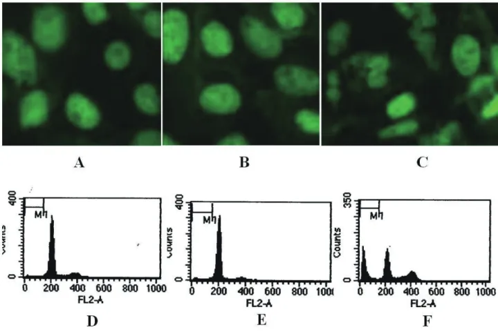
1. Induction of necrotic and apoptotic cell death Many cells exhibit bimodal responses, with necrotic cell death and apoptotic cell death. Necrosis is a rapid response, while apoptosis is a slow response (Eastman, 1993; Lieberthal et al, 1996). In order to evaluate the ef- fect of quercetin on necrotic and apoptotic death induced by H2O2, cells were treated with 0.5 mM H2O2 for 2 hrs in HBSS or were incubated for 18 h in normal culture medi- um following exposure of cells to 0.5 mM H2O2. Cells were stained with a fluorescent dye Hoechst 33258, which spe- cifically binds to DNA. The nuclei of untreated control cells showed normal morphology (Fig. 1A). The nuclei of cells
treated with 0.5 mM H2O2 for 2 hrs (Fig. 1B) could not be distinguished from those of control cells. In contrast, when cells were incubated in normal culture media for 18 hrs fol- lowing exposure of cells to 0.5 mM H2O2 for 2 h, the nuclei of cells were disrupted and fragmented, exhibiting typical features of apoptosis (Fig. 1C). Cell viability estimated by trypan blue exclusion was approximately 61 and 62% un- der these two conditions, respectively. Similar results were obtained from flow cytometric analysis (Fig. 1D-E).
2. Effect of quercetin on necrotic cell death
Fig. 2 shows the time course of H2O2-induced necrotic cell death. Cells were exposed to 1.0 mM H2O2, and cell vi- ability was determined at various time points (0-180 min).
Fig. 1. Induction of apoptosis by H2O2. Hoechst 33258 staining (A-C) and flow cytometry (D-F) of cells exposed to H2O2. Apop- tosis was measured in control (A, D), cells exposed to 1.0 mM H2O2 in HBSS for 2 hrs (B, E), and cells exposed to 0.5 mM H2O2 in HBSS for 2 hr and further incubated for 18 hr in normal culture medium (C,F).
The significant loss of cell viability was present 30 min after exposure of cells to H2O2 and increased up to 180 min (Fig.
2). When cells were exposed to 0.1-1.0 mM H2O2 for 2 h, loss of cell viability increased in a dose-dependent man- ner and the significant loss was observed at 0.3 mM H2O2.
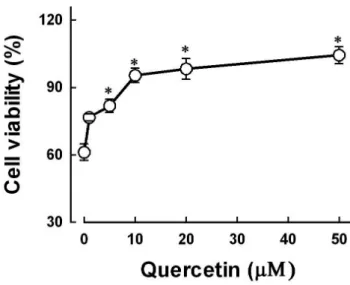
In order to examine the effect of quercetin on H2O2- induced necrotic cell death, cells were treated with 1.0 mM H2O2 in the presence of various concentrations of quercetin. As shown in Fig. 3, quercetin prevented H2O2- induced cell death in dose-dependent fashion and a sig-
nificant protection was present at 5 μM (81.80±2.99 vs.
61.20±3.67% in H2O2 alone). When quercetin concentra- tions were increased up to 10 and 50 μM, the cell viabil- ity was significantly different from the control. However, quercetin did not exert any effect in the viability of normal cells untreated with H2O2.
In order to define the underlying mechanism of cy- toprotective action of quercetin, effects of various agents including well-known antioxidants on necrotic cell death induced by H2O2 were examined. The results are summa- rized in Table 1. As expected, the H2O2 scavenger enzyme catalase prevented cell death. Similar protective effects were observed with the iron chelator DFO and the PARP inhibitor 3-AB, suggesting involvement of an iron-depen- dent mechanism and activation of PARP in H2O2-induced necrotic cell death. By contrast, antioxidants DPPD and Trolox did not affect the cell death, indicating that H2O2- induced cell death is not associated with lipid peroxida- tion. These results suggest that the cytoprotective effect of quercetin may be attributed to iron chelating ability rather Fig. 2. Effect of H2O2 on necrotic cell death. Cells were ex-
posed to 1.0 mM H2O2 for various time points (A) and various concentrations of H2O2 for 2 hrs (B). Cell viability was esti- mated by trypan blue exclusion assay. Data are mean±SEM of four experiments. *p <0.05 compared with control.
Fig. 3. Dose-dependent effect of quercetin on H2O2-induced necrotic cell death. Cells were exposed to 1.0 mM H2O2 for 2 hr in the presence or absence of various concentrations of quercetin. Cell viability was estimated by trypan blue exclu- sion assay. Data are mean±SEM of six experiments. *p <0.05 compared with the absence of quercetin.
than antioxidant action.
The organic hydroperoxide tBHP has been known as an established inducer of lipid peroxidation (Halliwell et al, 1992). In order to examine the efficiency of DPPD on lipid peroxidation-induced cell death, cells were treated with tBHP in the presence of DPPD. Unlike H2O2-induced cell death, tBHP-induced death was significantly prevented by DPPD (Table 1), indicating that tBHP causes necrotic cell death through a lipid peroxidation mechanism. If protec- tion by quercetin was resulted from H2O2 scavenging ac- tivity, tBHP-induced cell death could not be affected by quercetin. However, quercetin provided a significant pro- tection against tBHP-induced cell death. DFO also was ef- fective in preventing cell death (Table 1).
Table 1. Effects of various drugs on oxidant-induced cell death in T84 cells
Conditions Necrosis (%) Apoptosis (%)
Control H2O2
+ Catalase (500 U/mL) + DPPD (10 μM) + Trolox (1 mM) + DFO (1 mM) + 3-AB (1 mM) + Quercetin (10 μM)
tBHP
+ DPPD (10 μM) + DFO (1 mM) + Quercetin (10 μM)
5.54±0.28 46.49±4.56*
8.90±1.33† 49.35±0.76*
40.23±5.46*
15.38±2.68*† 10.10±1.42† 11.45±1.29†
39.29±2.33*
11.35±1.50*† 11.60±1.80*† 5.41±0.22†
6.21±0.44 38.42±2.34*
7.54±0.66† 34.26±1.04*
39.95±0.98*
36.02±0.74*
34.09±2.95*
15.16±0.57*†
35.27±1.85*
14.13±4.56*† 15.46±2.39*† 15.44±1.84*† Necrosis was induced by exposing cells to 1.0 mM H2O2 or t- butylhydroperoxide (tBHP) for 2 hrs in HBSS and apoptosis was induced by incubating cells for 18 hrs in normal culture medium following exposure of cells to 0.5 mM H2O or 0.3 mM tBHP in HBSS in the presence or absence of various drugs. Necrosis and apoptosis were estimated by trypan blue exclusion assay and flow cytometry, respectively. Data are mean±SEM of six determina- tions. *p <0.05 compared with control; †p <0.05 compared with oxidant alone.
In the present study, the PARP inhibitor 3-AB prevent- ed H2O2-induced necrotic cell death (Table 1), a result
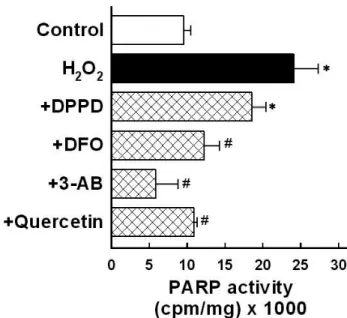
consistent with previous studies (Lee et al, 2001; Park et al, 2003). Therefore, we evaluated the effect of quercetin on PARP activation. Quercetin also inhibited the H2O2- induced activation of PARP similarly to DFO and 3-AB, but not DPPD (Fig. 4). These results suggest that H2O2 in- duces necrotic cell death by a mechanism dependent of PARP activation and independent of lipid peroxidation and that quercetin may exert the cytoprotective effect through inhibition of PARP directly or indirectly resulting from its iron chelating action.
3. Effect of quercetin on apoptotic cell death
Cells were exposed to various concentrations of H2O2 under experimental conditionsthat induce apoptosis and apoptotic cell death was estimated by flow cytometry.
H2O2 induced apoptosis in a dose-dependent manner over concentrations of 0.1-0.5 mM (Fig. 5A). Quercetin Fig. 4. Effects of various agents on H2O2-induced activa- tion of poly (ADP-ribose) polyerase (PARP). Cells were ex- posed to 1.0 mM H2O2 for 2 hr in the presence or absence of N,N’-diphenyl-p-phenylenediamine (DPPD, 10 μM), deferox- amine (DFO, 1 mM), 3-aminobenzamide (3-AB, 5 mM), and quercetin (20 μM). Data are mean±SEM of four experiments.
*p <0.05 compared with the absence of quercetin.
prevented H2O2-induced apoptotic cell death and its effect was dose-dependent (Fig. 5B), similarly to necrotic cell death.
In order to examine the role of lipid peroxidation and PARP in H2O2-induced apoptosis, effects of antioxidants and PARP inhibitor were evaluated. As shown Table 1,
H2O2-induced apoptosis was not prevented by the anti- oxidants DPPD and Trolox. The apoptosis was also not affected by DFO and 3-AB, unlike necrosis. These re- sults suggest that H2O2 induces apoptosis via mechanisms independent of lipid peroxidation and PARP activation.
Quercetin also prevented tBHP-induced apoptosis that is dependent of DPPD and DFO. These data indicate that quercetin can prevent the lipid peroxidation-dependent and -independent apoptosis.
Discussion
Since a growing body of evidence suggests that ROS are implicated in the pathogenesis of stress- and chemically- induced gastrointestinal injury (van der Vliet and Bast, 1992) potent antioxidants may serve as a possible preven- tive intervention for gastrointestinal injury. Indeed, previ- ous studies in mice and rats have reported that the dietary flavonoids such as quercetin protect colonic epithelial cells from carcinogenesis induced by azoxymethane. However, the precise molecular mechanism of quercetin cytoprotec- tion remains to be defined.
Although flavonoids such as quercetin have been re- ported to have the abilityto inhibit lipid peroxidation and iron chelating properties (Formica and Reglson, 1955).
they may also exhibit pro-oxidant activity. Previous stud- ies showed that quercetin exacerbated membrane damage resulting from menadione-induced oxidative stress.There- fore, quercetin may have multiple action mechanisms.
In the present study, quercetin prevented H2O2-induced necrotic cell death in a dose-dependent manner (Fig. 3).
Since flavonoids have free radical scavenging capacity (Fraga et al, 1987; Ratty and Das, 1988). and inhibits some biological processes triggered by H2O2 quercetin could prevent H2O2-induced cell death by directly scavenging H2O2 itself. However, quercetin also prevented cell death Fig. 5. Effect of quercetin on H2O2-induced apoptosis. (A)
Dose-dependent effect of H2O2 on apoptosis. Cells were ex- posed to various concentrations of H2O2 in HBSS for 2 h and further incubated for 18 h in normal culture medium. Apop- tosis was estimated by flow cytometry. Data are mean±SE of six experiments. (B) Dose-dependent effect of quercetin on H2O2-induced apoptosis. Apoptosis was induced as de- scribed in (A) with 0.5 mM H2O2 in the presence or absence of various concentrations of quercetin. Data are mean±SEM of six experiments. *p <0.05 compared with the respective control.
induced by tBHP, similarly to its effect on H2O2-induced cell death (Table 1).
Since the cytoprotective effects of flavonoids have been reported to be associated with antioxidant capacity the ef- fect of quercetin was compared with that of other well- known antioxidants. The necrotic cell death induced by H2O2 was not prevented by antioxidants DPPD and Trolox.
Although lipid peroxidation of cell membrane has been considered to be an evidence for oxidant-induced cell in- jury dissociation of H2O2-induced cell injury from lipid peroxidation has been reported in various cell types (Zager and Burkhart, 1998; Min et al, 2000; Park et al. 2005). Lipid peroxidation can be a result or an epiphenomenon of cell death rather than a cause of cell injury . These results sug- gest that H2O2-induced cell death is not mediated by lipid peroxidation in T84 cells. Therefore, these data strongly suggest that the protective effect of quercetin against H2O2- induced cell death may be attributed to a mechanism other rather than its antioxidant action.
Iron appears to be the critical in the cytotoxic effect of H2O2 in T84 cells as the iron chelator DFO was mark- edly protective (Table 1). These data indicate that H2O2- induced necrotic cell killing is resulted from an iron-de- pendent mechanism and hence the protection of quercetin may be attributed to its iron chelating activity. Indeed, pre- vious studies have demonstrated that the cytoprotection of flavonoids is related to iron chelating peroperties (Morel et al, 1993). Since PARP catalyzes the transfer of ADP-ribose from NAD to protein with the concomitant release of nic- otinamide, the activation of this enzyme results in deple- tion of NAD and a consequent reduction in ATP, which may be involved in the pathogenesis of oxidant-induced injury. In fact, PARP activation has been demonstrated to be involved in cell death, predominantly necrosis, caused by H2O2 Interestingly, H2O2 induces cell injury through a PARP activation-dependent and lipid peroxidation-inde- pendent mechanism, whereas tBHP-induced cell injury is
dependent of lipid peroxidation and independent of PARP activation in renal epithelial cells (Min et al, 2000; Park et al, 2003). In the present study, H2O2-induced necrotic cell death was prevented by the PARP inhibitor 3-AB (Ta- ble 1). Exposure of cells to H2O2 caused PARP activation, which was inhibited by DFO and 3-AB, but not DPPD (Fig. 4). These data suggest involvement of PARP activa- tion in H2O2-induced death in T84 cells. We did not de- termine whether quercetin inhibits PARP activity directly or indirectly by iron chelation in this study. However, inhibi- tion of PARP activation by quercetin is likely due to its iron chelating properties because many of the biological roles played by the flavonoids are associated with their iron- binding capabilities (Formica and Regelson, 1995).
Effects of quercetin on apoptosis are controversial. Quer- cetin facilitates apoptosis of tumor cells, in part through de- pression of an endogenous cytoprotective molecule, heat shock protein.The apoptosis-inducing action of querce- tin has been also observed in colonic cells . On the other hand, quercetin prevents apoptosis induced by oxidants in renal epithelial cells and fibroblasts (Yokoo and Kitamura, 1997), liver epithelial cells and mesangial cells. The precise molecular mechanism of pro- and anti-apoptotic effect of quercetin remains poorly understood. Furthermore, it is unclear whether the ability of quercetin to inhibit apoptosis is attributed to antioxidant capacity.
The present study showed that quercetin prevents H2O2-induced apoptosis in a dose-dependent manner (Fig. 5B). Unlike necrotic cell death, H2O2-induced apop- tosis was not affected by the iron chelator (DFO) and PARP inhibitor (3-AB). The data that the PARP inhibitor does not prevent H2O2-induced apoptosis are consistent with those reported in the intestinal epithelial cell line HT-29- 18-C1. Antioxidants also were not effective in preventing H2O2-induced apoptosis, whereas tBHP-induced apop- tosis was significantly prevented by antioxidant and iron chelator (Table 1). Taken together, these data suggest that
the protective effect of quercetin against H2O2-induced apoptosis is not associated with its antioxidant capacity in- cluding an iron chelating activity and a direct scavenging of H2O2. These results also indicate that quercetin prevents apoptosis via a mechanism different from that of necrosis
In conclusion, the present study demonstrated that quer- cetin prevented necrotic and apoptotic cell death induced by H2O2. The anti-necrotic effect of quercetin may be at- tributed to inhibition of PARP activation by its iron chelator activity. These results indicate that quercetin has the poten- tial for inhibiting necrotic and apoptotic death of T84 cells and exerts the cytoprotective effect against both cell death modes through a different mechanism. The results of the present study provide information that quercetin may be useful in treatment and prevention of gastrointestinal inju- ries mediated by oxidants.
REFERENCES
1. Ahlenstiel T, Burkhardt G, Kohler H, Kuhlmann MK. Biofla- vonoids attenuate renal proximal tubular cell injury during- cold preservation in Euro-Collins and University of Wiscon- sin solutions. Kidney Int. 2003, 63:554-563.
2. Cohen JJ, Duke, RC, Fadok VA, Sellins KS. Apoptosis and programmed cell death in immunity. Annu Rev Immunol.
1992, 10:267-293.
3. Eastman A, Apoptosis: a product of programmed and unpro- grammed cell death. Toxicol Appl Pharmacol. 1993, 121:160- 164.
4. Filipovic DM, Meng X, Reeves, WB.Inhibition of PARP pre- vents oxidant-induced necrosis but not apoptosis in LLC- PK1 cells. Am J Physiol.1999, 277:F428-F436.
5. Formica JV, Regelson W. Review of the biology of Querce- tin and related bioflavonoids. Food Chem Toxicol. 1995, 33:1061-1080.
6. Fraga CG, Martino VS, Ferraro GE, Coussio JD, Boveris A.Flavonoids as antioxidants evaluated by in vitro and in situ liver chemiluminescence. Biochem Pharmacol. 1987, 36:717- 720.
7. Halliwell B, Gutteridge, JMC, Cross CE.Free radicals, antioxi- dants, and human disease: Where are we now? J Lab Clin
Med. 1992, 119:598-620.
8. Ishikawa Y, Kitamura M. Anti-apoptotic effect of quercetin:
intervention in the JNK- and ERK- mediated apoptotic path- ways. Kidney Int. 2000, 58:1078-1087.
9. Forman HJ, and Cadenas E. In Oxidative stress and signal transduction. In: Janssen YMW, Timblin CR, Zanella CL, Jimenez LA, Mossman BT. Induction of gene expression by envirnmental oxidants associated with inflammation, fibro- genesis, and carcinogenesis. Ed, 1997, p387-414. Chapman
& Hall, Washington.
10. Keshavarzian A, Morgan G, Sedghi S, Gordon JH, Doria M, Role of reactive oxygen metabolites in experimental colitis.
Gut. 1990, 31:786-790.
11. Kostyuk VA, Potapovich AI, Antiradical and chelating effects in flavonoid protection against silica-induced cell injury.
Arch Biochem Biophys. 1998, 355:43-48.
12. Kuhlmann MK, Burkhardt G, Horsch E, Wagner M, Kohler H.
Inhibition of oxidant-induced lipid peroxidation in cultured renal tubular epithelial cells (LLC-PK1) by quercetin. Free Radic Res. 1998, 29:451-460.
13. Kvietys PR, Smith SM, Grisham MB, Manci EA. 5-Amino- salicylic acid protects against ischemia/reperfusion-induced gastric bleeding in the rat. Gastroenterol. 1988, 94:733-738.
14. Lee YW, Ha MS, Kim YK. H2O2-induced cell death in human glioma cells: role of lipid peroxidation and PARP activation.
Neurochem Res. 2001, 26:337-343.
15. Lieberthal W, Triaca V, Levine J. Mechanisms of death in- duced by cisplatin in proximal tubular epithelial cells: apop- tosis vs. necrosis. Am J Physiol.1996, 270: F700-708.
16. Lipkin M, Reddy B, Newmark H, Lamprecht SA. Dietary factors in human colorectal cancer. Annu Rev Nutr. 1999, 19:545-586.
17. Min SK, Kim SY, Kim CH, Woo JS, Jung JS, Kim YK. Role of lipid peroxidation and poly (ADP-ribose) polymerase activa- tion in oxidant-induced membrane transport dysfunction in opposum kidney (OK) cells. Toxicol. Appl. Pharmacol. 2000, 166:196-202.
18. Morel I, Lescoat G, Cogrel P, Sergent O, Pasdeloup N, Bris- sot P, Cillard P, Cillard J. Antioxidant and iron-chelating ac- tivities of the flavonoids catechin, quercetin and diosmetin on iron-loaded rat hepatocyte cultures. Biochem Pharmacol.
1993, 45:13-19.
19. Nunez MT, Osorio A, Tapia V, Vergara A, Mura CV. Iron- induced oxidative stress up-regulates calreticulin levels in intestinal epithelial (Caco-2) cells. J Cell Biochem. 2001, 82:660-665.
20. Ormerod MG, Collins MK, Rodriguez-Tarduchy G, Robert- son D. Apoptosis in interleukin-3-dependent haemopoietic
cells. Quantification by two flow cytometric methods. J Im- munol Methods. 1992, 153:57-65.
21. Park BG, Yoo CI, Kim HT, Kwon CH, Kim YK. Role of mitogen-activated protein kinases in hydrogen peroxide- induced cell death in osteoblastic cells. Toxicology 2005, 215:115-125.
22. Park SM, Jung HC, Koak IS, Na HY, Woo JS, Jung JS, Kim YK. Oxidant-induced cell death in renal epithelial cells:Differential effects of inorganic and organic hydroperox- ides. Pharmacol Toxicol. 2003, 92:43-50.
23. Pavlick KP, Laroux FS, Fuseler J, Wolf RE, Gray L, Hoffman J, Grisham MB. Role of reactive metabolites of oxygen and ni- trogen in inflammatory bowel disease. Free Radic Biol Med.
2002, 33:311-322.
24. Raffray M, Cohen GM, Apoptosis and necrosis in toxicology:
a continuum or distinct modes of cell death. Pharmacol Ther.
1997, 75:153-177.
25. Ratty AK, Das NP. Effects of flavonoids on nonenzymatic lip- id peroxidation: structure-activity relationship. Biochem Med Metab Biol. 1988, 39:69-79.
26. Samali A, Nordgren H, Zhivotovsky B, Peterson E, Orrenius S.A comparative study of apoptosis and necrosis in HepG2 cells: Oxidant-induced caspase inactivation leads to necro- sis. Biochem. Biophys. Res Commun. 1999, 255:6-11.
27. Sanchez de MedinaF, Vera B, Galvez J, Zarzuelo A.Effect of quercitrin on the early stages of hapten induced colonic in- flammation in the rat. Life Sci. 2002, 70:3097-3108.
28. Simmonds NJ, Rampton DS. Inflammatory bowel disease-a radical view. Gut. 1993, 34:865-868.
29. Vaananen PM, Meddings JB, Wallace JL. Role of oxygen- derived free radicals in indomethacin-induced gastric injury.
Am J Physiol. 1991, 261:G470-475.
30. van der Vliet A, Bast A. Role of reactive oxygen species in intestinal diseases. Free Radic Biol Med. 1992, 12:499-513.
31. Yokoo T, Kitamura M. Unexpected protection of glomerular mesangial cells from oxidant- triggered apoptosis by biofla- vonoid quercetin. Am J Physiol. 1997, 273: F206-212.
32. Zager RA, Burkhart KM. Differential effects of glutathione an- dcysteine on Fe2+, Fe3+, H2O2 and myoglobin-induced proxi- mal tubular cell attack. Kidney Int. 1998, 53: 1661-1672.