The Phosphodiesterase 4 Inhibitor
Roflumilast Protects against Cigarette Smoke Extract–Induced Mitophagy-Dependent Cell Death in Epithelial Cells
Sun Young Kyung, M.D., Ph.D.
1,*, Yu Jin Kim, M.D., Ph.D.
1,*, Eun Suk Son, Ph.D.
1,2,3, Sung Hwan Jeong, M.D., Ph.D.
1,3and Jeong-Woong Park, M.D., Ph.D.
1,31
Division of Pulmonary, Department of Internal Medicine, Gachon University Gil Medical Center, Incheon,
2Department of Biomedical Chemistry, Konkuk University, Chungju,
3Gachon Medical Research Institute, Gachon University Gil Medical Center, Incheon, Korea
Background: Recent studies show that mitophagy, the autophagy-dependent turnover of mitochondria, mediates pulmonary epithelial cell death in response to cigarette smoke extract (CSE) exposure and contributes to the development of emphysema in vivo during chronic cigarette smoke (CS) exposure, although the underlying mechanisms remain unclear.
Methods: In this study, we investigated the role of mitophagy in the regulation of CSE-exposed lung bronchial epithelial cell (Beas-2B) death. We also investigated the role of a phosphodiesterase 4 inhibitor, roflumilast, in CSE-induced mitophagy-dependent cell death.
Results: Our results demonstrated that CSE induces mitophagy in Beas-2B cells through mitochondrial dysfunction and increased the expression levels of the mitophagy regulator protein, PTEN-induced putative kinase-1 (PINK1), and the mitochondrial fission protein, dynamin-1-like protein (DRP1). CSE-induced epithelial cell death was significantly increased in Beas-2B cells exposed to CSE but was decreased by small interfering RNA-dependent knockdown of DRP1. Treatment with roflumilast in Beas-2B cells inhibited CSE-induced mitochondrial dysfunction and mitophagy by inhibiting the expression of phospho-DRP1 and -PINK1. Roflumilast protected against cell death and increased cell viability, as determined by the lactate dehydrogenase release test and the MTT assay, respectively, in Beas-2B cells exposed to CSE.
Conclusion: These findings suggest that roflumilast plays a protective role in CS-induced mitophagy-dependent cell death.
Keywords: Mitophagy; Roflumilast; Tobacco Use; Pulmonary Disease, Chronic Obstructive
Address for correspondence: Jeong-Woong Park, M.D., Ph.D.
Division of Pulmonary, Department of Internal Medicine, Gachon University Gil Medical Center, 21 Namdong-daero 774beon-gil, Namdong-gu, Incheon 21565, Korea
Phone: 82-32-460-8416, Fax: 82-32-469-4320, E-mail: [email protected]
*Sun Young Kyung and Yu Jin Kim contributed equally to this work.
Received: Oct. 10, 2017, Revised: Nov. 14, 2017, Accepted: Nov. 18, 2017
cc It is identical to the Creative Commons Attribution Non-Commercial License (http://creativecommons.org/licenses/by-nc/4.0/).
Copyright © 2018
The Korean Academy of Tuberculosis and Respiratory Diseases.
Introduction
Chronic obstructive pulmonary disease (COPD) is the fourth leading cause of death worldwide, with cigarette smoke (CS) identified as the main causative agent
1. COPD is characterized by progressive obstruction of the airways as- sociated with chronic inflammation of the peripheral airways and progressive destruction of the lung parenchyma
2, which contribute to the two major phenotypes of chronic bronchitis and emphysema, each with a different pathophysiology and symptoms
3. Chronic inflammation and increased oxidative stress may cause tissue destruction and impair lung mainte- nance and repair, which are associated with the pathogenesis of emphysema.
It is well known that mechanisms of programmed cell death (i.e., extrinsic and intrinsic apoptosis) contribute to the loss of lung structural cells in the pathogenesis of emphysema
4. We and other researchers previously demonstrated the activation of apoptosis signaling pathways in endothelial, epithelial, and fibroblast cells subjected to cigarette smoke extract (CSE) ex- posure
5-7. However, the molecular mechanisms of apoptosis and their significance to the etiology of emphysema remain only partially understood.
Recent studies using in vitro and in vivo models of CSE and CS exposure, respectively, as well as human lung tissue from COPD patients, have demonstrated a role for the cellular au- tophagy pathway (also called macroautophagy) in the patho- genesis of emphysema
8,9. Autophagy is a homeostatic process for the turnover of cytoplasmic proteins and organelles. Lung tissue derived from COPD patients or from mice chronically exposed to CS display increased expression levels of autoph- agy-related proteins and increased autophagosome numbers.
A genetic deletion study of crucial autophagy proteins, such as Beclin 1 and microtubule-associated protein-1 light chain-3B (LC3B), revealed the inhibition of CSE-induced lung epithelial cell death in response to CS exposure
8.
In addition to the general autophagy pathway, selective forms of autophagy may also be important in the pathogenic mechanisms of COPD. Of these, mitophagy provides a mech- anism for the selective autophagic degradation of mitochon- dria
8. According to previous reports, CSE caused mitochon- drial dysfunction by decreasing the mitochondrial membrane potential (Δψm) and increasing the production of mitochon- drial reactive oxygen species (mtROS). Furthermore, a genetic deficiency experiment of the mitophagy regulator protein, PTEN-induced putative kinase-1 (PINK1), and treatment with the mitophagy/fission inhibitor, Mdivi-1, demonstrated pro- tection against CSE-induced necroptosis and mitochondrial dysfunction in epithelial cells. In lung tissue of COPD patients, lung epithelial cells displayed increased expression levels of PINK1 and the necroptosis protein, receptor-interacting ser- ine/threonine protein kinase 3. These studies have also indi- cated mitophagy-dependent necroptosis in lung emphysema-
tous changes in response to CS exposure
9and suggest that the activation of mitophagy by CS exposure may promote the in- duction of necroptosis and lead to depletion of the functional mitochondrial pool during chronic CS exposure
9-11. Therefore, strategies targeting this pathway may lead to novel therapies for COPD and emphysema. However, the precise mechanism by which mitophagy contributes to lung injury and cell death in the CS exposure model remains unclear. Further studies are needed to improve our understanding of the role of mi- tophagy in the pathogenesis of emphysema.
Phosphodiesterase-4 (PDE4) inhibitors provide therapeutic benefits, particularly in patients with late-stage inflammation- dominant COPD. Phosphodiesterases (PDEs) are cyclic adenosine monophosphate (cAMP)- or cyclic guanosine monophosphate‒specific enzymes that are ubiquitously distributed in most human cells. Eleven PDE isozymes have been identified to date
12. Of these, PDE4, which hydrolyzes cAMP, is expressed in all lung structural cells, such as smooth muscle cells, airway epithelium, and inflammatory cells (i.e., neutrophils, lymphocytes, and macrophages)
13. Phase III clini- cal studies have shown that the PDE4 inhibitor roflumilast significantly improved clinical outcomes, such as post-bron- chodilator forced expiratory volume in 1 second, and reduced the exacerbation rate and dyspnea severity in the clinical phe- notype of chronic bronchitis
14. In vivo studies have confirmed that the PDE4 inhibitor roflumilast mitigates emphysema in chronic CS-exposed mice
15. In the current study, we examined the functional significance of mitophagy and its relationship with cell death in the context of CSE-induced lung epithelial cell injury. We also investigated the potential therapeutic ef- fects of roflumilast on mitophagy-dependent cell death in CSE-induced Beas-2B cells.
Materials and Methods
1. Chemicals and reagents
The MitoSOX Red Mitochondrial Superoxide Indicator Kit was purchased from Life Technologies (Carlsbad, CA, USA).
The TMRE Mitochondrial Membrane Potential Assay Kit was obtained from Abcam (Cambridge, UK). Dulbecco’s modi- fied Eagle’s medium (DMEM), fetal bovine serum (FBS), and penicillin-streptomycin solution were obtained from Wel- gene (Gyeongsan, Korea). Dimethyl sulfoxide (DMSO) and 3-[4,5-dimethylthiazol-2-yl]-2,5-diphenyl tetrazolium bromide (MTT) were purchased from Sigma-Aldrich (St. Louis, MO, USA). The following primary antibodies used in this study were purchased from Cell Signaling Technology (Beverly, MA, USA):
anti–dynamin-1-like protein (DRP1), anti–phospho-DRP1, an-
ti-PINK1, and anti-actin. The secondary antibodies, anti–rabbit-
horseradish peroxidase (HRP), and anti–mouse-HRP, were
purchased from Santa Cruz Biotechnology (Dallas, TX, USA).
2. Cell culture
Beas-2B human lung epithelial cells were purchased from Lonza Biologics (Cambridge, UK). Cells were cultured at 37°C in 5% CO
2in DMEM (Welgene) supplemented with 100 U/mL penicillin, 100 μg/mL streptomycin, and 10% FBS (Welgene).
Cells were also treated with 0.01% DMSO as a vehicle control.
In the indicated experiments, cells were pre-treated with roflu- milast (Santa Cruz Biotechnology) for 2 hours and exposed to 10% CSE for the indicated time intervals.
3. Preparation of CSE
Kentucky 3R4F research-reference filtered cigarettes (The Tobacco Research Institute, University of Kentucky, Lexing- ton, KY, USA) were consumed using a peristaltic pump (VWR International, Radnor, PA, USA). The filters were removed from the cigarettes before the experiments. Each cigarette was consumed to a 17-mm butt remaining for 7–8 minutes.
The smoke from eight cigarettes was bubbled through 40 mL of cell culture medium, and this solution, regarded as 100%
strength CSE, was adjusted to a pH of 7.45 and used within 15 minutes of preparation.
4. Cytotoxicity and viability assays
Lactate dehydrogenase (LDH) release was measured using a cytotoxicity detection kit (Roche, Indianapolis, IN, USA) ac- cording to the manufacturer’s protocol. After gentle agitation, 100 μL of culture medium was collected at various times and after different doses during the assay. Cell viability was mea- sured using blue formazan, which is metabolized from MTT by mitochondrial dehydrogenases that are active only in living cells. Beas-2B cells were seeded onto 48-well plates (1×10
4cells/well) and cultured in DMEM for the indicated time pe- riods. Cells were exposed to 10% CSE for the indicated time intervals. MTT reagent (5 mg/mL) was added to each well, and the plate was incubated for an additional 2 hours at 37°C.
The medium was then removed and the intracellular forma- zan product was dissolved in 300 μL DMSO. The absorbance of each well was measured at 570 nm on a microplate reader (Molecular Devices Emax, Sunnyvale, CA, USA). Optical den- sity values from untreated control cells were designated as 100%.
5. Flow cytometric analysis
mtROS generation was assayed using MitoSOX Red (510/580 nm ex/em), a mitochondrial superoxide indicator.
For the assay, a stock solution of MitoSOX Red was diluted in phosphate-buffered saline (PBS) and added to 10×10
6cells/
well at a final concentration of 5 μM, and incubated for 10 minutes at 37°C as described by the manufacturer. Samples
were then centrifuged for 5 minutes at 600 ×g, and the pellets were resuspended in 1 mL PBS and subsequently transferred to 5-mL fluorescence-activated cell sorting (FACS) tubes. The TMRE Mitochondrial Membrane Potential Kit uses tetrameth- ylrhodamine, ethyl ester (TMRE; 488/575 nm ex/em) to label active mitochondria. TMRE was added to cells in media at a final concentration of 200 nM and incubated for 20 minutes at 37°C. Samples were then centrifuged for 5 minutes at 600
×g, and the pellets were suspended in 0.2% bovine serum albumin in PBS and subsequently transferred to 5-mL FACS tubes. For the determination of mtROS and mitochondrial membrane potential (Δψm), the assays were performed on a FACScalibur system (BD Biosciences, San Jose, CA, USA).
6. Confocal microscopy
mtROS levels and mitochondrial membrane potential in CSE-induced Beas-2B cells were measured using MitoSOX Red and TMRE assays as described by the manufacturer. Briefly, cells were incubated in growth medium containing MitoSOX Red at a final concentration of 5 μM for 10 minutes at 37°C, protected from light. Cells were gently washed one time with warm PBS, and the slides were mounted using 4′,6-diamidino- 2-phenylindole (DAPI; Vector Laboratories, Burlingame, CA, USA). For each designated treatment, cells were incubated in culture medium with TMRE at a final concentration of 100 nM for 20 minutes at 37°C, protected from light. Cells were gently washed one time with warm PBS, and the slides were mounted using DAPI. The fluorescence of MitoSOX Red and TMRE was detected under a confocal microscope. Digital images were acquired on an inverted confocal laser-scanning microscope (LSM Pascal, Zeiss, Germany) at 2,048×2,048 pixels. Images were captured using either a 40× or 100× oil immersion objec- tive lens, and the optical section was <1 μm.
7. Western blot analysis
Proteins were extracted from Beas-2B cells using radioim-
munoprecipitation assay buffer (50 mM Tris-HCl, pH 8.0, 150
mM NaCl, 0.5% sodium deoxycholate, 0.1% sodium dodecyl
sulfate [SDS], and 1% NP-40) according to the manufacturer’s
protocol. Then, 20–40 μg of protein from each sample was
separated by 10%–15% SDS–polyacrylamide gel electropho-
resis and transferred to a polyvinylidene fluoride membrane
(Millipore, Bedford, MA, USA). The membrane was blocked
with 5% non-fat skim milk (w/v) for 60 minutes at room tem-
perature and incubated with the indicated primary antibody
(anti–cleaved-caspase-3, anti–cleaved-caspase-8, anti-DRP1,
anti–phospho-DRP1, anti-PINK1, or anti-β-actin) at 4°C over-
night. The membrane was incubated with a 1:3,000–1:5,000
dilution of HRP-conjugated secondary antibody for 1 hour at
room temperature. Each protein was detected using the West-
ern Blot Hyper HRP Substrate Kit (TaKaRa Bio, Shiga, Japan).
Band intensity was quantified by densitometric analysis using NIH ImageJ software.
8. Small interfering RNA transfection
Anti‒human small interfering RNAs (siRNAs) purchased from Bioneer (Daejeon, Korea) were used for DRP1 and PINK1 knockdown as described by the manufacturer. The target sequences for si-DRP1 included 1040816, 1040813, and 1040810, and those for si-PINK1 included 1116919, 1116915, and 1116923. The latter had the highest inhibition efficiency.
Synthetic sequence-scrambled siRNA was used as a negative control (non-targeting) siRNA. Cells were plated and cultured in growth media until cell confluency reached 70%–80% prior to siRNA transfection using Lipofectamine 2000 (Invitrogen, Shanghai, China) according to the manufacturer’s instruc- tions. Cells were harvested after 48 hours for Western blot
analysis.
9. Statistical analysis
Data are expressed as the mean±standard deviation from at least three independent experiments. One-way analysis of variance was used to determine the statistical significance between different groups. p-values less than 0.05 were consid- ered statistically significant.
Results
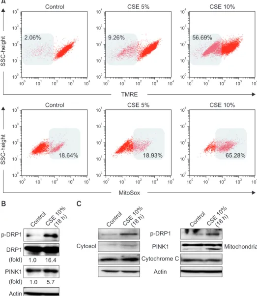
1. CSE causes mitochondrial dysfunction and mitophagy in Beas-2B epithelial cells
To investigate the effect of CSE on mitochondrial function
100 100 104
101 102 103 104 103
102
101
100 100 104
101 102 103 104 103
102
101
A
SSC-height
TMRE 100
100 104
101 102 103 104 103
102
101
Control CSE 5% CSE 10%
100 100 104
101 102 103 104 103
102
101
100 100 104
101 102 103 104 103
102
101
SSC-height
MitoSox 100
100 104
101 102 103 104 103
102
101
Control CSE 5% CSE 10%
2.06% 9.26% 56.69%
18.64% 18.93% 65.28%
B
p-DRP1 DRP1
PINK1
Actin
1.0 16.4
1.0 5.7 (fold)
(fold) Contro
l CSE
10%
(18h)
C
Cytosol
Cytochrome C p-DRP1
PINK1
Actin
Mitochondria Contro
l CSE
10%
(18h)
Contro l
CSE 10%
(18h)
Figure 1. Effects of cigarette smoke ex- tract (CSE) on mitochondrial dysfunc- tion and mitophagy in Beas-2B epithelial cells. Human bronchial epithelial (Beas- 2B) cells were treated with the indicated percent of CSE for 24 hours. (A) Flow cytometric analysis of CSE-treated Beas- 2B cells left unstained or labeled with tet- ramethylrhodamine, ethyl ester (TMRE) and MitoSOX Red. Cells were induced with 10% CSE for 18 hours. (B, C) The expression of mitophagy-related proteins was determined by Western blot analysis.
DRP1: dynamin-1-like protein; PINK1:
PTEN-induced putative kinase-1.
in human bronchial epithelial cells (Beas-2B), we evaluated the mitochondrial membrane potential (Δψm) and mtROS production using TMRE and MitoSOX assays, respectively.
CSE caused mitochondrial depolarization and a decrease in functional mitochondria in Beas-2B cells (Figure 1A). We also observed that CSE treatment increased mtROS production in Beas-2B cells in a dose-dependent manner (Figure 1A). To investigate whether CSE induces mitophagy in lung epithe- lial cells, we measured the expression of p-DRP1 and PINK1, regulator proteins of mitophagy. Compared to untreated cells, the expression of p-DRP1 and PINK1 was increased by 16.4- and 5.7-fold, respectively, in Beas-2B cells after CSE treatment (Figure 1B). These results suggest that CSE induces mitoph- agy in lung bronchial epithelial cells, which is associated with increased expression of mitophagy-related proteins. After CSE exposure, p-DRP1 and PINK1 expression was preferen- tially increased in the mitochondrial fraction of Beas-2B cells
(Figure 1C).
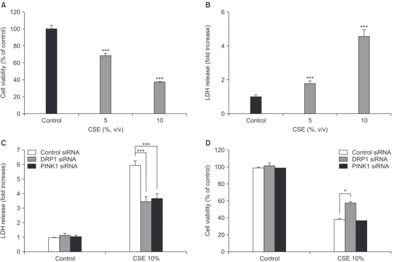
2. CSE treatment in Beas-2B epithelial cells causes mitophagy-regulated apoptotic cell death via upregulation of the mitochondria fission regulator, DRP1, and the mitophagy protein, PINK1
As shown in Figure 2A and B, cell viability, as determined by the MTT assay, and cell death, as determined by the LDH release assay, were effected in a dose-dependent manner after 18 hours of CSE exposure. To determine whether DRP1 and/or PINK1 play a role in CSE-induced cell death, DRP1 or PINK1 was knocked down in Beas-2B cells using DRP1- or PINK1-targeted siRNA. As shown in Figure 2C, control siRNA-transfected cells displayed increased LDH release after CSE treatment (5.71-fold relative to untreated cells), whereas knockdown of DRP1 or PINK1 in Beas-2B cells resulted in de-
120
100
80
60
40
20
0
Cellviability(%ofcontrol)
Control
***
A
5 10
***
CSE (%, v/v)
6
4
2
0
LDHrelease(foldincrease)
Control
***
B
5 10
***
CSE (%, v/v)
7 6 5 4 3 2 1 0
Control
C
CSE 10%
120
100
80
60
40
20
0
Cellviability(%ofcontrol)
D
LDHrelease(foldincrease)
Control siRNA DRP1 siRNA PINK1 siRNA
Control CSE 10%
*
Control siRNA DRP1 siRNA PINK1 siRNA
***
***
Figure 2. Effects of dynamin-1-like protein (DRP1) and PTEN-induced putative kinase-1 (PINK1) silencing on cigarette smoke extract (CSE)–
induced cell death in Beas-2B epithelial cells. Beas-2B cells were exposed to different concentrations of CSE for 18 hours. Cell viability was evaluated using the MTT assay and cytotoxicity was measured by the lactate dehydrogenase (LDH) release test. Beas-2B cells were pre-treat- ed with control, DRP1, or PINK1 siRNAs for 48 hours prior to treatment with 10% CSE for 18 hours. Cell cytotoxicity (C) and viability (D) were estimated by the LDH and MTT assays, respectively. The data shown represent the mean±SD derived from three determinations. *p<0.05 and
***p<0.001, compared with the control siRNA-transfected group.
creased LDH release after CSE treatment (3.42- and 3.55-fold, respectively) relative to untreated cells. Moreover, the MTT as- say revealed that knockdown of DRP1 decreased cellular cy- totoxicity following CSE exposure compared to control siRNA- infected cells, whereas knockdown of PINK1 did not affect cell viability (Figure 2D).
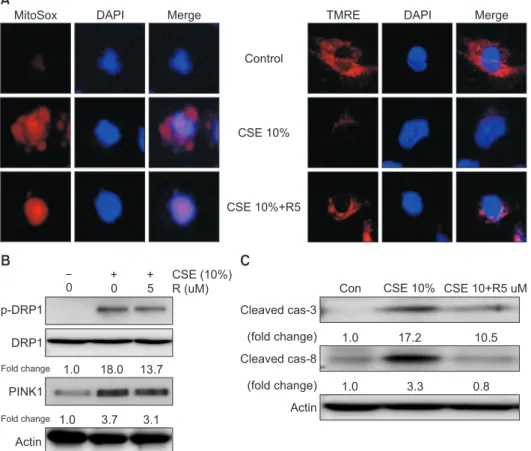
3. Roflumilast inhibited mitochondrial dysfunction and mitophagy in CSE-induced Beas-2B epithelial cells Next, we investigated the role of roflumilast in mitochondri- al dysfunction and mitophagy induced by CSE exposure using confocal microscopy and Western blot analysis. Roflumilast pretreatment markedly protected against mitochondrial dys- function, as evident by an increase in functional mitochondria and a decrease in mtROS production in CSE-treated cells compared with that observed in epithelial cells treated with CSE only (Figure 3A). Roflumilast also decreased the expres- sion of mitophagy regulator proteins, such as p-DRP1 and PINK1 (Figure 3B).
4. Roflumilast inhibited apoptotic cell death in CSE- induced Beas-2B epithelial cells
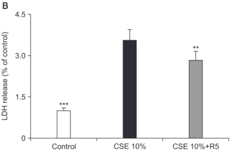
Roflumilast markedly decreased CSE-induced cytotoxicity,
as measured by the LDH release assay, and increased cell via- bility, as assessed by the MTT assay (Figure 4A, B). To explore the role of roflumilast in CSE-induced apoptotic cell death, we examined the effect of roflumilast on known apoptosis-related proteins, such as cleaved caspase-3 and -8. Treatment with ro- flumilast markedly inhibited the expression of these proteins (Figure 3C). The efficacy of roflumilast against CSE-induced cell cytotoxicity was apparent at the concentration of 5 μM roflumilast. DRP1 siRNA–transfected cells showed no more protection following roflumilast treatment in CSE-induced cell death; however, PINK1 siRNA–transfected cells exhibited increased protection following the addition of roflumilast (Fig- ure 4C).
Discussion
Traditionally, chronic inflammation, increased oxidative stress, and/or impaired lung repair processes have been known to contribute to the pathogenesis of emphysema, which is mainly caused by CS and biomass equivalent, and is characterized by an increase in alveolar wall cell death and/
or a failure of alveolar wall maintenance. There are no current therapies available that either prevent or cure the progression of emphysema
16,17. Preclinical studies have shown that PDE4
A
Control
CSE 10%
CSE 10%+R5
MitoSox DAPI Merge TMRE DAPI Merge
B
p-DRP1
DRP1
PINK1
Actin
Fold change 1.0 18.0 13.7
Fold change 1.0 3.7 3.1
0 0 5
CSE (10%)
R ( M)u Con CSE 10% CSE 10+R5 uM
Cleaved cas-3
Cleaved cas-8
Actin
C
1.0 17.2 10.5
1.0 3.3 0.8
(fold change)
(fold change)
Figure 3. Effects of roflumilast on mito- chondrial dysfunction and mitophagy in cigarette smoke extract (CSE)–induced Beas-2B epithelial cells. Beas-2B cells were pre-treated for 2 hours with 5 μM roflumilast and exposed to 10% CSE for 4 hours. (A) Detection of mitochon- drial dysfunction (×400). Cells labeled with tetramethylrhodamine, ethyl ester (TMRE) or MitoSOX Red were incubated with control or 5 μM roflumilast and treated with 10% CSE for 4 hours. (B) The expression of mitophagy-related proteins was determined by Western blot analysis.
(C) The expression of apoptosis-related proteins was determined by Western blot analysis. DRP1: dynamin-1-like protein;
PINK1: PTEN-induced putative kinase-1;
R5: roflumilast 5 μM.
inhibitors consistently reduce the accumulation of neutrophils in bronchoalveolar lavage fluid following short-term exposure to CS in mice
18. Thus, PDE4 inhibitors have been exploited primarily for their anti-inflammatory effects in the treatment of COPD, especially in the chronic bronchitis-dominant phe- notype. However, several studies that investigated the effects of PDE4 inhibitors on the development of emphysema
19have mainly focused on apoptosis
20. We also previously investigat- ed the effects of a PDE4 inhibitor in CSE-induced apoptosis of lung structural cells, and revealed that the PDE4 inhibitor ro- lipram protects lung fibroblasts from CSE-induced apoptosis by inhibiting the activation of caspase-3 and -8
21. Autophagy, a cellular pathway for the degradation of damaged organelles and proteins, has gained increasing importance in human dis- eases, both as a modulator of pathogenesis and as a potential therapeutic target
22,23. In addition, autophagy can influence most fundamental processes, including inflammation and immune responses, the host defense, metabolic pathways, and programmed cell death. Some studies have suggested that autophagy promotes lung epithelial cell death, airway dysfunction, and ultimately emphysema in response to CS exposure
9,24-26. Furthermore, recent studies have reported that CS exposure in epithelial cells promotes the autophagy-de-
pendent turnover of mitochondria (mitophagy) via mitochon- drial dysfunction, as evident by a decrease in mitochondrial membrane potential and an increase in mtROS production
9. Here, we confirmed that CSE causes mitochondrial dysfunc- tion using TMRE and MitoSOX assays. We also demonstrated that roflumilast markedly inhibited mitochondrial dysfunction by increasing functional mitochondria and decreasing mtROS production.
In general, mitophagy is a cellular response that is com- monly associated with mitochondrial dysfunction
8. PINK1 and Parkin represent well-recognized regulators of mitophagy in neural cells and tissues
27. There are two altered mitochon- drial dynamics in terms of mitophagy (i.e., fusion or fission), which play key roles in the response of cells to exogenous stress. Mitochondrial fission, a process regulated by DRP1, is needed to trigger mitophagy
28. The phosphorylation of DRP1 promotes DRP1 recruitment to mitochondria
29. In adult car- diac myocytes, overexpression of the dominant-negative form of DRP1 results in decreased mitochondrial fission and mi- tophagy
29. Mild oxidative stress specifically triggers mitophagy in a DRP1-dependent manner
11. Recent studies have applied Mdivi-1, a pharmacological inhibitor of DRP1, for the function- al study of mitophagy
30-33. We confirmed that CSE increased
120
100
80
60
40
20
0
Cellviability(%ofcontrol)
Control
*
A
CSE 10% CSE 10%+R5
4.5
3.0
1.5
0
LDHrelease(%ofcontrol)
Control
**
B
CSE 10% CSE 10%+R5
7.0 6.0 5.0 4.0 3.0 2.0 1.0 0
Control
C
CSE 10%
LDHrelease(foldincreaseofcontrol)
Control siRNA DRP1 siRNA PINK1 siRNA
***
***
***
CSE 10%+R5
*
**