The C
a2+-activated K
+(BK) Channel-opener NS 1619 Prevents Hydrogen Peroxide- induced Cell Death and Mitochondrial Dysfunction in Retinal Pigment Epithelial Cells
Jae Hoon Kang and Jae Suk Woo*
Department of Physiology, Pusan National University School of Medicine, Yangsan 50612, Korea Received September 13, 2017 /Revised October 19, 2017 /Accepted October 23, 2017
Potassium channel openers (KCOs) produce physiological and pharmacological defense mechanisms against cell injuries caused by oxidative stress of diverse origins. Openings of mitochondrial and plas- malemmal K+ channels are involved in the defense mechanisms. This study tested whether NS 1619, an opener of large-conductance BK channels, has a similar beneficial influence on the pigment epi- thelial cells of retinas. The human retinal pigment epithelial cell line ARPE-19 was exposed to H2O2-induced oxidative stress in the absence and presence of NS 1619. The degrees of the cells' in- juries were assessed by analyzing the cells' trypan-blue exclusion abilities and TUNEL staining. NS 1619 produced remarkable protections against cell injuries caused by H2O2. It prevented apoptotic and necrotic cell deaths. The protective effect of NS 1619 was significantly diminished when the cells were treated with NS 1619 in combination with the BK channel-blocker paxilline. NS 1619 significantly ame- liorated cellular ATP deprivations in H2O2-treated cells. It helped mitochondria preserve their func- tional integrity, which was estimated by their MTT reduction abilities and mitochondrial membrane potential. In conclusion, it was suggested that NS 1619 had a beneficial effect on mitochondria in re- gards to preserving their functional integrity under oxidative stress, and it produces defense mecha- nisms against oxidant-induced cell injuries in ARPE-19 cells.
Key words : Cell death, KCOs, mitochondria, oxidative stress, RPE cells
*Corresponding author
*Tel : +82-51-510-8072, Fax : +82-51-510-8011
*E-mail : [email protected]
This is an Open-Access article distributed under the terms of the Creative Commons Attribution Non-Commercial License (http://creativecommons.org/licenses/by-nc/3.0) which permits unrestricted non-commercial use, distribution, and reproduction in any medium, provided the original work is properly cited.
Journal of Life Science 2017 Vol. 27. No. 11. 1349~1356 DOI : https://doi.org/10.5352/JLS.2017.27.11.1349
Introduction
The retinal pigment epithelium (RPE) supports overlying photoreceptor cells and regulates transport across the blood- retina barrier. The relationship between the RPE and photo- receptor cells is crucial to sight. It has been demonstrated that dysfunction of the RPE can result in the death of visual cells and blindness [26]. Oxidative stress plays an important role in diverse types of RPE pathophysiology including age- related macular degeneration [24].
Large conductance Ca2+-activated (BK) channels show in- creased open provability by a decrease in membrane poten- tial or rise in the cellular Ca2+ concentration ([Ca2+]i). These characteristics of the BK channels allow them to serve as an important physiological regulator of the membrane po- tential or Ca2+ concentration in cells and mitochondria [16].
Wimmers [31] have shown the RPE cell line ARPE-19 as well
as freshly isolated RPE cells express functionally active BK channels.
Maintenance of appropriate level of intracellular ATP is essential for optimal cellular function. Intracellular ATP is supplied primarily by through oxidative phosphorylation in mitochondrial machinery. Conditions that result in de- creased oxygen supply or accumulation of reactive oxygen species (ROS) can compromise mitochondrial energetic ma- chinery and lead to cell injury. Moreover, mitochondria itself is a principal source of ROS during ischemic or ischemia/re- perfusion challenges [12, 30]. Although a lot of trials to find pharmacological tools to improve cell protection mecha- nisms have been suggested, no effective pharmacological tools that help mitochondria to preserve their function or morphological integrity have been developed.
It is now well documented that potassium channel open- ers (KCOs) can provide effective defense mechanisms to pro- tect myocardium against ischemic or oxidative stress-in- duced cell injuries. ATP-sensitive K+ (KATP) channels [28] as well as BK channels [1] are believed to play important roles to provide the protection mechanism. Initially, the protective effects provided by KCOs were believed to be due to activa- tion of BK or KATP channels in plasma membranes. However, later studies demonstrated that the KCOs-induced defense
mechanism could not be entirely attributed to the plasma- lemmal K+ channels, suggesting that interaction with addi- tional cellular targets could mediate the action of KCOs. The overriding candidate is mitochondrial membranes which al- so express BK and KATP channels [14].
NS 1619 (1,3-dihydro-1-[2-hydroxy-5-(trifluoromethyl) phenyl]-5-(trifluoromethyl)- 2H-benzimidazol-2-one) is a close chemical relative of NS 004 (5-trifluoromethyl-(5- chloro-2-hydroxyphenyl)-1,3-dihydro-2H-benzimidazole- 2-one), a prototype of benzimidazole derivative. It was shown to hyperpolarize vascular smooth muscle cells by di- rectly activating the BK channels [7]. Later studies have shown that it renders the ischemic heart resistant to the pro- gression of necrosis and hence, facilitate survival of my- ocardial cells [1]. However, the pharmacological effects and action mechanism in other tissues are still to be elucidated.
In this study, we examined the effect of NS 1619 on oxidative injuries in RPE cells.
Materials and Methods
Cell culture
ARPE-19 cells (ATCC, Manassas, VA, USA) were cultured on plastic flasks in DMEM/F12 supplemented with 10% FBS and antibiotics streptomycin (50 μg/ml) and penicillin G (50 IU/ml). When cells reached confluency (approximately 4-5 days after seeding) cells were detached using 0.05% trypsin solution containing 0.53 mM EDTA. The subsequent cell sus- pension was reseeded at one-sixth of the initial density.
Induction of oxidant-induced injury
Experiments were carried out with cells that were grown on 12-well plates for 3 to 4 days when cells form confluent monolayers. Culture media bathing the cell monolayer were washed out, and cells were incubated in serum-free media with hydrogen peroxide (H2O2), t-butylhydroperoxide (t- BHP), or menadione for 3 hr at 37oC, unless otherwise indicated.
Induction of chemical hypoxia
To induce chemical hypoxia, cells were deprived of glu- cose and treated with the inhibitor of mitochondrial electron transport, antimycin A (20 μM) as described by Hagar et al. [13].
Trypan blue exclusion assay
Dead cells become unable to exclude trypan blue and get
stained by the dye. Accordingly, assessment of cellular abil- ity to exclude trypan blue provides an excellent indicator for the measurement of cell viability [2]. After treatment with experimental agents, cells were further incubated in HBSS containing 4% trypan blue for 30 min. Trypan blue-stained cells were counted as dead cells under light microscopy.
Tunel assay
Cells were incubated for 3 hr with 0.1 mM H2O2 in se- rum-free media to initiate apoptotic signal. Cells were then transferred to fresh media and further incubated for 18 hr.
Tunel staining analysis was carried out to assess the degree of apoptosis. With this procedure, necrotic cell death did not exceed 5% of the whole cell population when analyzed by trypan blue exclusion ability.
MTT reduction assay
Mitochondria in intact cells reduce 3-(4,5-dimethyl-2-thia- zyl)-2,5-diphenyl- 2H-tetrazolium bromide (MTT) to for- mazan [19]. Thus, production of formazan as a result of MTT reduction is an excellent indicator to assess the functional integrity of mitochondria. After treatment with experimental agents, MTT (62.5 μg/ml) was added to each culture well and cells were incubated for 30 min. After removal of super- natant by aspiration, crystalized formazan was dissolved in DMSO. Spectrophotometry (absorbance at 570 nm) was per- formed to determine the concentration of formazan.
Assay of ATP content
Luciferin-luciferase assay [18] was used to determine cel- lular ATP content. After treatment with experimental agents, cells were solubilized in a mixture of 0.5% Triton X-100 (500 μl) and 0.6 M perchloric acid (100 μl). The solubilized sample was diluted with potassium glutamate buffer which is com- posed of 10 mM potassium glutamate and 4 mM MgSO4
(pH 7.4). Luciferin-luciferase (100 μl of 20 mg/ml) was add- ed to each suspension, and light emission was determined with a luminometer. Biorad protein assay kit was used to determine the protein content in the cell preparations.
Measurement of mitochondrial membrane potential Changes in mitochondrial membrane potential were measured by fluorocytometry using DiOC6(3). Cell-permeable DiOC6(3) incorporates into mitochondria depending upon the transmembrane voltage across the inner mitochondrial
H2O2 (mM)
Fig. 1. Effect of NS 1619 on H2O2-induced cell injury. RPE cells were incubated for 3 hr at 37oC in serum-free media con- taining indicated concentrations of H2O2 with or without NS 1619 (10 μM). Trypan blue exclusion ability was de- termined to assess cell viability. Means ± S.E. (n=5).
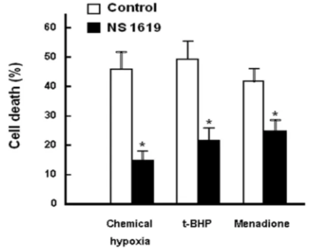
Fig. 2. Effect of NS 1619 on cell injuries caused by different types of oxidative stresses. RPE cells were treated with chemical hypoxia, 0.5 mM t-butylhydroperoxide (t-BHP) and menadione (2 mM) for 3 hr at 37oC with or without NS 1619 (10 μM). Trypan blue exclusion ability was de- termined to assess cell viability. Chemical hypoxia was induced by glucose deprivation in combination with an- timycin A (20 μM). Means ±S.E. (n=5). *p<0.01 vs. control.
membrane. When there is a disturbance in the transmem- brane voltage across the inner mitochondrial membrane, it is reflected by decreased cellular DiOC6(3) fluorescence.
Cells were loaded with DiOC6(3) by incubation for 20 min at 37oC in PBS containing 50 nM DiOC6(3). After wash-out three times cells were suspended in PBS, and then flur- ocytometeric analysis was carried out.
Chemicals
NS 1619, NS 1619, 5-hydroxydecanoic acid, and paxilline were obtained from Research Biochemicals International (Natick, MA). DiOC6(3) was obtained from Molecular Probes (Eugene, OR, USA). Other chemicals were purchased from Sigma-Aldrich Korea Co. (Seoul, Korea).
Data analysis
Data were presented as means ± SE. The data were ana- lyzed by ANOVA. Duncans multiple comparison test was carried out when necessary. We considered the difference as statistically significant when p value was less than 0.05.
Results
Protective effect of NS 1619 on H2O2-induced cell death
Fig. 1 depicts effects of different concentrations of NS 1619 on cell viability assessed by trypan blue exclusion. After treatment for 3 hr with different concentrations of H2O2, cell death rate increased in a dose-dependent manner. At 0.5 mM, H2O2 resulted in cell death by 63.8%. In the presence
of the BK channel opener NS 1619, H2O2-induced cell death was significantly reduced.
Effect of NS 1619 on different types of oxidative cell injuries
In the results presented in Fig. 2, it was examined whether NS 1619 could prevent cell injuries induced by different types of oxidative stress. Menadione and t-butylhydroper- oxide (t-BHP) were adopted as models for oxidative agents.
In addition, chemical hypoxia was induced by deprivation of glucose combined with treatment with antimycin A, an inhibitor of mitochondrial respiratory chain. These maneu- vers all caused cell death assessed by trypan blue exclusion assay. In the presence of NS 1619, cell death was signifi- cantly prevented regardless of the cause of oxidative injuries.
These results suggest that NS 1619 might have protective effects against cell injuries caused by different types of oxi- dative stress.
Effects of KCOs and potassium channel blockers on H2O2-induced cell death
In Fig. 3, effects of NS 1619 and diazoxide were examined in combination with the BK and KATP channel blockers, pax- illine and 5-hydroxydecanoate [22]. Diazoxide as well as NS 1619 was effective to ameliorate cell death determined by trypan blue exclusion ability. Paxilline antagonized the pro-
Fig. 3. Effects of different K+ channel openers and blockers on RPE cell death caused by H2O2. RPE cells were incubated for 3 hr at 37oC in serum-free media containing 0.5 mM H2O2 with or without of NS 1619 (NS, 10 μM) and diazo- xide (Diaz, 10 μM) in combination with paxilline (Pax, 10 μM) or 5-hydroxydecanoic acid (5-HD, 10 μM). Trypan blue exclusion ability was determined to assess cell viability. Means ± S.E. (n=4). *p<0.01 vs. NS alone,
#p<0.01 vs. Diaz alone.
A
B
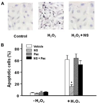
Fig. 4. Protection by NS 1619 against apoptotic cell death caused by H2O2. RPE cells were incubated for 3 hr at 37oC in serum-free media containing 0.1 mM H2O2 with or with- out NS 1619 (NS, 10 μM) and Paxilline(Pax, 10 μM), and were then transferred to the fresh media. After further incubation for 18 hr, Tunel staining was performed to assess apoptosis. Means ± S.E. (n=6). *p <0.01 vs. Vehicle.
tective effect of NS 1619, whereas 5-hydroxydecanoate an- tagonized the protective effect of diazoxide, respectively.
Effect of NS 1619 on H2O2-induced apoptotic cell death
We examined whether NS 1619 has a protective effect to ameliorate apoptotic events. We treated RPE cells for 3 hr in HBSS containing 0.1 mM H2O2. In these cell populations, necrotic cell death estimated by trypan blue staining did not exceed 5% of the whole cell population. Dead cells floating in the media caused by necrotic injury were removed by aspiration. Cells were then transferred to fresh media and incubated further for 18 hr. Tunel staining was then per- formed for assay of apoptosis. Apoptotic cells could be dis- criminated by their condensed or fragmented nuclei in the micrographs of Tunel staining as shown in Fig. 4A. In H2O2-treated preparations 61.4.±6.8% of cells were counted as Tunel-positive (Fig. 4B). The number of apoptotic cells was remarkably reduced in cells with NS 1619. In the pres- ence of paxilline, the protective effect of NS 1619 was sig- nificantly inhibited.
Effect of NS 1619 on H2O2-induced changes in MTT reduction
To examine whether NS 1619 could prevent H2O2-induced
mitochondrial dysfunction, changes in mitochondrial func- tion was examined using a MTT reduction assay. When we treated cells with 0.5 mM H2O2, MTT reduction ability de- creased to 43.2% of the control cells (Fig. 5). When we treated the cells with NS 1619 together, the H2O2-induced effect on MTT reduction was significantly attenuated. These results indicate that NS 1619 provide a defense mechanism to mi- tochondria to maintain their functional integrity under oxi- dative stress induced by H2O2.
Effect of NS 1619 on cellular ATP content Deprivation of cellular ATP precedes irreversible injury process occurring upon ischemic- or oxidative stress-in- duced tissue damage [9]. Therefore, it was investigated whether NS 1619 could ameliorate H2O2-induced ATP deprivation. The results in Fig. 6 represent the effect of NS 1619 on changes in ATP concentration in H2O2-treated cells.
In H2O2-treated cells, cellular ATP content was diminished to lower than 10% of the control level in 3 hr. When cells were pre-treated with NS 1619, ATP depletion by H2O2 was remarkably delayed. These results, together with those from MTT assay, suggest that NS 1619 exerts a beneficial influence
Fig. 5. Effects of NS 1619 and paxilline on H2O2-induced de- crease in mitochondrial MTT reduction ability. RPE cells were incubated for 3 hr at 37oC in serum-free media containing 0.5 mM H2O2 for 3 hr with or without NS 1619 (NS, 10 μM) and Paxilline (Pax, 10 μM), and degree of MTT reduction was determined. Means ± S.E. (n=6.
*p<0.01 vs. Vehicle (Veh), #p<0.01 vs. NS.
Time in H2O2 (min)
Fig. 6. Effect of NS 1619 on cellular ATP content. H2O2 (0.1 mM)-induced decrease in cellular ATP content was monitored in the presence and absence of NS 1619 (10 μM). Mean ± S.E. (n= 4).
A
Control H2O2 H2O2+NS
B
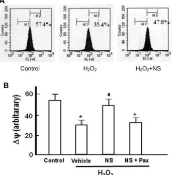
Fig. 7. Effect of NS 1619 on H2O2-induced changes in mitochon- drial membrane potential. RPE cells were treated with H2O2 (0.1 mM) for 3 hr in the presence and absence of NS 1619 (NS, 10 μM) and paxilline (Pax, 10 μM), and analyzed by fluorocytometric analysis of DiOC6(3) fluo- rescence. A, Representative graphs of flurocytormetric analysis. B, Summarized data of 4 independent experi- ments. Mean ± S.E. (n=4). *p<0.01 vs. control, #p<0.01 vs.
Vehicle.
on mitochondria to protect their energy production machi- nery against irreversible injuries under oxidative stress.
Effect of NS 1619 on mitochondrial membrane potential In the results summarized in Fig. 7, effects of NS 1619 on mitochondrial membrane potential were presented. As shown in fluorocytometric analysis in Fig. 7A and Fig. 7B, NS 1619 effectively prevented H2O2-induced disruption of the mitochondrial membrane potential. These results dem- onstrate that NS 1619 ameliorates functional and structural deterioration of mitochondria and it could provide a pro- tection mechanism against H2O2-induced cell death in the
RPE cells.
Discussion
Cell death processes appear to be distinguished as three morphologically distinct modes. They include necrotic cell death, apoptosis (programmed cell death) and autophagocy- totic cell death [3, 23]. The present study suggested that H2O2
could trigger different modes of cell death processes de- pending on its concentration. In RPE cell preparations treat- ed with relatively higher concentration (0.5 mM) of H2O2, necrotic cell death seemed to be dominant. On the other hand, in cell preparations treated with relatively lower con- centration (0.1 mM) of H2O2 apoptotic cell death was ob- served after further overnight incubation in fresh media.
With the later procedure,necrotic cell death estimated by trypan blue staining did not exceed 5% of the whole cell population.
This study suggested that NS 1619 might produce an ef- fective protection mechanism against RPE cell injury in- duced by H2O2. It ameliorated RPE cell death induced by
different types of oxidative agents as well as H2O2 (Fig. 1 and Fig. 2). It was effective to prevent both the necrotic and apoptotic cell death. These results imply that the beneficial effect of NS 1619 to prevent RPE cell death should be ex- plained in connection with the common pathways associated with both the apoptotic and necrotic cell death (Fig. 4). NS 1619 and diazoxide have been described as relatively specific openers of mitochondrial BK and KATP channels, respectively [29]. The present study demonstrated that diazoxide, an opener of mirochondrial KATP channels, also provided a sim- ilar protective effect against H2O2-induced RPE cell death (Fig. 3). It suggested that whether the K+ channel type is BK and KATP does not have influence on the efficacy of the defense mechanism provided by the opening of mitochon- drial K+ channel. The protective effects NS 1619 and diazo- xide disappeared almost completely when cells were pre-treated with their structurally related K+ channel block- ers paxilline and 5-hydroxydecanoate, respectively (Fig. 3 and Fig. 4). These blockers did not affect the protective ef- fects provided by structurally unrelated openers, respec- tively, suggesting that the opening of mitochondrial K+ chan- nels is a crucial process in NS1619- as well as diaxoxide- induced protection mechanism.
KCOs were first introduced in the early 1980s. They pro- duce their pharmacological effect by facilitation of ion flow through their target K+ channels. It was followed by consec- utive identification of many other drugs as a member of KCOs [6, 8]. Initially, KCOs which include pinacidil, croma- kalim, diazoxide and nicorandil etc. were introduced as medications for hypertension (pinacidil and cromakalim) or angina (nicorandil). These actions are not irrational when we recognize that the increased opening provability of K+ channels leads to hyperpolarization of smooth muscle mem- branes, inhibition of Ca2+ entry through voltage-gated Ca2+
channels, and hence promotion of vasodilation [5, 10, 20, 25].
Recently, accumulating evidence has demonstrated that the pharmacological actions of KCOs are not confined to vas- odilatation of peripheral and coronary vessels. KCOs have been reported to provide a protection mechanism which mimics the endogenous protection mechanism described as
‘ischemic preconditioning’ [4, 15, 27, 28]. It is a paradoxical phenomenon in which repeated exposure to brief periods of ischemia provide the heart with a beneficial effect to be- come resistant to later lethal ischemia [21]. Recruitment of such defense mechanism pharmacologically leads to open- ing of new prospects for preventing or ameliorating cardiac
damages as a consequence of coronary artery disease. Now it has become evident that protective action of KCOs is not confined to the heart but also functioning in other organs including the neuron [11].
In the heart, opening of plasmalemmal BK channels in- duces hyperpolarization of membrane potential, reduces cel- lular Ca2+ and thereby, alleviates myocardial work load [1].
In addition to this initially proposed protection mechanism, mitochondrial membrane which also possesses BK channels was suggested as an additional important cellular target that could mediate the defense mechanism provided by NS 1619.
Protective effect of NS 1619 has also been described in neu- ron cells. NS 1619 has been shown to induce immediate and delayed preconditioning [11]. However, these neuro- protective effects were suggested to be independent of BK channels and rather to be the consequence of ROS gen- eration, activation of the PI3K pathway, and inhibition of caspase activation [11]. In the present study, as RPE cells are non-excitable cells, the role of plasmalemmal BK chan- nels are not considered to be importantly involved in the defense mechanism provided by NS 1619. We did not exam- ine the role of ROS and PI3K in this study
Several evidence in this study indicates that prevention of mitochondrial dysfunction during oxidative stress is a key event essential for the protection mechanism provided by NS 1619 against H2O2-induced RPE cell injury. It provided mitochondria with beneficial effects to retain MTT reduction ability under H2O2-induced oxidative stress (Fig. 5). In addi- tion, it remarkably prevented H2O2-induced ATP depriva- tion (Fig. 6). These results suggested that NS 1619 produces a beneficial effect on mitochondria to protect their metabolic machinery against irreversible injuries under oxidative stress.
Formation of mitochondrial permeability transition (MPT) pores and cytochrome c release through these MPT pores has been suggested to be importantly associated with oxida- tive stress-induced cell injury [17]. Cytochrome c released into cytosol triggers activation of caspase pathways that are essential events for the progression process of apoptosis.
Formation of MPT pore results in loss of selective perme- ability in the inner mitochondrial membrane which leads to deterioration of mitochondrial membrane potential. The present study demonstrated that NS 1619 ameliorated the H2O2-induced disruption of mitochondrial membrane poten- tial (Fig. 7). It also seems to be importantly related to the protection mechanism of NS 1619 against apoptotic cell
death. In conclusion, it is suggested that NS 1619 prevents functional and structural deterioration of mitochondria un- der oxidative stress and it could provide a defense mecha- nism that renders the RPE cells resistant to H2O2-induced cell injury.
Acknowledgement
This work was supported for 2 years by Pusan National University Research Grant.
References
1. Bentzen, B. H., Olesen, S. P., Rønn, L. C. and Grunnet, M.
2014. BK channel activators and their therapeutic perspectives. Front. Physiol. 5, 389.
2. Bjorkerud, S. and Bondjers, G. 1972. Endothelial integrity and viability in the aorta of the normal rabbit and rat as evaluated with dye exclusion tests and interference contrast microscopy. Atherosclerosis 15, 285-300.
3. Clarke, P. G. 1990. Developmental cell death: morphological diversity and multiple mechanisms. Anat. Embryol. 181, 195- 213.
4. Cole, W. C., McPherson, C. D. and Sontag, D. 1991. ATP- regulated K+ channels protect the myocardium against is- chemia/reperfusion damage. Circ. Res. 69, 571-581.
5. Daut, J., Maier-Rudolph, W., von Beckerath, N., Mehrke, G., Gunther, K. and Goedel-Meinen, L. 1990. Hypoxic dilation of coronary arteries is mediated by ATP-sensitive potassium channels. Science 247, 1341-1344.
6. Duty, S. and Weston, A. H. 1990. Potassium channel openers.
Pharmacological effects and future uses. Drugs 40, 785-791.
7. Edwards, G., Niederste-Hollenberg, A., Schneider, J., Noack, T., and Weston, A. H. 1994. Ion channel modulation by NS 1619, the putative BKCa channel opener, in vascular smooth muscle. Br. J. Pharmacol. 113, 1538-1547.
8. Edwards, G. and Weston, A. H. 1990. Structure-activity rela- tionships of K+ channel openers. Trends. Pharmacol. Sci. 11, 417-422.
9. Farber, J. L., Kyle, M. E. and Coleman, J. B. 1990. Mechanisms of cell injury by activated oxygen species. Lab. Invest. 62, 670-679.
10. Foster, C. D., Fujii, K., Kingdon, J. and Brading, A. F. 1989.
The effect of cromakalim on the smooth muscle of the guin- ea-pig urinary bladder. Br. J. Pharmacol. 97, 281-291.
11. Gáspár, T., Katakam, P., Snipes, J. A., Kis, B., Domoki, F., Bari, F. and Busija, D. W. 2008. Delayed neuronal pre- conditioning by NS1619 is independent of calcium activated potassium channels. J. Neurochem. 105, 1115-1128.
12. Gunter, T. E., Gunter, K. K., Sheu, S. S. and Gavin, C. E.
1994 Mitochondrial calcium transport: physiological and pathological relevance. Am. J. Physiol. 267, C313-C339.
13. Hagar, H., Ueda, N. and Shah, S. V. 1996. Endonuclease
induced DNA damage and cell death in chemical hypoxic injury to LLC-PK1 cells. Kidney Int. 49, 355-361.
14. Inoue, I., Nagase, H., Kishi, K. and Higuti, T. 1991. ATP-sen- sitive K+ channel in the mitochondrial inner membrane.
Nature 352, 244-247.
15. Kersten, J. R., Gross, G. J., Pagel, P. S. and Warltier, D. C.
1998. Activation of adenosine triphosphate-regulated potas- sium channels: mediation of cellular and organ protection.
Anesthesiology 88, 495-513.
16. Latorre, R., Castillo, K., Carrasquel-Ursulaez, W., Sepulveda, R. V., Gonzalez-Nilo, F., Gonzalez, C. and Alvarez, O. 2017. Molecular determinants of BK channel functional diversity and functioning. Physiol. Rev. 97, 39-87.
17. Lemasters, J. J., Nieminen, A. L., Qian, T., Trost, L. C., Elmore, S. P., Nishimura, Y., Crowe, R. A., Cascio, W. E., Bradham, C. A., Brenner, D. A. and Herman, B. 1998. The mitochondrial permeability transition in cell death: a com- mon mechanism in necrosis, apoptosis and autophagy.
Biochim. Biophys. Acta 1366, 177-196.
18. Lyman, G. E. and DeVincenzo, J. P. 1967. Determination of picogram amounts of ATP using the luciferin-luciferase en- zyme system. Anal. Biochem. 21, 435-443.
19. Morgan, D. M. 1998. Tetrazolium (MTT) assay for cellular viability and activity. Methods. Mol. Biol. 79, 179-183.
20. Murray, M. A., Boyle, J. P. and Small, R. C. 1989. Cromaka- lim-induced relaxation of guinea-pig isolated trachealis: an- tagonism by glibenclamide and by phentolamine. Br. J.
Pharmacol. 98, 865-874.
21. Murry, C. E., Jennings, R. B. and Reimer, K. A. 1986. Precon- ditioning with ischemia: a delay of lethal cell injury in ische- mic myocardium. Circulation 74, 1124-1136.
22. Robertson, D. W. and Steinberg, M. I. 1990. Potassium chan- nel modulators: scientific applications and therapeutic pro- mise. J. Med. Chem. 33, 1529-1541.
23. Schwartzman, R. A. and Cidlowski, J. A. 1993. Apoptosis:
the biochemistry and molecular biology of programmed cell death. Endocr. Rev. 14, 133-151.
24. Sparrow, J. R., Hicks, D. and Hamel, C. P. 2010. The retinal pigment epithelium in health and disease. Curr. Mol. Med.
10, 802-823.
25. Standen, N. B., Quayle, J. M., Davies, N. W., Brayden, J.
E., Huang, Y. and Nelson, M. T. 1989. Hyperpolarizing vaso- dilators activate ATP-sensitive K+ channels in arterial smooth muscle. Science 245, 177-180.
26. Stern, J. and Temple, S. 2015. Retinal pigment epithelial cell proliferation. Exp. Biol. Med (Maywood). 240, 1079-1986.
27. Szewczyk, A. and Marban, E. 1999. Mitochondria: a new target for K+ channel openers? Trends Pharmacol. Sci. 20, 157- 161.
28. Testai, L., Rapposelli, S. and Calderone, V. 2007. Cardiac ATP- sensitive potassium channels: a potential target for an anti-ischaemic pharmacological strategy. Cardiovasc.
Hematol. Agents. Med. Chem. 5, 79-90.
29. Testai, L., Rapposelli, S., Martelli, A., Breschi, M. C. and Calderone, V. 2015. Mitochondrial potassium channels as pharmacological target for cardioprotective drugs. Med. Res.
초록:망막 색소상피세포에서 산화성 세포 손상과 미토콘드리아기능 저해에 미치는 NS 1619의 보호 효과
강재훈․우재석*
(부산대학교 의학전문대학원 생리학교실)
K+ 통로 개방제들은 심근, 뇌, 골격근 등에서 세포막 혹은 미토콘드리아 내막에 존재하는 큰 전도성의 Ca2+-의
존성 K+ (BK) 통로 및 ATP-조절성 K+ 통로(ATP-sensitive K+ channels, KATP)에 작용하여 허혈성 혹은 산화성 세 포 손상을 완화하는 효과가 있는 것으로 보고되어 있다. 본 연구에서는 망막 색소 상피세포주인 ARPE-19 세포를 실험 모델로 하여 큰 전도성의 BK 통로 개방제인 NS 1619가 유사한 보호 효과를 나타낼 수 있는지, 또한 그 작용 기전이 무엇인지를 확인하고자 하였다. AREE-19 세포를 여러 형태의 산화 스트레스에 노출시켜 세포 손상을 유 발하고 그 손상의 정도 및 이에 미치는 NS 1619의 효과를 trypan blue 배출능, Tunel 염색 분석을 통하여 측정하 였다. NS 1619는 여러 형태의 산화 스트레스에 의한 괴사성 및 apoptosis에 의한 세포 손상을 효과적으로 방지하 였으며 그 보호 효과는 BK 통로 봉쇄제인 paxilline 의해 차단되었다. NS 1619는 H2O2에 의한 세포내 ATP 고갈을 현저히 완화시켰으며, 또한 MTT 환원능으로 측정한 미토콘드리아의 기능을 보호하는 효과를 보였다. 유세포형광
분석법을 이용한 실험에서 NS 1619는 H2O2에 의한 미토콘드리아 막전압의 소실을 유의하게 방지하였다. 이상의
결과들을 종합하면 NS 1619는 망막 색소 상피세포에서 산화성 세포 손상을 방지하는 효과를 나타내며 그 기전에 미토콘드리아 기능에 대한 보호 작용이 연관되어 있는 것으로 사료된다.
Rev. 35, 520-553.
30. Vakifahmetoglu-Norberg, H. Ouchida, A. T. and Norberg, E. 2017. The role of mitochondria in metabolism and cell
death. Biochem Biophys. Res. Commun. 482, 426-431.
31. Wimmers, S., Karl, M. O. and Strauss, O. 2007. Ion channels in the RPE. Prog. Retin. Eye. Res. 26, 263-301.