190 서 론
간에 발생한 미분화성 배아육종은 매우 드문 원발성 간 암으로 주로 5세에서 20세 사이의 소아나 청소년기에 발생 한다. 호발 시기는 5∼10세 사이로 6세 이하에서 호발하는
간아 세포 종 또는 10세 이후에 호발 하는 간세포성 간암과 차이를 보인다. 간에서 발생하는 소아의 원발성 간암 중 간 아세포종, haemangioendothelioma, 간세포성 간암에 이어 네 번째로 흔한 악성 종양이지만, 성인에서는 매우 드물게 발 생한다. 2000년까지 43예가 보고되었으며 국내에서는 1예 가 보고되었다. 특이 증상이 없고, 일반적인 종양 표지자가 정상소견을 보여 조기진단이 힘들고, 종양의 빠른 성장으 로 인하여 불량한 예후를 보인다. 치료방법에 대한 원칙은 정해져 있지 않으나, 근치적 절제 후 장기 생존 예가 보고되 고 있으며, 최근 화학 요법과 방사선 요법의 병행요법이 시 도되고 있다. 본 연구에서는 42세 여자 환자에서 발생한 간 의 미분화성 배아 육종으로 부분 간 절제술 후 16개월경에 국소 재발하여, 재발종양 절제 시행 후 39개월 간 생존하고 있는 1예를 경험하여 보고하는 바이다.
증 례
42세 여자가 20일전부터 시작된 우상복부 동통과 소화불 량을 주소로 내원하였다. 과거력상 10년 전 우측유방 섬유 선종으로 절제 생검술을 시행하였고, 가족력상 어머니가
책임저자:최동욱, 서울특별시 노원구 공릉2동 215-4 ꂕ 139-706, 원자력의학원 원자력병원 외과 Tel: 02-970-1218, Fax: 02-978-2005
E-mail: dwchoi@kcch.re.kr
본 논문의 요지는 2003년 대한간담췌학회 춘계학술대회에서 구연 발표하였음.
A Case of Long-Term Survivor after Surgical Resec- tion for Undifferentiated Embryonal Sarcoma of the Liver in Adult
Young Min Choi, M.D., Dong Wook Choi, M.D., Sang Beom Kim, M.D., Yoo Chul Kim, M.D.1 and Sun Hoo Park, M.D.2
Departments of Surgery, 1Internal Medicine and 2Pathology, Korea Cancer Center Hospital, Seoul, Korea
An undifferentiated (embryonal) sarcoma of the liver (USL) is a rare and highly malignant hepatic neoplasm of mesen- chymal origin. This tumor almost exclusively affects pediatric patients, with a poor prognosis. The highest incidence is noted in pediatric patients, usually from 5∼10 years of age, but only 43 cases of USL have been reported in adults worldwide since it was first as a clinicopathological entity.
With a USL in adults, most patients are known to have died within 1 year of diagnosis. The absence of specific symp- toms, rapid tumor growth, normality of common tumor mark- ers and the consequential delay in diagnosis often result in significant enlargement of the tumor, with a poor prognosis.
Various therapeutic modalities have been attempted in adult patients, but only a few long-term survivors have been reported. Herein, our experience of a 42-year-old female patient who is still alive, 55 months after the first resection for a USL is reported. (Korean J HBP Surg 2004;8:190-194) ꠏꠏꠏꠏꠏꠏꠏꠏꠏꠏꠏꠏꠏꠏꠏꠏꠏꠏꠏꠏꠏꠏꠏꠏꠏꠏꠏꠏꠏꠏꠏꠏꠏꠏꠏꠏꠏꠏꠏꠏꠏꠏꠏꠏꠏꠏꠏꠏꠏꠏꠏꠏꠏꠏ Key W ords: Undifferentiated Embryonal Sarcoma
Surgical Resection
중심 단어: 미분화성 배아 육종, 수술적 절제
부분 간 절제술 후 장기 생존한 성인의 미분화성 배아 육종 1예
원자력병원 외과, 1내과, 2병리과
최영민․최동욱․김상범․김유철1․박선후2
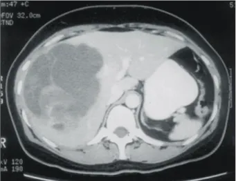
Fig. 1. Abdominal CT scan shows well dermarcated, multiple sepa- rated cystic mass with papillary growing solid portion.
난소암으로 사망한 병력외에 특이사항은 없었다. 신체 증 후는 혈압 120/80 mmHg, 맥박수 분당 80회, 호흡수 분당 20회, 체온은 36.5oC였으며, 이학적 검사상 우상복부에 간 이 2 횡지 정도 촉진되었고 다른 이상소견은 관찰되지 않았 다. 검사실 소견상 말초 혈액 검사는 혈색소 13.1 g/dl, 혈색 치 42%, 백혈구 5,670/mm3, 혈소판 293,000/mm3이었고, 혈 액 화학 검사, 혈청 전해질 검사 및 소변 검사는 정상이었 다. 간염 항원 항체 검사상 HBsAg (-), Anti-HBs Ab (-), IgG-HBc Ab (+)이고, 종양표지자는 CEA 1.28 ng/ml, CA19-9 13.96 U/ml, AFP 2.44 ng/ml으로 정상범위였다. 복부 전산화 단층촬영 상 간 우엽에 15 cm 크기의 유두상 고형부
분을 가진 낭성 종괴가 발견되었고(Fig. 1), 혈관 조영술 상 종괴로 혈류를 공급하는 수많은 비정상 혈관들이 보이고 종괴에 의해 정상 혈관이 변위된 양상이 보였으며 담도성 낭성 종양으로 추정하였다. 수술 소견상 13×10×10 cm 크 기의 종괴가 주변 장기에 침습이나 전이된 소견은 보이지 않고, 간우엽 전체를 침범하고 있었으며 간 우삼구역 절제 술을 시행하였다(Fig. 2).
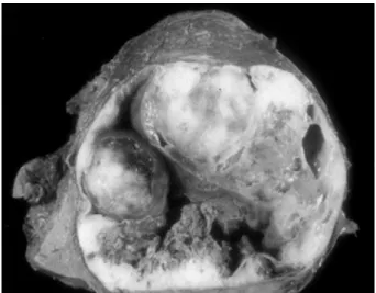
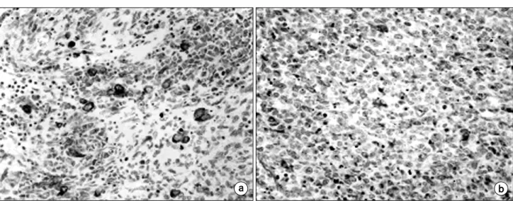
병리학적 검사 상 육안 소견은 경계가 잘 지어진 낭종성 종괴였으며, 중앙조직의 괴사와 출혈이 있으면서 점액성 종양이 부분적으로 보였다(Fig. 3). 광학 현미경적 검사 상 세포학적 다양성이 심한 방추형 세포와 비정형 세포의 증
Fig. 4. Microscopic finding of tumor. (a) At low magnification, the center of the tumor is hypercellular with high vascularity. Peripheral area is myxoid with moderate cellurarity (H&E, ×100). (b) The tumor cell is ovoid and pleomorphic with scanty cytoplasm. Nucleus is hyperchromatic with prominent nucleoli. Mitosis is frequently seen (H&E, ×400).
Fig. 2. Postoperative findings. (Rt.Trisegmentectomy) was done. Fig. 3. Gross finding shows the cut section of hepatic mass with multi-septated cyst containing central necrosis, hemorrhage and myxoid material.
식이 관찰되었으며 기관 특이적 분화는 보이지 않았다(Fig.
4). 면역세포화학 검사상 pas, vimentin, desmin, myoglobin 등 에 양성 반응을 보여 미분화성 배아 육종으로 진단되었다 (Fig. 5). 수술 후 합병증은 없었으며 술 후 13일째 퇴원하였 다. 술 후 보조적 화학 항암 요법이나 방사선 치료는 시행하 지 않았고, 3개월 간격으로 복부 초음파 검사와 복부 전산 화 단층 촬영을 실시하여 정기적으로 추적 관찰하였다. 수 술 후 16개월 째 전산화 단층 촬영상 간 절제면 부위에 우측 대장과 우측 신장을 침범하는 13 cm 크기의 재발성 종괴가 발견되었고(Fig. 6), 2차 수술을 시행하였다. 수술 소견상 종 괴가 간곡부 결장, 우측 신장을 침범하고 횡경막과의 유착 이 심하였으나 간과는 유착이 없어 재발 종괴와 우측 신장, 우측 대장의 병합 절제가 가능하였다. 병리 육안 소견상 13
×10×9 cm 크기의 종괴가 광범위한 출혈과 괴사 소견을 보이며 대장과 신장을 침윤하는 양상을 보였고, 광학 현미 경 소견과 면역 세포 화학 검사상 재발성 미분화성 배아 육종으로 진단되었다. 재발 종양 절제술 후 3개월 간격으로 추적검사를 실시였고 2차 수술 후 39개월 간 재발의 증후 없이 생존하고 있다.
고 찰
간에 발생한 미분화성 배아육종 (Undifferentiated embryonal sarcoma of the liver: USL)은 1948년 Stout에 의해 malignant mesenchymoma로 처음으로 기술되었다.1 이후 embryonal sarcoma, malignant mesenchymoma, fibromyxosarcoma 등의 명 칭으로 다양하게 불리어졌으나,2 1978년 Stocker와 Ishak 등 이 간에 발생한 미분화성 배아육종을 임상병리학적으로 정 의하였다.3 소아에서는 간에 발생한 악성 신생물 중 13%의 빈도를 보여, 간아세포종, haemangioendothelioma, 간세포 암에 이어 네 번째 빈도를 보이며,4 15세 이상의 성인에서는 전 세계적으로 43예가 보고된 드문 질환이다.4
인종, 지리학적 분포 등 역학적으로 관련된 인자는 아직 규명된 것이 없다.4 병력상 특이 증상은 없으나 복통, 황달, 빈혈, 촉진되는 복부 종괴 등을 주소로 내원 하며, 발열, 체 중감소, 구토 등의 비특이적 증상이 동반되기도 한다.2 검사 실 소견으로는 AST, ALT 등의 간기능 검사의 이상 소견을 보이는 경우도 있으나 대부분 정상 소견을 보인다.4,5 대부 분 초음파와 전산화 단층 촬영 등의 방사선 영상학적 방법 으로 발견된다. 초음파 검사에서는 고형 부분과 낭종성 부 분이 혼재되어 있는 구획화 된 큰 종괴로 나타나고, 전산화 단층 촬영에서 종괴는 간의 실질과 비교해서 조영증강이 덜 되며 여러 개의 낭종을 포함하는 소견을 보인다. 종괴의 바깥쪽은 섬유화 된 가성 피막으로 이루어져있어서 일부에 Fig. 5. Immunohistochemical staining of tumor. (a) Vimentin are strong and diffusely positive. (b) Desmin are strong but focal positive.
Fig. 6. Abdominal CT scan (after 13 months) showed about 12 cm sized heterogenous low attenuating mass invading right kidney and perirenal space.
서 조영증강이 되는 경우도 있다.6 혈관 조영술상 대개 무혈 류성 종괴, 또는 저혈류성 종괴의 소견을 보이나 고혈류성 종괴로 보이는 경우도 보고되고 있다.3,4,6 Tokunaga 등4은 1999년까지 성인에서 총 43예의 간의 미분화성 배아 육종 이 보고되었으며, 이 중 간우엽에 발생한 경우가 26예, 간 좌엽에 발생한 경우가 6예, 양 엽에 발생한 경우가 2예로, 기술되지 않은 경우가 9예로 간 우엽에 발생하는 경우가 많았다고 보고하고 있다.
술 후 병리 조직학적으로 간의 미분화성 배아 육종은 확 진이 가능하다. 병리 조직학적으로는 mesenchymal 기원으 로 강력히 추정되며,7,8 세포학적 다양성이 심한 방추형세포 또는 비정형 세포의 증식이 관찰된다. 면역조직화학적 염 색 결과에서도 alpha-1-anti-trypsin, vimentin 등의 mesenchymal antigen에 양성 반응을 보인다.7,9 일부에서는 cytokeratin에 양성 소견을 보고하기도 한다.10 cytokeratin은 일반적으로 상피세포에서 기원한 경우 양성소견을 보인다고 알려져 있 으나, 최근 mesenchyma 기원 세포에서도 양성 소견을 보인 다고 보고되고 있다.11
소아에서는 치료 후 3년 생존율이 37.5%로 보고되기도 하였으나,12 성인에서 치료와 예후에 대한보고는 많지 않다.
수술적 방법과 화학 항암요법이나 방사선 요법 등을 포 함한 다양한 비수술적 방법이 시도되고 있으며, 타 장기의 전이, 림프절 전이, 복막강으로의 파괴성 출혈이 없다면 근 치적인 외과적 절제술이 장기간의 생존을 기대할 수 있는 유일한 치료 방법으로 알려져 있다. 대부분의 환자에서 간 경변이 동반되지 않아 대량 간절제가 시도될 수 있으며, 수 술적 절제율은 70% 정도로 알려져 있다.4 화학항암 요법과 방사선 요법의 단독 치료는 효과가 낮은 것으로 알려져 있
다.3,4,13 1980년대 후반까지 수술이나 병합요법에도 불구하
고 불량한 예후를 보여 대부분의 환자가 조직 생검이나 비 치유적 절제 후 1개월에서 30개월 사이에 원발 질환의 진행 이나 원격 전이로 사망하였으며, 근치적 절제 후에도 3∼36 개월 사이에 사망하였다.14 초기 보고에서도 2년 무병 생존 율은 10% 미만으로 보고되었으나,14 1990년 이후 수술 술기 의 발전과 술 후 관리의 개선 및 cisplatin, adriamycin, actinomycin, vincristin, ifosfamide 등을 이용한 복합 화학 요 법 또는 방사선 요법의 병행으로 향상된 성적들이 보고되 고 있다.14 McFadden 등10은 수술 전 화학항암요법, 외과적 절제술과 술 후 방사선 병합요법으로 성공적으로 종양이 완전 괴사된 후 20개월 간 생존한 성인 환자의 경우를 보고 하였다. 수술 후 가장 흔한 재발의 양상은 간내 재발이며, 복강 내 파종이나 흉강내의 원격 전이 등의 형태로 나타난 다.4,15 재발 시에도 절제가 가능하다면 절제하는 것이 생존 율을 높일 수 있으며, 수술 후 2년 내에 재발이 없으면 장기 생존을 기대할 수 있다고 보고되고 있다.14 저자들은 성인에 서 발생한 간의 미분화성 배아육종으로 근치적 부분 간 절 세술 후 16개월경 국소 재발하여, 재 절제 시행 후 39개월
간 재발의 증후 없이 생존하고 있는 1예를 보고하는 바이다.
결 론
간의 미분화성 배아육종은 주로 소아에서 발생하며 성인 에서는 드문 간의 원발성 종양으로 성인에서는 좋지 않은 예후를 보이고 있다. 근치적 외과적 절제술로 장기 생존을 기대 할 수 있으며, 재발시에도 절제가 가능하다면 적극적 인 재절제가 고려되어야 한다. 최근 복합 화학 병합요법과 방사선 요법의 사용으로 생존율의 향상이 기대되고 있으 며, 앞으로 전향적인 다기관 연구가 필요하다고 생각된다.
저자들은 성인에서 발생한 간의 미분화성 배아육종으로 근 치적 부분 간 절제술 후 16개월경 국소 재발하여, 재 절제 시행 후 39개월 간 재발의 증후 없이 장기 생존하고 있는 42세 성인 여자 환자에서 발생한 간의 미분화성 배아육종 의 수술적 치료 경험 1예를 보고하는 바이다.
참 고 문 헌
1) Mattilla S, Keskitalo E, Makinen J. Primary non-differentiated sarcoma of the liver. Acta Chir Scand 1974;140:303-307.
2) Johnson JA 3rd, White JG, Thompson AR. Undifferentiated (embryonal) sarcoma of the liver in an adults. Am Surg 1995;61:285-287.
3) Stocker JT, Ishak KG. Undifferentiated (embryonal) sarcoma of the liver: Report of 31 cases. Cancer 1978;42:336-348.
4) Yukihino T, Jummei R, Toshitaka H, et al. Hepatic undifferen- tiated (embryonal) sarcoma in an adult: A case report review of the literature. Eur J Gastroenterol Hepatol 2000;12:1247-1251.
5) Forbes A, Portmann B, Johnson P, Williams R. Hepatic sar- coma in adults a review of 25 cases. Gut 1987;28:668-674.
6) Ross PR, Olmsted WW, Dachman AH, Goodman ZD, Ishak, KG, Hartman DS. Undifferentiated (embryonal) sarcoma of the liver: Radiologic-pathologic correlation. Radiology 1986;
160:141-145.
7) Barwick KW, Rosai J. Malignant mesechymoma. In: Ackerman's Surgical Pathology. 8th ed. St Louis, Missouri: Mosby-Year Book; 1996. 918-919.
8) Keating S, Taylor GP. Undifferentiated (embryonal) sarcoma of the liver: Ultrastructural and immunohistiochemical simi- larities with malignant fibrous histiocytoma. Hum Pathol 1985;
16:693-699.
9) Abramowsky CR, Cebelin M, Choudhury A, Izant RJ. Undif- ferentiated (embryonal) sarcoma of the liver with alpha-1- antitrypsin deposits: Immunohistiochemical and ultrastructural studies. Cancer 1980;45:3108-3113.
10) Mcfadden DW, Kelly DJ, Sigmund DA, Barrett WL, Dickson B, Aron BS. Embryonal sarcoma of the liver in an adult treated with preoperative chemotherapy, radiation therapy and hepatic lobectomy. Cancer 1992;60:39-44.
11) Mienetten M, Rapola J. Immunohistiochemical spectrum of rhabdomyosarcoma and rhabdomyosarcoma-like tumours: Ex- pression of cytokeratin and the 68-kD neurofilament protein.
Am J Surg Pathol 1988;155:127-132.
12) Leuschner I, Schmidt D, Harms D. Undifferentiated (embryonal) sarcoma of the liver in childhood: Morphlogy, flow cytometry, and literature review. Hum Pathol 1990;20:68-76.
13) Tanner AR, Bolton PM, Powell LW. Primary sarcoma of the liver. Gastroenterology 1978;74:121-123.
14) Masanori U, Makoto I, Minoru Y, et al. Treatment of ruptured undifferentiated sarcoma of the liver in children: A report of two cases and review of the literature. J Hepatobiliary Pancreat Surg 2001;8:87-91.
15) Lack EE, Schloo BL, Azumi N, Travis WD, Grier HE, Kozakewich HPW. Undifferentiated (embryonal) sarcoma of the liver. Clinical and pathologic study of 16 cases with em- phasis on immunohistiochemical features. Am J Surg Pathol 1991;15:1-16.