저 악성도 중심성 연골육종의 광범위 소파술 후 종양학적 및 기능적 예후
Oncologic and Functional Outcomes of Extended Curettage for Low-Grade Central Chondrosarcoma
조완형 • 이수용 • 송원석 • 공창배 • 조상현 • 전대근
원자력병원 정형외과
목적: 저 악성도 중심성 연골육종의 치료방법 중 광범위 소파술 후 종양학적 및 기능적 결과에 대하여 알아보고자 하였다.
대상 및 방법: Grade I으로 진단되어 광범위 소파술을 시행하고 보조 요법으로 과산화수소에 의한 화학적 소작술을 시행한 후 24개월 이 상 추시가 가능하였던 16명의 연골육종 환자를 대상으로 하여 환자의 특성 및 국소 재발 및 원격 전이, 술후 기능 평가를 시행하였다.
결과: 5년 무병 생존율은 87.5%였다. 평균 추시 기간은 82.7개월이었으며 최종 추시까지 질환으로 인한 사망은 없었다. 술후 16명 환자의 평균 MSTS는 27.5 (91.6%)로 측정되었다.
결론: 저 악성도 중심성 연골육종의 치료에 있어서 철저한 광범위 소파술과 보조요법을 병행하는 것은 광범위 절제술에 비하여 생존율에 영향을 주지 않으면서 광범위 절제술로 인한 합병증과 기능 소실을 피할 수 있는 방법이라 판단된다.
색인단어: 중심성 연골육종, 저 악성도, 병소내 소파술
접수일 2010년 9월 1일 게재확정일 2011년 6월 7일 교신저자 전대근
서울시 노원구 공릉동 215-4, 원자력병원 정형외과 TEL 02-970-1242 or 2176, FAX 02-970-2403 E-mail [email protected]
Copyright © 2011 by The Korean Orthopaedic Association
“This is an Open Access article distributed under the terms of the Creative Commons Attribution Non-Commercial License (http://creativecommons.org/licenses/by-nc/3.0/) which permits unrestricted non-commercial use, distribution, and reproduction in any medium, provided the original work is properly cited.”
대한정형외과학회지:제 46권 제 5호 2011
서 론
연골육종은 원발성 악성 골종양 중 골육종 다음으로 호발하는 종 양으로 30대 이후에 주로 발생되며 예후가 골육종에 비해 비교 적 양호한 악성 종양이다.1) 위치에 따라 골수강 속에 생기는 중심 성과 뼈의 표면에 생기는 말초성으로 분류하며 조직병리학적 분 화도와 절제연이 예후와 가장 관련이 있는 것으로 알려져 있다.2,3) 저자에 따라 5년에서 10년 생존율을 저 악성도인 경우 85%에서 100%까지, 고 악성도인 경우 20%에서 40%까지 보고하고 있다.4-7) 연 골육종은 항암 치료나 방사선 치료에 대한 반응도가 낮으므로,8) 고 악성도인 경우 일차치료로 광범위 절제연을 갖는 수술적 치료 가 적절한 치료로 받아들여지고 있으며 이에 대하여는 큰 이견이 없다.9) 하지만 저 악성도 연골육종의 경우에는 아직 논란이 많은
상태이다.10-12) 저 악성도 연골육종의 경우 조직검사상 양성 연골 병변과 감별이 어려우며,13,14) 광범위 절제술로 종양학적으로는 좋 은 결과를 얻을지라도 수술의 범위가 크고 재건술이 필요하여 합 병증이나 기능소실의 문제가 발생할 수 있다.15) 이에 따라 사지에 발생한 저 악성도 연골육종의 치료로 골소파술을 선택하는 경향 이 있다.10-12,16,17)
골소파술은 병소내 절제술에 속하며 단순 소파술과 광범위 소 파술로 나눌 수 있다. 광범위 소파술은 거대 세포종에서 주로 쓰 이던 방법으로 단순 소파술을 시행한 후 고속 천공술을 이용하 여 모든 방향으로 약 1-2 cm 가량의 광범위 소파술을 시행한 후 페놀 소작, 액화 질소 동결, 전기 소작 등의 보조 요법을 시행하는
것이다.18,19) 저자들은 저 악성도 중심성 연골육종환자에서 광범위
소파술과 보조 요법으로 과산화수소에 의한 화학적 소작술을 병 행하여 시행한 경우의 종양학적 결과와 기능적 결과를 분석하고 자 하였다.20-22)
대상 및 방법
1986년 4월부터 2009년 4월까지 본원에 내원한 183명의 연골육종 환자 중 Grade I으로 진단되어 광범위 소파술을 시행하고 보조 요 법으로 과산화수소에 의한 화학적 소작술을 시행한 16예의 환자 를 대상으로 후향적 연구를 시행하였다. 16예 중 외부에서 수술 적 치료를 시행한 후 본원으로 전원되어 재소파술을 시행한 경우 가 2예였으며 그 중 술전 항암 치료와 방사선 치료를 시행한 경우 가 1예였다. 16예의 환자를 대상으로 환자의 특성 및 국소 재발, 원격 전이, 술후 기능평가를 시행하였다. 술후 기능평가 방법은 Musculoskeletal Tumor Society의 평가 기준을 이용하였다. 생존율
분석은 Kaplan-Meier법을 사용하였다.
결 과
16예 중 남자가 7예, 여자가 9예였으며, 평균연령은 44세(21-60)였 다. 추시 기간은 평균 82.7개월(25.6-223.3)이었다. 발생부위별로 는 대퇴골 8예, 상완골 4예, 경골 2예, 요골, 치골이 각각 1예였다.
Enneking의 병기는 15예가 IA이었으며 1예가 IB였다. 수술적 치 료는 16예 모두에서 광범위 소파술을 시행하였으며 소파술 후 골 결손 부위에 대하여 11예에서 골시멘트 충전술을 시행하였고, 4 예에서 골이식을, 1예에서 골시멘트 충전술시 내고정을 시행하여
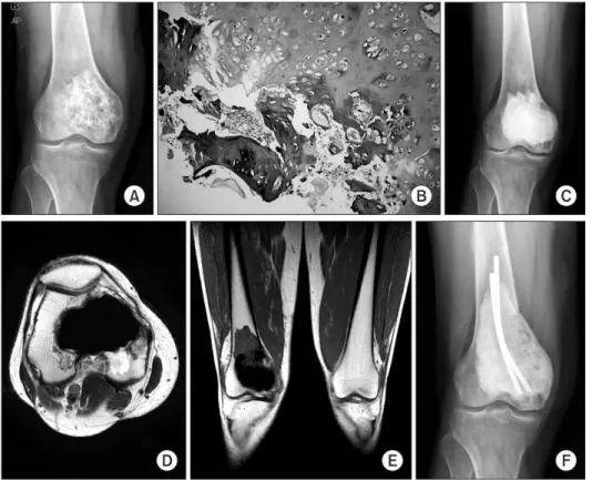
Figure 1. (A) Plain radiograph of a 52-year-old woman (Case 1) shows centrally located faint radiolucent and calcified lesion in distal metaphysis. (B) She was treated with curettage and bone cementation. (C) Histology of case 1 shows low cellularity and chondroid matrix (H&E, ×200). (D) After 7 months, pathologic fracture developed. (E) Open reduction & internal fixation was performed.
Figure 2. (A) Initial plain radiograph of a 51-year-old man (Case 8) shows mixed osteolytic and sclerotic lesion with calcifications in distal epi-metaphysis of right femur. (B) Histology of case 8 shows low cellularity and chondroid matrix (H&E,
×200). (C, D, E) He was treated with curettage and bone cementation, and after 14 months, local recurrence developed. (F) Re-curettage and bone cementation with ender-nail was performed.
골 결손 부위를 재건하였다.
수술적 치료에 따른 합병증은 수술 부위의 골절이 1예 있었는 데 술후 7개월째 발생하였으며 관혈적 정복 및 내고정술을 시행 하여 골 유합을 얻을 수 있었다(Fig. 1).
1. 국소재발
총 3예(18.8%)에서 국소 재발이 발생하였으며, 술후 국소 재발까 지 기간은 평균 61개월(13.0-156.0)이 소요되었다. 위치는 근위 및 원위 대퇴골에 각 1예, 치골이 1예였으며 근위와 원위 대퇴골에 발생한 2예에서는 병변의 위치가 관절면에 가까이 있어 철저한 소파술이 어려웠던 경우였고(Fig. 2), 치골에 발생한 1예의 경우 외부에서 술전 항암 요법과 방사선 치료를 시행하고 소파술 후 골이식을 시행한 경우로 본원 내원 후 재소파술 후 골시멘트 충 전술을 시행한 경우였다.
2. 원격전이
16예 모두에서 원격 전이는 관찰되지 않았다.
3. 종양학적 결과
평균 추시기간은 82.7개월(25.6-223.3)이었으며, 최종 추시까지 사 망한 예는 없었다. 5년 무병 생존율은 87.5%이었다.
4. 기능적 결과(Musculoskeletal Tumor Society Score;
MSTS score)
16예의 평균 MSTS는 27.5점(25-30)으로 91.6% (83.3-100)로 측정 되었으며 하지에서 평균 27.1점(90.3%), 상지에서 28.6점(95.3%), 기타 부위에서 26점(86.6%)이었다(Table 1).
고 찰
연골육종은 골육종에 비해 예후가 비교적 양호한 악성 종양으로 조직병리학적 분화도와 절제연이 예후와 가장 관련이 있는 것으 로 알려져 있다.2,3) 고 악성도 연골육종의 치료시 광범위 절제연의 확보가 필수적임에는 이견이 없으나,2,9,23) 저 악성도 중심성 연골 육종의 경우 병소내 소파술만 시행하거나 국소 병행요법을 함께 시행하는 경우, 변연 절제술, 전절제술 후 재건술을 시행하는 방 법 등 다양한 치료법이 알려져 있다.13,14) 그러나, 병리학적으로 양 성 연골 병변과 감별이 어려우며, 재발이나 전이율이 적고, 광범 위 절제술을 선택한 경우 발생할 수 있는 수술 부위의 기능 장애 등을 고려할 때 적절한 치료 방법이 무엇인지에 대해서는 논란의 여지가 있다.10-12,16,17,23)
저자들이 광범위 소파술을 시행한 후 보조 요법으로 과산화수소에 의한 화학적 소작술을 병행하여 치료한 저 악성도 중심성 연골육종 환자 16예에서 사망이나 전이는 발생 하지 않았으며 87.5%의 5년 무병생존율을 보였다.
본 연구에서 총 3예(18.75%)에서 국소 재발이 발생하였으며 이 Table 1. Demographic characteristics of 16 cases of low-grade chondrosarcoma
Case No Sex/Age Stage Location Recurrence
(months) Complication MSTS score MSTS
score (%) Final F/U Final status
1 F/52 IA Distal femur - Postoperative fracture 28 93.3 48.3 CDF2 M/49 IA Distal femur - - 26 86.6 104.9 CDF
3 F/38 IA Proximal femur 13.0 - 25 83.3 100.1 NED
4 F/47 IA Distal femur - - 29 96.6 25.6 CDF
5 M/24 IA Distal femur - - 28 93.3 101.6 CDF
6 F/60 IA Femur shaft - - 26 86.6 62.8 CDF
7 F/50 IA Proximal femur - - 28 93.3 150.4 CDF
8 M/51 IA Distal femur 14.0 - 28 93.3 33.3 NED
9 F/51 IA Proximal humerus - - 29 96.6 67.9 CDF
10 M/38 IA Proximal humerus - - 30 100.0 62.5 CDF
11 F/46 IA Proximal humerus - - 28 93.3 60.0 CDF
12 F/42 IB Proximal humerus - - 29 96.6 51.5 CDF
13 M/35 IA Pubis 156 - 26 86.6 223.3 NED
14 M/21 IA Distal radius - - 27 90.0 70.8 CDF
15 M/48 IA Proximal tibia - - 26 86.6 30.1 CDF
16 F/52 IA Proximal tibia - - 27 90.0 130.1 CDF
CDF, continuous disease free; NED, no evidence of disease.
중 2예는 근위 및 원위 대퇴골의 골간단부의 병변으로써, 광범위 소파술을 시행할 경우 관절면으로의 천공이 우려되어 다른 증례 에서처럼 광범위 소파술을 시행하지 못한 경우였으며, 치골에 발 생한 1예는 외부에서 소파술 및 골이식을 시행한 후 본원 내원하 였으나 불충분한 소파술을 시행하여 본원에서 재소파술을 시행 한 경우였다. 본 연구에서 국소 재발이 발생한 3예 모두 병변의 발생 부위가 철저한 광범위 소파술을 시행하기 어려운 위치에 있 는 경우로 이로 인해 국소 재발이 발생한 것으로 생각된다.
저 악성도 연골육종에서 병소내 소파술을 시행하는 경우 광범 위 절제술을 시행하는 경우에 비해 충분한 절제연을 가지지 못하 는 단점이 있다. 이러한 점을 보완하기 위하여 병변 주변으로 약 1-2 cm 가량의 광범위 소파술을 시행하게 되며, 액화 질소를 이 용한 동결 치료을 해주는 것이 도움이 된다는 보고가 있다.24,25) 본 연구에서는 액화 질소를 대체하여 과산화수소를 이용하였다.
수술적 치료에 따른 합병증으로는 1예에서 수술 부위의 골절 이 술후 7개월째 발생하였으며 관혈적 정복 및 내고정술을 시행 하여 골 유합을 얻을 수 있었다. 소파술 후 병적 골절을 예방하기 위한 내고정술의 적응증은 아직 명확하게 정립되어 있지 않으나 하지에 발생한 중심성 저등급의 연골 육종의 경우에는 종양의 침 범이 상대적으로 광범위하여 개방창이 큰 경우나 종양이 피질골 을 침식하여 피질골이 약해진 경우에는 내고정술을 고려하여야 한다.
본 연구에서 평균 MSTS는 27.5점(25-30)으로 91.6% (83.3-100) 이었으며 하지에서 평균 27.1점(90.3%), 상지에서 28.6점(95.3%), 기타 부위에서 26점(86.6%)으로 병변의 부위에 따라 차이를 보였 으나 모든 환자는 일상 생활이 가능하였고 기능적으로 양호한 결 과를 보여주었다.26)
연골육종의 경우 병리학적으로 양성 골종양과 저 악성도 중심 성 연골육종과의 감별이 어려워 조직검사상 양성으로 진단되어 병소내 소파술을 시행한 후 최종 진단은 저 악성도 연골육종으로 진단되는 경우도 종종 발생하는 것과 저 악성도 연골육종이 재발 할 경우 악성도가 높아지는 경우가 있는 것을 고려한다면 조직
검사상 양성 골종양으로 진단된 경우에 있어서 임상적으로 저 악 성도 중심성 연골육종이 의심될 경우에는 광범위 소파술을 시행 하고 보조 요법을 병행하는 것도 적절한 치료 방법이 될 수 있을 것으로 생각된다.14)
다양한 치료법을 적용한 시행한 저 악성도 연골육종의 5년 생 존율은 문헌에 따라 85-100%까지 보고되고 있다.4-7) 많은 저자들 은 병소내 소파술이 광범위 절제술에 비하여 생존율에 영향을 주 지 않으면서 광범위 절제술로 인한 합병증과 기능소실을 피할 수 있는 적합한 방법임을 보고하고 있다.10-12,16,17) 하지만, 병소내 소파 술이 광범위 절제술에 비해 높은 국소 재발과 원격 전이를 보인 다는 보고도 있어 아직까지 적절한 치료법에 대하여는 논란의 여 지가 있다(Table 2).6,20,27-29)
Evans 등은 병리학적 등급 1, 2, 3에 따 라 5년 생존율은 90%, 81%, 43%, 10년 생존율은 83%, 64%, 29%로 보고하였으며, 원격 전이도 등급 1, 2, 3에 따라 0%, 10%, 71%로 보고하였다.5) 본 연구 결과 5년 무병 생존율은 87.5%로 관찰되어, 광범위 소파술이 저 악성도 연골육종의 치료법으로 적절한 방법 이라고 생각된다.
결 론
본 연구의 결과 저 악성도 중심성 연골육종의 치료에 있어서 철 저한 광범위 소파술과 보조 요법을 병행하는 것은 광범위 절제술 에 비하여 생존율에 영향을 주지 않으면서 광범위 절제술로 인한 합병증과 기능 소실을 피할 수 있는 방법이라 판단된다. 이러한 치료법의 적용을 위해 정확한 병리학적 진단이 필수적이며 주의 깊은 추시관찰이 필요할 것으로 생각된다.
참고문헌
1. Campanacci M. Bone and soft tissue tumors. 2nd ed. Wien:
Springer-Verlag; 1999. 283.
2. Bruns J, Elbracht M, Niggemeyer O. Chondrosarcoma of Table 2. Comparative summery of oncologic results of low-grade chondrosarcoma
Study Number of cases Mean follow up (months) Adjuvant therapy Recurrence Metastasis MSTS Complication
Ahlmann et al. 10 39 Cryotherapy 0 (0%) 0 (0%) 27.0 1 (10%)
Bauer et al. 23 78 PMMA 2 (9%) 0 (0%) NA NA
Leerapun et al. 13 64 Phenol 1 (8%) 1 (8%) NA NA
Van der Geest et al. 55 60 Cryotherapy 0 (0%) 0 (0%) 28.0 NA
Souna et al. 15 96 Cryotherapy 0 (0%) 0 (0%) 27.9 0 (0%)
Aarons et al. 17 43 Phenol, PMMA 1 (6%) 0 (0%) 29.5 1 (6%)
Etchebehere et al. 11 64 PMMA 0 (0%) 0 (0%) NA 4 (36%)
Schreuder et al. 9 26 Liquid nitrogen 0 (0%) 0 (0%) 28.8 1 (11%) Current study 16 82.7 H2O2, PMMA 3 (18.8%) 0 (0%) 27.5 1 (6.3%)
bone: an oncological and functional follow-up study. Ann Oncol. 2001;12:859-64.
3. Murphey MD, Walker EA, Wilson AJ, Kransdorf MJ, Temple HT, Gannon FH. From the archives of the AFIP: imaging of primary chondrosarcoma: radiologic-pathologic correlation.
Radiographics. 2003;23:1245-78.
4. Eriksson AI, Schiller A, Mankin HJ. The management of chondrosarcoma of bone. Clin Orthop Relat Res. 1980;(153):
44-66.
5. Evans HL, Ayala AG, Romsdahl MM. Prognostic factors in chondrosarcoma of bone: a clinicopathologic analysis with emphasis on histologic grading. Cancer. 1977;40:818-31.
6. Fiorenza F, Abudu A, Grimer RJ, et al. Risk factors for survival and local control in chondrosarcoma of bone. J Bone Joint Surg Br. 2002;84:93-9.
7. van Loon CJ, Veth RP, Pruszczynski M, Wobbes T, Lemmens JA, van Horn J. Chondrosarcoma of bone: oncologic and fun- ctional results. J Surg Oncol. 1994;57:214-21.
8. Lee FY, Mankin HJ, Fondren G, et al. Chondrosarcoma of bone: an assessment of outcome. J Bone Joint Surg Am. 1999;
81:326-38.
9. Healey JH, Lane JM. Chondrosarcoma. Clin Orthop Relat Res. 1986;(204):119-29.
10. Ahlmann ER, Menendez LR, Fedenko AN, Learch T. Influ- ence of cryosurgery on treatment outcome of low-grade chon- drosarcoma. Clin Orthop Relat Res. 2006;451:201-7.
11. Bauer HC, Brosjö O, Kreicbergs A, Lindholm J. Low risk of recurrence of enchondroma and low-grade chondrosarcoma in extremities. 80 patients followed for 2-25 years. Acta Or- thop Scand. 1995;66:283-8.
12. McLoughlin GS, Sciubba DM, Wolinsky JP. Chondroma/
Chondrosarcoma of the spine. Neurosurg Clin N Am. 2008;
19:57-63.
13. Alho A, Skjeldal S, Melvik JE, Pettersen EO, Larsen TE. Th e clinical importance of DNA synthesis and aneuploidy in bone and soft tissue tumours. Anticancer Res. 1993;13:2383-7.
14. Weiner SD. Enchondroma and chondrosarcoma of bone:
clinical, radiologic, and histologic diff erentiation. Instr Course Lect. 2004;53:645-9.
15. Gosheger G, Gebert C, Ahrens H, Streitbuerger A, Winkel- mann W, Hardes J. Endoprosthetic reconstruction in 250 pa- tients with sarcoma. Clin Orthop Relat Res. 2006;450:164-71.
16. Normand AN, Cannon CP, Lewis VO, Lin PP, Yasko AW. Cu-
rettage of biopsy-diagnosed grade 1 periacetabular chondro- sarcoma. Clin Orthop Relat Res. 2007;459:146-9.
17. Schreuder HW, Pruszczynski M, Veth RP, Lemmens JA.
Treatment of benign and low-grade malignant intramedul- lary chondroid tumours with curettage and cryosurgery. Eur J Surg Oncol. 1998;24:120-6.
18. Suk SI, Lee CK, Ahn JK, et al. Orthopaedics. 6th ed. Seoul:
Newest Medicine Company; 2006.311.
19. McDonald DJ, Sim FH, McLeod RA, Dahlin DC. Giant-cell tumor of bone. J Bone Joint Surg Am. 1986;68:235-42.
20. Nicholson NC, Ramp WK, Kneisl JS, Kaysinger KK. Hydro- gen peroxide inhibits giant cell tumor and osteoblast metabo- lism in vitro. Clin Orthop Relat Res. 1998;(347):250-60.
21. Gortzak Y, Kandel R, Deheshi B, et al. Th e effi cacy of chemical adjuvants on giant-cell tumour of bone. An in vitro study. J Bone Joint Surg Br. 2010;92:1475-9.
22. Aarons C, Potter BK, Adams SC, Pitcher JD Jr, Temple HT. Ex- tended intralesional treatment versus resection of low-grade chondrosarcomas. Clin Orthop Relat Res. 2009;467:2105-11.
23. Ozaki T, Lindner N, Hillmann A, Rödl R, Blasius S, Winkel- mann W. Infl uence of intralesional surgery on treatment out- come of chondrosarcoma. Cancer. 1996;77:1292-7.
24. Marcove RC. A 17-year review of cryosurgery in the treat- ment of bone tumors. Clin Orthop Relat Res. 1982;(163):231- 4.
25. Mohler DG, Chiu R, McCall DA, Avedian RS. Curettage and cryosurgery for low-grade cartilage tumors is associated with low recurrence and high function. Clin Orthop Relat Res.
2010;468:2765-73.
26. Enneking WF, Dunham W, Gebhardt MC, Malawar M, Pritchard DJ. A system for the functional evaluation of re- constructive procedures after surgical treatment of tumors of the musculoskeletal system. Clin Orthop Relat Res. 1993;
(286):241-6.
27. Henderson ED, Dahlin DC. Chondrosarcoma of the bone -- a study of two hundred and eighty-eight cases. J Bone Joint Surg Am. 1963;45:1450-8.
28. Rizzo M, Ghert MA, Harrelson JM, Scully SP. Chondrosarco- ma of bone: analysis of 108 cases and evaluation for predictors of outcome. Clin Orthop Relat Res. 2001;(391):224-33.
29. Tsuchiya H, Ueda Y, Morishita H, et al. Borderline chon- drosarcoma of long and fl at bones. J Cancer Res Clin Oncol.
1993;119:363-8.
Oncologic and Functional Outcomes of Extended Curettage for Low-Grade Central Chondrosarcoma
Wan-Hyeong Cho, M.D., Soo-Yong Lee, M.D., Won-Seok Song, M.D., Chang-Bae Kong, M.D., Sang-Hyun Cho, M.D., and Dae-Geun Jeon, M.D.
Department of Orthopedic Surgery, Korea Cancer Center Hospital, Seoul, Korea
Purpose: We analyzed the oncological and functional outcome of extended curettage for low-grade central chondrosarcoma.
Materials and Methods: We retrospectively reviewed 16 patients with Grade I central chondrosarcomas treated with extended
curettage and adjuvant therapy of chemical cauterization with H2O2 at a minimum follow up of 24 months.Results: Five-year disease free survival rate was 87.5%. Mean follow up was 82.7 months and no patient developed metastases or
died of disease. The mean fi nal Musculoskeletal Tumor Society functional score was 27.5 (91.6%).Conclusion: Extended curettage is an effective treatment strategy for low-grade central chondrosarcoma with improved functional
outcomes and lower complication rates compared to wide resection and reconstruction.Key words: central chondrosarcoma, low-grade, curettage
Received September 1, 2010 Accepted June 7, 2011 Correspondence to: Dae-Geun Jeon, M.D.
Department of Orthopedic Surgery, Korea Cancer Center Hospital, 215-4, Gongneung-dong, Nowon-gu, Seoul 139-709, Korea