Differential Diagnosis of Gallbladder Wall Thickening by Two Phase Spiral CT: Gallbladder Carcinoma versus Cholecystitis1
Sun Park, M.D., Soon Gu Cho, M.D., Mi Young Kim, M.D., Je Hong Woo, M.D.2, Seok Hwan Shin, M.D.2, Kyung Hee Lee, M.D., Chang Hae Suh, M.D.
Purpose: To determine whether an analysis of two-phase spiral CT features provides a sound basis for differential diagnosis between gallbladder carcinoma and cholecystitis.
Materials and Methods: We reviewed a total of 89 cases of gallbladder carcinoma (n=35) or cholecystitis (n=54) in patients who had undergone two-phase spiral CT. For this, a GE Highspeed Advantage scanner (GE Medical Systems, Milwaukee, U.S.A.) was used. A total of 120ml of contrast material was injected at a rate of 2-3 ml/sec. Arterial and venous phase scans were obtained 35 and 65 seconds, respectively, after the initiation of contrast infusion.
All cases of gallbladder carcinoma and 468 of cholecystitis (of a total of 482) were confirmed by histopathology. We reviewed the two phase spiral CT features, analyzing and assessing thickness of the lesion, the enhancement pattern seen during the arterial and the venous phase, invasion of the liver, pericholecystic fat infiltration, dilatation of intrahepatic ducts, and other associated findings.
Results: Mean wall thickness was 12.6 mm in the gallbladder carcinoma group, and 7.2 mm in the cholecystitis group. The common enhancement patterns seen in gallbladder carcino- ma were 1) a highly enhanced thick inner wall layer during the arterial phase which became iso attenuated with adjacent liver parenchyma during the venous phase (16/35; 45.7%), and 2) a highly enhanced thick inner wall layer during both the arterial and the venous phase (8/35; 22.9%). The most common enhancement pattern in cholecystitis cases was an iso at- tenuated thin inner wall layer during both the arterial and the venous phase (44/54; 81.5%).
Findings of intrahepatic mass formation by direct invasion (9/35), lymph node enlargement (12/35), and metastasis to other organs (7/35) occurred only in cases of gallbladder carcinoma.
Dilatation of intrahepatic ducts was more frequent in cases of gallbladder carcinoma (18/35, 51.4%) than of cholecystitis (10/54, 18.5%). The incidence of pericholecystic fat infiltration and fluid collection was not significantly different between the gallbladder cancer and chole- cystitis groups.
Conclusion: Gallbladder carcinoma and cholecystitis varied in terms of wall thickness, en- hancement pattern, and intrahepatic ductal dilatation, as seen on two phase spiral CT.
Findings of liver invasion, lymph node enlargement and distant metastasis strongly suggest- ed gallbladder carcinoma. These results suggested that gallbladder carcinoma and cholecysti- tis can be distinguished by analysis of their two phase spiral CT features.
Index words : Cholecystitis
Gallbladder, neoplasms
Computed tomography (CT), helical
1Departments of Radiology and 2Surgery, Inha University College of Medicine
This paper was presented during the educational exhibition session of the annual meeting of RSNA, held on 26 November, 2000.
Received January 5, 2001 ; Accepted February 22, 2001
Address reprint requests to : Soon Gu Cho, M.D., Department of Radiology, Inha University Hospital, 7-206 3rd St., Shinheung-dong, Choong-gu, Inchon 400-711, Korea.
Tel. 82-32-890-2767 Fax. 82-32-890-2743 E-mail: [email protected]
Because the procedures involved in the prognosis and management of gallbladder carcinoma and cholecystitis differ, the differential diagnosis of these diseases is im- portant. The clinical manifestations of gallbladder carci- noma are nonspecific and often indistinguishable from those of cholecystitis, and for this reason the accurate preoperative diagnosis of gallbladder cancer is difficult:
differential is frequently, achieved by radiologists (1-7).
Many previous investigators have reported the imaging features of gallbladder carcinoma and cholecystitis (1- 7). Computed tomography (CT) is often helpful in the preoperative diagnosis of the former, the most common CT finding being a slightly enhancing mass in the region of the gallbladder that replaces most or all of this organ (2). Less common configurations are irregular or smooth wall thickening and intraluminal masses. Both types ex- hibit mild to moderate contrast enhancement and are difficult to distinguish from complicated cholecystitis (2). The ultrasonographic (US) findings of gallbladder carcinoma are not different to those of CT, and include thickening of the gallbladder wall, a fungating tumor, and a mass filling the gallbladder. As for the differential diagnosis, chronic cholecystitis, which mimics the flat- type, and benign polypoid lesions, resembling the fun- gating mass-type, are important (8). Elvin et al. (9) sug- gested that the existence of a low echoic zone, represent- ing edema in the gallbladder wall, indicates cholecysiti- tis rather than carcinoma. Despite the efforts of previous investigators, the early diagnosis of gallbladder carcino- ma and differentiation between malignancy and an in- flammatory process remain problematic. Compared with incremental CT, spiral CT offers the advantages of faster scan times, smaller interscan intervals, and opti- mization of the use of contrast medium (10). To date, no previous published investigation has focused on differ- ential diagnosis between gallbladder carcinoma and an inflammatory process of the gallbladder by means of two-phase spiral CT. The purpose of this study was to determine the two-phase spiral CT features of gallblad- der carcinoma and cholecystitis, and whether the two diseases can be successfully differentiated through analysis of their two-phase spiral CT features.
Materials and Method
Forty-one consecutive patients with suspected gall- bladder carcinoma and 482 with cholecystitis visited our institution between March 1997 and May 2000. In 26 of the 41 with suspected carcinoma and 468 of the 482
with suspected cholecystitis, the respective conditions were confirmed by histopathologic examination after cholecystectomy, and in the remaining 15 with suspect- ed carcinoma, this was confirmed by biopsy. Thirty-five of the 41 with confirmed gallbladder carcinoma and 54 of the 468 with confirmed cholecystitis (a total of 46 males and 43 females, mean age±SD = 60±12) under- went two-phase spiral CT, and these 89, in whom the respective conditions had previously been pathological- ly proven, were included in our series. The remaining 420, whose condition had been pathologically con- firmed but who did not undergo spiral CT were exclud- ed from our study.
For two-phase spiral CT a GE Highspeed Advantage scanner (GE Medical Systems, Milwaukee, Wis., U.S.A.) was used (5-mm collimation and 5 mm/sec table move- ment; scan time per section, 1 sec; scan collimation, 5 mm; scan pitch, 1), and 120 ml of iohexol (300 mg I/ml) (Omnipaque; Nycomed Ireland Ltd., Cork, Ireland) was mechanically injected into the antecubital vein at a rate of 2-3 ml/sec. Transaxial CT images, obtained in all cas- es with patients in a supine position and from the dome of the diaphragm to the symphysis pubis, were recon- structed with 5-7mm overlapping intervals. The arteri- al and the venous phase began 35 and 65 seconds, re- spectively, after the injection of contrast material.
All patients fasted for at least six hours prior to CT.According to the histopathological test results of the 54 cholecystitis patients, five were suffering from acute cholecysitits, 47 from chronic cholecysitits, and two from xanthogranulomatous cholecystitis. In all patients with gallbladder carcinoma, adenocarcinomaa was histopathologically proven.
We retrospectively reviewed the two-phase spiral CT findings, paying special attention to thickness and type of lesion, and the enhancement pattern seen on arterial and venous phase scans. We also analyzed associated findings such as invasion of the liver, pericholecystic fat infiltration, dilatation of intrahepatic ducts, enlargement of lymph nodes to more than 1cm in long diameter, the presence of gallstones and ascites, distant metastasis, and pericholecystic fluid collection. On the basis of cate- gories established by previous investigators (3, 8, 11- 13), lesions were classified as having diffuse or focal wall thickening, as a polypoid mass, or as a mass replac- ing the gallbladder. The thickness of the gallbladder wall was measured at its most hypertrophied portion, while its enhancement pattern was classified by com- paring the degree of enhancement of thickened wall, as
seen during the arterial and the venous phase, with that of adjacent liver parenchyma. Two-phase spiral CT scans were reviewed by two radiologists, whose deci- sions were reached by consensus. For statistical analy- sis, Student t test and x2-square test were used, together with an SPSS-PC v9.0 program on a personal computer.
Results
Mean wall thickness in the gallbladder carcinoma and the cholecystitis group was 12.6 mm and 7.2 mm, re- spectively, the greater thickness found in the former group being statistically significat (p<0.05). The various enhancement patterns and types of wall thickening seen in gallbladder carcinoma and cholecystitis are shown in Table 1.
For the former, the most common enhancement pat- terns were 1) a highly enhanced thick inner-wall layer, seen during the arterial phase and becoming iso-attenu- ated with adjacent liver parenchyma during the venous phase (16/35; 45.7%) (Fig. 1), and 2) a highly enhanced thick inner-wall layer seen during both the arterial and the venous phase (8/35; 22.9%) (Fig. 2). For cholecystitis, the most common enhancement pattern was an iso-at- tenuated thin inner-wall layer, seen during both the ar- terial and the venous phase (44/54; 81.5%) (Fig. 3).
Findings of intrahepatic mass formation by direct in- vasion (9/35), lymph node enlargement (12/35), and metastasis to other organs (7/35) were demonstrated on- ly in cases of gallbladder carcinoma, while dilatation of intrahepatic ducts was more frequently seen in cases of gallbladder carcinoma (18/35, 51.4%) than of cholecysti-
tis (10/54, 18.5%). The incidence of pericholecystic fat infiltration and fluid collection, and ascites, was not sig- nificantly different between the gallbladder cancer and the cholecystitis group. Gallstones were more frequent in cholecystitis patients than in those with gallbladder carcinoma (Table 2).
Discussion
Carcinoma of the gallbladder has a low overall preva- lence, but it is the most common malignancy of the bil- iary tract and the fifth most common in the alimentary tract (3, 11-15). The prognosis for patients with gall- bladder carcinoma is bleak: 88% die within one year of diagnosis, and only 4% live five years. If tumor invasion is confined to the mucosa, the survival rate is excellent,
A B
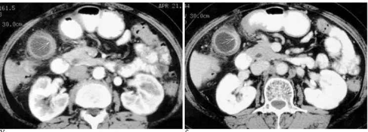
Fig. 1. Gallbladder carcinoma
Two phase spiral CT scan shows highly enhanced focal thick inner wall layer(arrow) on arterial phase scan (A) which becomes iso-attenu- ation on venous phase scan (B).
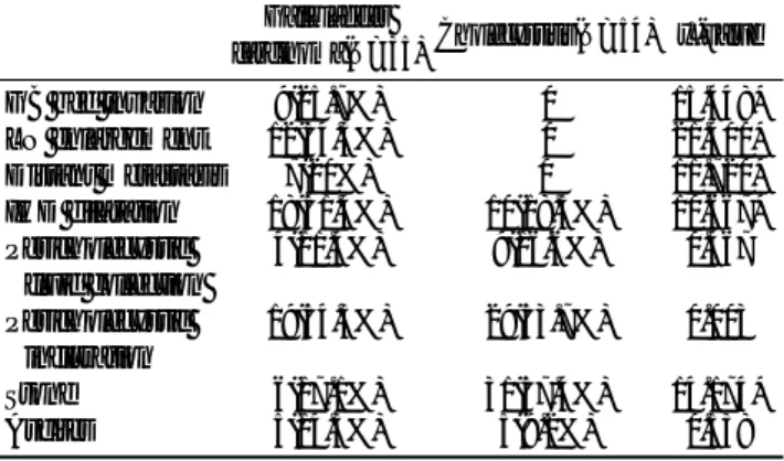
Table 1. Enhancement Patterns of Wall Thickening in Gallbladder Carcinoma and Cholecystitis
Arterial phase Venous phase Gallbladder carcinoma* Cholecystitis*
High Iso 16 (45.7%) 3 (5.6%)0
High High 08 (22.9%) 7 (13.0%)
High Low 1 (2.9%) 0
Iso Iso 05 (14.2%) 44(81.5%)
Iso Low 1 (2.9%) 0
Low Iso 2 (5.7%) 0
Low Low 2 (5.7%) 0
Total 35 (100%). 54 (100%)
Abbreviations: High, high attenuation; Iso, iso attenuation; Low, low attenuation.
*Difference from enhancement pattern in gallbladder carcinoma group and cholecystitis group was significant (x2-square test, p<0.005)
but serosal and adventitial involvement is lethal. When disease extends through the serosa, palliative surgery to bypass biliary obstruction is recommended. Radical tu- mor resection in this setting is associated with a high op- erative mortality rate and few long-term survivors (13).
A general awareness of the radiologic features of gall- bladder carcinoma will enhance the preoperative diag- nosis.
The clinical features of gallbladder carcinoma include constant right upper quadrant pain, malaise, weight loss, and jaundice (3, 13, 15). The risk of gallbladder car- cinoma is greater in patients with gallstones. The preva- lence of gallstone in cases of gallbladder carcinoma has been variously reported as around 55.6% (16), 80%- 90% (17), 73-98% (18), and 75-90% (19). A porcelain
A B
Fig. 3. Acute cholecystitis
Two phase spiral CT scan shows iso-attenuated thin inner wall layer (arrow) on both the arterial (A) and venous phase (B) scans.
A B
Fig. 2. Gallbladder carcinoma.
Two phase spiral CT scan shows thickened inner wall layer with high attenuation (arrow) on both the arterial (A) and venous phase (B) scans.
Table 2. Findings Associated with Gallbladder Carcinoma and Cholecystitis
Gallbladder
carcinoma(N=35) Cholecystitis(N=54) x2-value
GB bed invasion 9(25.7%) 0 15.448*
LN enlargement 12(34.3%) 0 21.400*
Distant metastasis 7(20%) 0 11.720*
IHD dilatation 18(51.4%) 10(18.5%) 10.667*
Pericholecystic 4(11.4%) 9(16.6%) 0.467 fluid collection
Pericholecystic 19(54.3%) 29(53.7%) 0.003 infiltration
Stone 6(17.1%) 31(57.4%) 14.174*
Ascites 5(14.3%) 5(9.2%) 0.538
Abbreviations: GB, gallbladder;
LN, lymph node;
IHD, intrahepatic duct.
* p<0.05
gallbladder is another predisposing factor and an esti- mated 22% of patients with this condition develop carci- noma (20, 21). In the present study, six cases (6/35;
17.1%) of gallstones were identified radiologically in the gallbladder carcinoma group. This low prevalence of as- sociated gallstone is thought to be due to bias in the se- lection of patients. That is, if diffuse gallbladder wall thickening with gallstones had been detected by US, acute cholecystitis might have been diagnosed in such patients and cholecystectomy performed before two- phase spiral CT was undertaken. These cases were ex- cluded from our series.
Gallbladder carcinoma as seen on cross-sectional im- ages may follow one of three mafor patterns: 1) a mass obscuring or replacing the gallbladder; 2) focal or diffuse thickening of the gallbladder wall; 3) a polypoid mass originating in the gallbladder wall and projecting into the lumen (3, 8, 11-13). Several previous investigators reported that among these three types, focal or diffuse thickening of the gallbladder wall was the least common presentation (3, 13, 22). Piehler et al. (23), however, re- ported that gallbladder carcinoma grossly appears most often as diffuse or localized thickening of the wall and less frequently as a polypoid or papillary projection into the lumen. The results of our study also showed that fo- cal or diffuse thickening of the gallbladder wall was the most common type of gallbladder carcinoma (22/35, 62.9%).
The differential diagnosis of infiltrating gallbladder carcinomas includes complicated cholecystitis, hepato- cellular carcinoma, and metastasis to the gallbladder fos- sa. Clinically and radiologically, gallbladder carcinoma can be difficult to differentiate from cholecystitis with pericholecystic fluid and abscess (13). Diffuse thicken- ing of the wall, secondary to uniform infiltration by a tu- mor, can be similar in appearance to chronic cholecysti- tis, and this appearance is nonspecific (3). Focal thicken- ing of the wall may indicate the involvement of cancer.
Focal thickening is difficult to differentiate from an area of fibrosis associated with chronic cholecystitis or an area of adenomatous hyperplasia. However, a wall infil- trated by cancer is typically thicker and more irregular than one thickened by inflammation, and thicker and more irregular wall has suggested malignancy (22, 23).
Smathers et al. (1), on the other hand, reported that smoothness versus irregularity of the gallbladder wall was not a reliable differentiating feature between carci- noma and benign conditions. The results of our study showed that thicker and more irregular wall thickening
was more frequent in patients with gallbladder carcino- ma than in those with cholecystitis.
A small polypoid carcinoma can be indistinguishable from a cholesterol polyp, adenoma, or adherent stone.
Chijiiwa et al. (24) claimed that where a polypoid lesion exceeds 10 mm in size, the lesion is solitary, and the pa- tient is older than 60, carcinoma should be strongly sus- pected. An intraluminal mass within the gallbladder, with a fungate appearance and irregular border, is strongly suggestive of gallbladder cancer.
Such a mass, however, could also represent either a cholesterol or inflammatory polyp, or a benign tumor.
Furukawa et al. (25) demonstrated that small polypoid lesions of the gallbladder were invariably detected by enhanced CT, but this was not the case with cholesterol and hyperplastic polyps. In two of our patients choles- terol polyps were pathologically confirmed after chole- cystectomy, but had not been demonstrated by two- phase spiral CT.
Although US is widely accepted for imaging of the gallbladder, CT is thought to complement its findings, and awareness of the two-phase spiral CT features of gallbladder diseases is thus useful for diagnosis. In our study, the finding of highly enhanced thick inner wall layer, seen during the arterial phase, which became iso- or hyper-attenuated with adjacent liver parenchyma during the venous phase, strongly suggested malignan- cy, and iso-attenuated thin inner wall layer, during both the arterial and the venous phase was a more frequent feature of cholecystitis. Thus, awareness of the enhance- ment patterns of the focal or diffuse thickening type of gallbladder carcinoma and cholecystitis, as seen on two- phase spiral CT, appears to be helpful in differentiating these two different disease entities.
The most common route of dissemination for gallblad- der carcinoma is direct invasion of the liver. This can be explained by noting that the hepatic surface of the gall- bladder is drained by vessels that communicate with ad- jacent hepatic veins; spread through this route leads to involvement of the adjacent liver (26). Spread to lymph nodes around the cystic and pericholedochal nodes is al- so common (27). Other structures that may be involved are lymph nodes in the region of the porta hepatis, he- patic and common bile ducts, pancreas, colon, and duo- denum. In our series, direct invasion of the liver was ap- parent in nine of the 35 gallbladder carcinoma patients (25.7%), and twelve of the 35 (34.3%) had demonstrated lymph node enlargement cystic (n=7), pericholedochal (n=5), porta hepatis (n=4), pancreatoduodenal (n=3).
Direct or lymphatic spread of malignancy to the porta hepatis results in obstruction of the biliary tree, which in manifested as jaundice. Although this is one of the earliest clinical manifestations of the disease, it unfortu- nately signifies an advanced stage of malignancy.
In this study, 18 patients (51.4%) presented with di- latation of the biliary tree due to tumor invasion of the common bile duct (n=5) and at the bifurcation level of the intrahepatic duct (n=4), and to lymph node enlarge- ment in the porta hepatis (n=8) and pericholedocal re- gion (n=1).
In summary, the results of this study showed that if a hyper-attenuated thick inner-wall layer of gallbladder was seen during the arterial phase, and this became iso- or hyper-attenuation during the venous phase, malig- nancy was strongly suggested; if, on the other hand, an iso-attenuated thin inner wall layer was seen during both the arterial and venous phase, this suggested chole- cystitis rather than gallbladder cancer. Findings of liver invasion, regional lymph node enlargement, and distant metastasis strongly suggested gallbladder carcinoma.
In conclusion, awareness of these specific two-phase spiral CT features should be valuable in differentiation between an inflammatory process and gallbladder carci- noma. Two-phase spiral CT is thus useful for differential diagnosis between these two disease entities.
References
1. Smathers RI, Lee JKT, Heiken JP. Differentiation of complicated cholecystitis from gallbladder carcinoma by computed tomogra- phy. AJR Am J Roentgenol 1984;143:255-259
2. Abraham HD. Benign and malignant tumors of the gallbladder. In:
Arnold CF, Abraham HD, eds. Radiology of the liver, biliary tract, and pancreas. 1st ed. St. Louis, Mo: Mosby-Year Book Inc., 1994:
555-576
3. Seyed AR, Nasser ST, Mabmood KR, et al. Imaging of gallbladder carcinoma. Radiographics 1994;14:291-306
4. Maeyama R, Yamaguchi K, Noshiro H, et al. A large inflammatory polyp of the gallbladder masquerading as gallbladder carcinoma. J Gastroenterol 1998;33:770-774
5. Onoyama H, Yamamoto M, Takada M, et al. Diagnostic imaging of early gallbladder cancer: retrospective study of 53 cases. World J Surg 1999;23:708-712
6. Pablo RR, Zachary DG. Xathogranulomatous cholecystitis versus gallbladder carcinoma. Radiology 1997;203:10-12
7. Chun KA, Ha HK, Yu ES, et al. Xanthogranulomatous cholecysti- tis: CT features with emphasis on differentiation from gallbladder carcinoma. Radiology 1997;203:93-97
8. Tsuchiya Y. Early carcinoma of the gallbladder: Macroscopic fea- tures and US finidings. Radiology 1991;179:171-175
9. Elgin A, Erwald R, Muren C, Mare K. Gallbladder carcinoma: di- agnostic procedures with emphasis on ultrasound diagnosis. Ann Radiol 1989;32:282-287
10. Zeman RK, Baron RL, Jeffrey RB, Klein J, Siegel MJ, Silverman PM. Helical body CT: evaluation of scanning protocols. AJR Am J Roentgenol 1998;170:1427-1438
11. Shelley NW, Mordecai K, Helen M. Sonography and computed to- mography in the diagnosis of carcinoma of the gallbladder. AJR Am J Roentgenol 1984;142:735-739
12. Yuji I, Tsutomu A, Koki Y, Shigeri F, Naobumi Y, Akira T.
Computed tomongraphy of gallbladder carcinoma. Radiology 1980;
137:713-718
13. Richard MG, Marc SL. Neoplasms of the gallbladder and biliary tract.
In: Ellen MW, Ann SF, Pereles FS, Richard MG, eds. Textbook of gastrointestinal radiology. 2nd ed. Philadelphia, Pa: W.B. Saunders Company, 1994:1360-1374
14. Frezza EE, Mezghebe H. Gallbladder carcioma: A 28 year experi- ence. Int Surg 1997;82:295-300
15. Thomas RK, Timothy RC. Carcinoma of the gallbladder. Am J Surg 1982;4:737-741
16. 정혜경, 이경환, 김홍길, 이 열, 윤종섭. 담낭암의 초음파 및 전산화 단층촬영 소견. 대한방사선의학회지 1988;24:1075-1080
17. Yum HY, Fink AH. Sonographic findings in primary carcinoma of the gallbladder. Radiology 1980;134:693-696
18. Kane RA, Jacobs R, Katz J, Costello P. Porcelain gallbladder: ultra- sound and CT appearance. Radiology 1984;152:137-141
19. Robbins SL. The liver and biliary tract. In: Robbins SL, Cotran RS, Kumar V, eds. Robbins pathologic basis of disease. 4th ed.
Philadelphia, Pa:Saunders, 1989:911-980
20. Polk HC. Carcinoma and the calcified gallbladder. Gastroenterology 1966;50:582-585
21. Berk RN, Armbuster TG, Saltzstein SL: Carcinoma in the porcelain gallbladder. Radiology 1973;106:29-31
22. Lane J, Buck JL, Zeman RK. Primary carcinoma of the gallbladder:
a pictorial essay. Radiographics 1989;9:209-227
23. Jeffrey RB, Laing FC, Wong W, Calen PW. Gangrenous cholecysti- tis: diagnosis by ultrasound. Radiology 1983;148:219-221
24. Chijiiwa K, Tanaka M. Polypoid lesion of the gallbladder:
Indications of carcinoma and outcome after surgery for malignant polypoid lesion. Int Surg 1994;79:106-109
25. Furukawa H, Takayasu K, Ushio K, et al. CT evaluation of small polypoid lesions of the gallbaldder. Hepatogastroenerology 1995;42:
800-810
26. Fultz PJ, Skucas J, Weiss SL. Comparative imaging of gallbladder cancer. J Clin Gastroenterol 1988;6:683-692
27. Tetsuya O, Yoshio S, Kazuhiro T, Katsuyoshi H, Terukazu M.
Carcinoma of the gallbladder: CT evaluation of lymphatic spread.
Radiology 1993;189:875-880
대한방사선의학회지 2001;44:497-503
이중 시기 나선식 CT를 이용한 담낭벽 비후의 감별진단: 담낭암 대 담낭염1
1인하대학교 의과대학 방사선과학교실
2인하대학교 의과대학 일반외과학교실
박 선・조순구・김미영・우제홍2・신석환2・이경희・서창해
목적: 담낭암과 담낭염의 이중 시기 나선식 CT소견을 알아보고, 그 소견 분석을 통한 담낭염과 담낭암의 감별진단 여부를 알아보고자 하였다.
대상과 방법: 담낭암 혹은 담낭염으로 진단된 89명의 환자들의 이중 시기 나선식 CT 소견을 후향적으로 분석하였
다. 그중 담낭암 환자가 35명이었고 담낭염 환자가 54명이었다. 이중 시기 나선식 CT는 GE Highspeed Advantage(GE Medical systems, Milwaukee, U.S.A.) 기기를 이용하여 시행하였다. 총 120 ml의 조영제를 2-3 ml/초의 속도로 주입하였고, 동맥기와 정맥기 영상을 조영제 주입 시작 후 각각 35초와 65초에 얻었다. 전예에서 담낭암과 담낭염은 병리조직학적으로 확진 되었다. 이중 시기 나선식 CT소견은 병변의 두께와 형태, 동맥기와 정 맥기 영상에서의 조영증강 양상, 간으로의 침습 여부, 담낭 주위 지방 침윤, 간내 담관의 확장, 그리고 다른 연관 소 견들을 평가하여 분석하였다.
결과: 담낭벽의 평균 두께는 담낭암 군에서 12.6 mm 이었고, 담낭염 군에서 7.2 mm 였다. 담낭암의 흔한 조영증 강 양상은 1) 동맥기에 강한게 조영 증강된 두꺼운 내벽이 정맥기에 인접 간 실질과 동등 감쇠로 변화하는 경우 (16/35; 45.7%), 2) 동맥기와 정맥기 모두에서 강하게 조영증강된 두꺼운 내벽을 보이는 경우 (8/35; 22.9%) 였 고, 담낭염의 가장 흔한 조영증강 패턴은 동맥기와 정맥기 모두에서 간 실질과 동등 감쇄로 조영되는 얇은 내벽을 보이는 경우였다 (44/54; 81.5%). 종양의 직접 침습에 의한 간내 종괴 형성 (9/35), 림프절 종대 (12/35), 그리고 다른 장기로의 전이 (7/35)등의 소견은 담낭암의 예에서만 발견되었다. 간내 담관의 확장은 담낭염 (10/54, 18.5%) 보다 담낭암 (18/35, 51.4%)의 예에서 더 흔하였다. 담낭주위 지방 침윤과 담낭주위 액체 저류소견의 빈도는 담낭 암과 담낭염 군간에 유의한 차이가 없었다.
결론: 담낭암과 담낭염은 이중 시기 나선식 CT소견상 벽 두께, 조영 증강 페턴, 그리고 간내 담관 확장에서 차이를 보였고, 간 침습, 림프절 증대, 그리고 원격 전이등의 소견은 담낭암을 강력히 시사하는 소견으로 생각된다. 이러한 결과들은 담낭암과 담낭염이 이중 시기 나선식 CT 소견의 분석을 통해 서로 구분될 수 있음을 시사하는 것으로 생 각된다.