online©ML Comm
0LABORATORY INVESTIGATION0
Copyright ⓒ 2007 Journal of Korean Neurotraumatology Society 99
J Kor Neurotraumatol Soc 2007;3:99-102 ISSN 1738-8708
백서의 척수손상에서 인체 중간엽 줄기세포의 치료효과 및 적정주입시간에 관한 연구
울산대학교 의과대학 서울아산병원 신경외과학교실1, 성균관대학교 의과대학 삼성서울병원 신경외과학교실2
양구현
1·김은상
2·민중기
1·노성우
1·임승철
1·전상룡
1Therapeutic Effect and Optimal Injection Time by Human Mesenchymal Stem Cell at Spinal Cord Injury of Rat
Ku Hyun Yang, MD1, Eun-Sang Kim, MD, PhD2, Joong Kee Min, MS1,
Sung-Woo Roh, MD, PhD1, Seung-Chul Rhim, MD, PhD1 and Sang Ryong Jeon, MD, PhD1
1Department of Neurological Surgery, Asan Medical Center, College of Medicine, University of Ulsan, Seoul, Korea
2Department of Neurosurgery, Samsung Medical Center, SungKyunKwan University School of Medicine, Seoul, Korea
Objective: Human mesenchymal stem cell (MSC) is known to have therapeutic effect in spinal cord injury (SCI), but the optimal therapeutic time window and survival pattern is not well known. The authors evaluated the treatment effect of MSC in the respect of injection time and survival rate in spinal cord injured rats. Methods: In experiment I, SCI is performed by New York State University (NYU) Impactor in 30 rats. Human MSCs were injected intramedullarily with 5 groups. The animals were grouped with Group 1 (injury only), Group 2 (immediate MSC injection), Group 3 (injection after 3 days), Group 4 (injection after 1 week), Group 5 (injection after 2 weeks), and Group 6 (injection after 4 weeks). The Basso Beattie Bresnahan (BBB) locomotor scale was evaluated during 8 weeks after SCI in all the animals, and then the spinal cords were stained with H&E and antinucleic acid staining (Antinuclei®). In experiment II, MSC were injected at 1 week after SCI. After that, cord tissue were stained with antinucleic acid at 1 week (n=5), 2 weeks (n=5), and 3 weeks (n=5) after injection. Results: In experiment I, BBB score improvement at 8th week after SCI were statistically significant at Group 4, 5, 6 compared to Group 1. The cavity size were not significantly different in each group. In the antinucleic acid staining, the MSCs could not be observed within the cords. In experiment II, stained cell numbers at each group were signifi- cantly different. Conclusion: In this study, the authors observed that neurological recovery after SCI in rats were improved by human MSCs which were injected intramedullarily at 1, 2, or 4 weeks after SCI and the injected cells could not survive over 4 weeks. Therefore, the neuroprotective effect by MSCs is supposed to be ocurred in the early period of injection time when the MSCs survive. (J Kor Neurotraumatol Soc 2007;3:99-102)
KEY WORDS: Mesenchymal stem cell·Spinal cord injury·Neuroprotective effect.
서 론
중추신경계의 치료에 관해 연구되고 있는 세포는 배아줄 기세포, 중간엽 줄기세포, 제대혈 줄기세포 등 여러 종류가 있다.14) 그 중 골수유래 중간엽 줄기세포(mesenchymal
stem cell: MSC)는 척수손상(spinal cord injury: SCI) 에서 치료효과를 나타내는 것으로 알려져 있다.19)
하지만 줄기세포를 적용할 경우 이러한 손상 후에 세포 주입을 할 때의 최적 치료 시점이나 이식된 부위에서의 세포생존 기간 등에 대해서는 잘 알려져 있지 않다. 본 Address for correspondence: Sang Ryong Jeon, MD, PhD
Department of Neurological Surgery, Asan Medical Center, College of Medicine, University of Ulsan, 388-1 Pungnap-2 dong, Songpa-gu, Seoul 138-736, Korea
Tel: +82-2-3010-3562, Fax: +82-2-476-6738, E-mail: [email protected] 본 연구는 아산생명과학연구소의 연구비 (2006-241) 지원에 의하여 이루어졌음.
Therapeutic Effect and Optimal Time of Human Mesenchymal Stem Cell
100 J Kor Neurotraumatol Soc 2007;3:99-102
연구는 이러한 점에 대해 알아보고자 백서를 대상으로 시 행되었다.
대상 및 방법
실험은 줄기세포 주입시기에 따른 신경학적 회복의 차이 를 관찰하기 위한 실험 Ⅰ과 주입된 줄기세포의 생존기간을 관찰하기 위한 실험 Ⅱ로 나누어 시행하였다. 실험 Ⅰ에서 백서의 척수손상은 New York State University (NYU) Impactor로 30마리에 행하였다. 실험쥐는 230~280 g Spraque-Dawley (female)를 사용하였다. 0.4 g/kg Chlo- ral hydrate 혹은 40 mg/kg zoletil을 사용한 복막강 전신 마취하에서 제 9번 흉추에서 후궁절제술을 한 후 10 g im- pact rod를 25 mm 높이에서 낙하시켜 완전 척수손상을 유발하였다. 중간엽 줄기세포는 인체에서 채취하여 배양한 이종세포를 사용하였고 무균처리된 세포를 Hamilton syr- inge (30 G)에 1×10⁶ cells/15 μl를 척수내에 주입하 였다. 5개 그룹으로 나누었고 그룹 1은 척수손상만을 가 하였고 (Group 1, n=5), 그룹 2는 손상직후 세포주입 (Group 2, n=5), 그룹 3은 손상 후 3일에 주입(Group 3, n=5), 그룹 4는 손상 후 일주일째 주입 (Group 4, n=5), 그룹 5는 손상 후 이주일째 (Group 5, n=5), 그룹 6은 손상 후 사주일째에 주입하였다 (Group 6, n=5). 신경학 적 회복의 정도는 BBB점수 (Basso Beattie Bresnahan locomotor rating scale)로 측정하였고 손상 후 8주일까 지 측정하였다. 이후 4% paraformaldehyde로 조직을 고 정하고 손상부분을 중심으로 2 cm의 척수를 채취하였다.
척수조직을 10% formalin 용액에서 24시간 동안 재고정 후 paraffin block을 제작하였다. 이후 척수조직을 분리하 여 Hematoxylin and Eosin (H&E)과 antinucleic acid staining (Antinuclei®)으로 염색하여 척수조직의 변화 및 줄기세포의 존재여부를 관찰하였다.
실험 Ⅱ는 15마리의 실험쥐에서 동일한 방법으로 척수손 상을 가한 후 손상 후 일주일째 중간엽 줄기세포를 동일한
방법으로 주입하고, 주입 일주일 후 (Group A, n=5), 이주 일 후 (Group B, n=5), 삼주일 후 (Group C, n=5)에 antinucleic acid (Antinuclei®)를 사용하여 척수의 중심선 에서 외측 150 μm 종단면에서 100배 확대시야하의 줄기 세포 수를 관찰하였다.
통계는 SPSS 8.0 standard version으로 analysis of variance (ANOVA) test를 이용하였다.
결 과
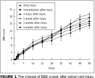
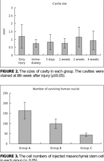
실험 Ⅰ에서 8주째 BBB 점수는 그룹 1, 2, 3, 4, 5, 6에 서 각기, 14.3±1.3, 16.0±0.6, 15.2±0.9, 19.0±0.7, 17.3±1.0, 17.6±1.3이었다 (Table 1, Figure 1). 손상 만을 가한 그룹 1에 비교하여 손상 후 1, 2, 4주째에 중간 엽 줄기세포를 주입한 그룹 4, 5, 6에서 BBB 점수의 상승 이 통계학적 의미가 있었다 (p<0.05). 손상을 가한 부위에 서 관찰되는 척수조직내의 공동은 크기가 각기 그룹 1, 2, 3, 4, 5, 6에서 1.18±0.8, 0.73±0.2, 0.82±0.5, 0.75±
0.3, 1.14±0.6, 0.93±0.6 mm2였고 이들의 차이는 통계 학적인 의미가 없었다 (p>0.05)(Figure 2). Antinucleic
TABLE 1. The change of BBB scores after spinal cord injury
Days after injury
0 1 2 3 7 14 21 28 35 42 49 56
Group 1 0 0.1 0.9 1.3 2.7 5.8 7.2 08.1 09.8 11.2 13.0 14.3
Group 2 0 0.2 1.1 2.0 3.2 5.7 7.3 10.4 12.2 13.5 14.6 16.0
Group 3 0 0.0 0.6 1.5 3.9 6.0 8.2 09.6 11.1 12.6 14.0 15.2
Group 4 0 0.0 0.6 1.5 3.1 7.4 9.0 10.4 12.7 14.6 17.8 19.0
Group 5 0 0.0 0.8 1.7 3.3 6.1 7.7 09.7 12.2 13.8 15.4 17.3
Group 6 0 0.0 0.6 1.8 3.3 5.8 7.6 09.1 12.6 15.0 16.8 17.6
BBB: Basso Beattie Bresnahan
0 3 6 9 12 15 18 21
0 7 14 21 28 35 42 49 56
Days
BBB score
Only injury
Immediately after injury
1 week after injury 2 weeks after injury 4 weeks after injury 3 days after injury
FIGURE 1. The change of BBB scores after spinal cord injury.
BBB: Basso Beattie Bresnahan.
Ku Hyun Yang, et al.
www.neurotrauma.or.kr 101 acid staining을 하였을 때 모든 그룹에서 중간엽 줄기세
포는 관찰되지 않았다. 실험 Ⅱ에서 줄기세포 주입 후 1, 2, 3주째에 관찰한 그룹 A, B, C에서 세포수가 100배 확대 시야에서 각기 163.4±40.7, 99.0±23.0, 43.8±8.8개가 관찰되었고 이는 통계학적인 의미가 있었다 (p<0.05) (Figure 3).
고 찰
최근의 줄기세포 연구가 발전하면서 중추신경계에서 세 포치료에 적용할 수 있는 세포의 종류로는 신경줄기세포 (neural stem cells), 배아줄기세포(embryonic stem cells), 조혈줄기세포(hematopoietic stem cells), 중간엽 줄기세 포(mesenchymal stem cells), 제대혈 조혈줄기세포(he- matopoietic stem cells from umbilical cord blood) 등 여러 종류가 있다.5,11,12,15) 줄기세포를 척수조직내에 주입할 경우, 최대 효과를 나타내는 시기에 대해서는 자세히 연구 된 바가 없다. 본 연구에서는 줄기세포를 손상 직후나 3일 째에 주입한 경우 손상만 가한 경우와 통계적 치료효과의
차이는 없는 것으로 나타났고 손상 일주일과 이주일뿐만 아 니라 만성기로 볼 수 있는 손상 4주째1,9)에 주입한 경우에 는 줄기세포에 의한 치료효과가 있는 것으로 나타났다. 그 리고 통계적 의미는 없지만 아급성기로 볼 수 있는 손상 일주일째에 그 치료효과는 가장 높은 것으로 보인다. 이는 아마도 급성기에는 활발히 진행되는 염증반응과 세포사멸 등의 이유로 줄기세포가 생존하기 힘들고 따라서 신경재생 효과가 일어나지 않기 때문으로 보인다.8) 문헌에 따르면 백 서의 손상된 척수에 신경줄기세포 및 전구세포를 주입했 더니 손상직후 주입보다는 9일 후 주입한 동물에서 더 오 랜기간 신경회복이 진행되는 것을 관찰한 보고도 있다.16) 실험 Ⅰ에서 척수조직내 주입된 줄기세포가 antinucleic acid 염색에서 관찰되지 않은 것은 적어도 4주 이상 지난 시점에서 생존하고 있는 세포가 없기 때문으로 추측되고 실 험 Ⅱ에서 1주, 2주, 3주 기간에 따라 의미 있게 세포수가 감소하는 것은 줄기세포들이 척수조직 내에서 시간이 흐를 수록 사멸해 가기 때문으로 보인다. 그러나 세포생존 기간 이 지난 8주까지 백서에서 신경학적 회복이 진행된 것은 줄 기세포 생존 이후에도 축삭재생에 유리한 환경이 만들어졌 기 때문으로 추측된다. Nishimo 등도 제대혈 조혈줄기세 포가 조직내에서 3주간 생존했지만 6주간의 관찰에서 신 경회복은 지속되었다고 보고했다.14)
줄기세포가 척수손상에 의한 마비에서 치료 효과를 나타 내는 기전은 이들 주입된 줄기세포들이 새로운 신경세포로 분화하여 기능을 나타낸다기 보다는 myelin 분비를 하는 희돌기세포나 교세포로 분화하거나 cytokine 등을 분비하 여 내재된 줄기세포의 활성화, 혹은 척수공동을 기계적으로 채워서 손상된 축삭이 하방으로 자랄 수 있게 하는 기계적 가교역할, 혹은 척수공동의 섬유질을 용해시켜 기계적 장 벽을 완화시키거나 급성기에서 세포사멸, 괴사를 감소시 키고 혈관재생을 활성화하는 역할에 의한 것으로 추측된 다.2-4,6,7,10,13,17,18)
그러므로 주입된 세포가 영구적으로 생 존하거나 분열, 증식하지 않더라도 단기간에 축삭재생의 환 경을 만들어서 이렇게 활성화된 축삭재생 과정이 지속되 는 것으로 여겨진다.
골수유래 중간엽 줄기세포가 척수손상에 대한 치료효과 가 있고 또한 부작용이 거의 없음을 본 연구로 알 수가 있 으나 세포의 생존기간에 한계가 있고 인체에 적용하기 위 해서는 치료효과의 정도를 향상시켜야 할 것으로 보이므로 이를 위한 추가적 방법이 고려되어야 할 것이다. 예를 들면 세포가 조직내에서 오랫동안 존재할 수 있도록 하기위한 지지체(scaffold)를 병용해 주입하거나 세포와 함께 trophic factor (Growth factor 혹은 neurotrophic factor 등)를
Cavity size
0 0.5 1 1.5 2 2.5 3
Only injury Imme-
diately 3 days 1 weeks 2 weeks 4 weeks
mm2
FIGURE 2. The sizes of cavity in each group. The cavities were stained at 8th week after injury (p>0.05).
Number of surviving human nuclei
0 50 100 150 200 250
Group A Group B Group C
FIGURE 3. The cell numbers of injected mesenchymal stem cell in each group (p<0.05).
Therapeutic Effect and Optimal Time of Human Mesenchymal Stem Cell
102 J Kor Neurotraumatol Soc 2007;3:99-102
병행해서 주입하거나, 궁극적으로 이들 factor를 분비하는 능력을 향상시킨 유전자변형 줄기세포를 이용하는 방법 등 이 있을 것이다.
결 론
본 실험에서 백서의 척수손상 후 인체의 골수유래 중간 엽 줄기세포를 척수내 직접 주입하는 방법이 아급성기나 만 성기인 손상 후 1, 2, 4주째 주입하는 경우에 의미 있는 신 경회복 효과가 있음이 관찰되었다. 또한 척수조직내에 주 입된 세포는 적어도 3주까지 생존하고 4주 이후에는 생존 하지 않음이 관찰되어 생존기간인 비교적 초기시기에 축 삭재생의 환경을 조성하는 것으로 추측되어 향후 줄기세포 의 기능을 향상시키는 추가적 방법이 연구되어야 할 것으 로 보인다.
중심 단어: 중간엽 줄기세포·척수손상·신경보호효과.
REFERENCES
1) Basso DM, Beattie MS, Bresnahan JC. A sensitive and relia- ble locomotor rating scale for open field testing in rats. J Neurotrauma 12:1-21, 1995
2) Chen J, Zhang ZG, Li Y, Wang L, Xu YX, Gautam SC, et al.
Intravenous administration of human bone marrow stromal cells induces angiogenesis in the ischemic boundary zone after stroke in rats. Circ Res 92:692-699, 2003
3) Chen X, Katakowski M, Li Y, Lu D, Wang L, Zhang L, et al.
Human bone marrow stromal cell cultures conditioned by trau- matic brain tissue extracts: growth factor production. J Neu- rosci Res 69:687-691, 2002
4) Chong ZZ, Kang JQ, Maiese K. Hematopoietic factor erythro- poietin fosters neuroprotection through novel signal transduc- tion cascades. J Cereb Blood Flow Metab 22:503-514, 2002 5) Chopp M, Zhang XH, Li Y, Wang L, Chen J, Lu D, et al. Spinal
cord injury in rat: treatment with bone marrow stromal cell transplantation. Neuroreport 11:3001-3005, 2000
6) Dame C, Wolber EM, Freitag P, Hofmann D, Bartmann P, Fandrey J. Thrombopoietin gene expression in the developing human central nervous system. Brain Res Dev Brain Res 143:
217-223, 2003
7) Fedoroff S, Berezovskaya O, Maysinger D. Role of colony stimulating factor-1 in brain damage caused by ischemia. Neu- rosci Biobehav Rev 21:187-191, 1997
8) Fitch MT, Doller C, Combs CK, Landreth GE, Silver J. Cellu- lar and molecular mechanisms of glial scarring and progressive cavitation: in vivo and in vitro analysis of inflammation-indu- ced secondary injury after CNS trauma. J Neurosci 19:8182- 8198, 1999
9) Hill CE, Beattie MS, Bresnahan JC. Degeneration and sprouting of identified descending supraspinal axons after contusive spinal cord injury in the rat. Exp Neurol 171:153-169, 2001 10) Hofstetter CP, Schwarz EJ, Hess D, Widenfalk J, El Manira A,
Prockop DJ, et al. Marrow stromal cells form guiding strands in the injured spinal cord and promote recovery. Proc Natl Acad Sci U S A 99:2199-2204, 2002
11) Koshizuka S, Okada S, Okawa A, Koda M, Murasawa M, Hashimoto M, et al. Transplanted hematopoietic stem cells from bone marrow differentiate into neural lineage cells and promote functional recovery after spinal cord injury in mice. J Neuro- pathol Exp Neurol 63:64-72, 2004
12) McDonald JW, Liu XZ, Qu Y, Liu S, Mickey SK, Turetsky D, et al. Transplanted embryonic stem cells survive, differentiate, and promote recovery in injured rat spinal cord. Nat Med 5:
1410-1412, 1999
13) Mehler MF, Rozental R, Dougherty M, Spray DC, Kessler JA.
Cytokine regulation of neuronal differentiation of hippocampal progenitor cells. Nature 362:62-65, 1993
14) Nishio Y, Koda M, Kamada T, Someya Y, Yoshinaga K, Okada S, et al. The use of hematopoietic stem cells from human um- bilical cord blood to promote restoration of spinal cord tissue and recovery of hindlimb function in adult rats. J Neurosurg Spine 5:424-433, 2006
15) Ogawa Y, Sawamoto K, Myata T, Miyao S, Watanabe M, Na- kamura M, et al. Transplantation of in vitro-expanded fetal neural progenitor cells results in neurogenesis and functional recovery after spinal cord contusion injuryin adult rats. J Neu- rosci Res 69:925-933, 2002
16) Okada S, Ishii K, Yamane J, Iwanami A, Ikegami T, Katoh H, et al. In vivo imaging of engrafted neural stem cells: its appli- cation in evaluating the optimal timing of transplantation for spinal cord injury. FASEB J 19:1839-1841, 2005
17) Sasaki M, Honmou O, Akiyama Y, Uede T, Hashi K, Kocsis JD.
Transplantation of an acutely isolated bone marrow fraction repairs demyelinated adult rat spinal cord axons. Glia 35:26-34, 2001
18) Storch A, Paul G, Csete M, Boehm BO, Carvey PM, Kupsch A, et al. Long-term proliferation and dopaminergic differen- tiation of human mesencephalic neural precursor cells. Exp Neurol 170:317-325, 2001
19) Wu S, Suzuki Y, Ejiri Y, Noda T, Bai H, Kitada M, et al. Bone marrow stromal cells enhance differentiation of cocultured neurosphere cells and promote regeneration of injured spinal cord. J Neurosci Res 72:343-351, 2003