Received : January 24, 2011 Revised : February 16, 2011 Accepted : February 22, 2011
Hyung-Jin Chung, M.D.
Department of Orthopedic Surgery, Sanggye Paik Hospital, Inje University College of Medicine, 761-1 Sanggye-dong, Nowon-gu, Seoul 139-707, Korea
Tel: +82-2-950-1026 Fax: +82-2-934-6342 E-mail: [email protected]
* 본 논문의 요지는 2010년도 대한족부족관절학회 추계학술대회에서 발표되었음.
* 본 논문은 2008년도 인제대학교 학술연구조성비 보조에 의한 것임.
J Korean Foot Ankle Soc. Vol. 15. No. 1. pp.13-17, 2011
족관절 골절과 동반된 전하 경비 인대 견열 골절의 치료
인제대학교 의과대학 상계백병원 정형외과 정형진 배서영 김만영
Treatment of Anteroinferior Tibiofibular Ligament Avulsion Fracture Accompanied with Ankle Fracture
Hyung-Jin Chung, M.D., Su-Young Bae, M.D., Man-Young Kim, M.D.
Foot and Ankle Center, Department of Orthopedic Surgery, Sanggye Paik Hospital, Inje University College of Medicine, Seoul, Korea
=Abstract=
Purpose: To evaluate the clinical results of anteroinferior tibiofibular ligament avulsion fracture accompanied ankle fractures treated with anatomical reduction and internal fixation.
Materials and Methods: From January 2007 to April 2010, 30 cases with anteroinferior tibiofibular ligament avulsion fracture that treated with anatomical reduction and internal fixation were analyzed. The average follow-up period was 26 months (minimum 6 months). We have reviewed the bony union, complication and subjective satisfaction according to the fracture classification and method of internal fixation.
Results: Among 30 cases, 28 cases were occurred in Lauge-Hansen classification supination-external rotation type, one case was fracture-dislocation and one case was Maisonneuve fracture. We have performed internal fixation with Mini screw in 11 cases, K-wire in 10 cases, repair in six cases and Mini screw & K-wire in three cases. In all cases bony union was completed. two cases in Mini screw, one case in K-wire, two cases in repair and one case in Mini screw & K-wire revealed LOM of ankle joint. Skin irritation and superficial peroneal nerve irritation happened in one case each. Other cases show good subjective satisfaction.
Conclusion: Anteroinferior tibiofibular ligament avulsion fracture accompanied with ankle fracture is a good clinical outcome with internal fixation. So we should not miss out the anteroinferior tibiofibular ligament avulsion fracture in radiologic evaluation or operation room.
Key Words: Ankle, Anteroinferior tibiofibular ligament avulsion fracture, Internal fixation
서 론
족관절의 안정성과 기능은 격자상의 골구조와 주변 인대 들에 의해 유지된다. 족관절 골절은 인대 손상을 포함한 주 변 연부 조직 손상과 동반되는 경우가 많으므로1) 족관절 골 절시 골 손상에 대한 정확한 해부학적 정복과 내고정 외에 주변 인대 손상에 대한 정확한 평가와 치료가 필요하다. 전 하 경비 인대 견열 골절은 족관절 골절 시 동반되어 원위 경비 인대 결합부의 이개 등 족관절 안정성에 영향을 줄 수
있다. 이에 대해 1875년 Wagstaffe2)가 최초로 2예를 보고 한 이후 1979년 Pankovich3)가 Wagstaffe의 분류를 수정하 여 보고하였고, 국내에서는 2002년 Park 등4)에 의해 기술 되었지만 이후 이에 대한 보고가 드문 상태이다.
본 연구는 족관절 골절과 동반된 전하 경비 인대 견열 골 절의 발생빈도, 분류, 족관절 골절 분류와의 상관관계 및 치 료 후 임상적, 방사선학적 결과에 대해 알아보고자 하였다.
대상 및 방법
1. 연구 대상
2007년부터 2010년 사이 본원에서 수술한 249예의 족관 절 골절 환자 중 전하 경비 인대 견열 골절 환자 30예를 대 상으로 하였다. 환자의 성별은 남자 10예, 여자 20예였고, 평균 연령은 53세(20~81세)였으며, 평균 추시 기간은 26개 월(6~44개월)이었다.
수술 전 족관절 전후방, 측면 및 격자 단순 방사선 사진 을 통해 골절을 진단하였으며, 족관절 골절의 양상은 Lauge-Hansen 분류법5)으로, 전하 경비 인대 견열 골절의 양상은 변형 Wagstaffe 분류법3,4)으로 분류하였다. 단순 방 사선 사진을 통한 전하 경비 인대 견열 골절이 의심되지만 확진이 어려운 경우에는 컴퓨터 단층촬영 검사를 시행하였 고, 수술 중 소견을 통해서도 전하 경비 인대 견열 골절을 진단할 수 있었다.
족관절 외과 골절 외에 원위 경비 관절의 이개에 대해 외 과 내측면과 경골 후과 외측 사이의 간격, 외과 내측면과 경골 전방 결절과의 중첩을 단순 방사선 사진에서 측정하 였으며,6-8) 내측 관절의 간격에 대해서도 측정하였다.
2. 수술 방법
족관절의 외과 및 동반된 내과 또는 후과 골절에 대해 관 혈적 정복과 내고정을 시행하였고, 1 mm 이상의 전위를 보 였던 전하 경비 인대 견열 골절에 대해 11예에서 mini screw, 10예에서 K-강선을 이용하여 고정하였고, 골편이 3 mm 미만으로 작거나 분쇄상으로 내고정이 어려웠던 6예에서 전하 경비 인대를 흡수성 봉합사를 이용하여 봉합하였다. 3 예에서 Miniscrew와 K-강선 모두를 이용하여 고정하였다.
이후 원위 경비 인대 결합의 손상이 의심되는 경우 수술 중 부하 검사(Cotton test)를 시행하였으며, 이를 통해 원위 경 비 인대 결합부 손상이 진단된 3예에 대해서는 유관 나사를 이용하여 원위 경비 인대 결합부를 고정하였다.9,10) 수술 후
모든 예에서 단하지 석고 고정을 하였으며, 평균 4주경에 관 절 운동 및 부분 체중 부하를 시작하였으며 이후 점진적인 체중 부하를 통해 평균 7주경에 전 체중 부하를 하였다.
3. 평가 방법
방사선학적 결과의 판정은 최종 추시 단순 방사선 사진 을 통해 골절의 정복 상태, 유합 정도 및 원위 경비 관절의 이개 및 내측 관절 간격에 대해 분석하였고, 임상적 결과의 판정은 미국정형외과족부족관절학회의 족관절-후족부 점수 와 합병증 여부에 대해 평가하였다.
결 과
1. 방사선학적 결과
전체 족관절 골절 249예 중 전하 경비 인대 견열 골절은 30예(12%)에서 진단되었으며, 이 중 Lauge-Hansen 분류법 회외-외회전형과 동반된 경우가 28예로 대부분을 차지하였 고, Lauge-Hansen 분류법 회내-외회전형과 족관절 골절-탈 구에 동반된 경우가 1예씩 있었다. 회내-외회전형 1예는 Masionneuve 골절이었다. 전하 경비 인대 견열 골절의 변 형 Wagstaffe 분류상 2형이 23예(77%)로 가장 많았고, 1형 이 1예(3%), 3형이 4예(13%), 4형이 2예(6%)였다.
최종 추시 시 전예에서 골절부위 동통 없었으며, 단순 방 사선 검사상 족관절 골절 및 전하 경비 인대 견열 골절 부 위의 골 연속성 확인되었다. 경골-비골 간격은 평균 2.5 mm (1.2~6.4 mm)에서 2.4 mm (1.2~3.8 mm)로 내측 관절 간 격은 평균 4.0 mm (2.3~10.7 mm)에서 3.2 mm (2.0~5.0 mm)로, 경골-비골 중복은 평균 6.3 mm (2.8~12.7 mm)에 서 6.8 mm (5.0~11.5 mm)로 변화를 보여 원위 경비 관절 이개의 호전을 확인하였다.
2. 임상적 결과
임상적 결과는 미국정형외과족부족관절학회의 족관절- 후족부 점수상 90점 이상을 우수, 80~89점을 양호, 70~79 점 보통, 60점 이하 불량으로 판단하였고,11) 23예에서 우수, 6예에서 양호의 결과를 보였으며, 족관절 초기 수상 정도가 심했던 삼과 골절 1예에서 보통의 결과를 보였다. 6예에서 족관절의 신전 제한(평균 9.2도)의 운동 범위의 감소 소견 을 보였고, 천비골 신경 및 내고정물의 자극에 의한 동통이 각각 1예 있었다.
Figure 1. Ankle fracture with an avulsion fragment by the anterior inferior tibiofibular ligament (X-ray).
Figure 3. Ankle fracture with an avulsion fragment by the anterior inferior tibiofibular ligament (x-ray, CT).
Figure 2. AITFL avulsion fragment was fixed with mini screw.
3. 증례보고
1) 증례 1
58세 남자로 Lauge-Hansen 분류법 회외-외회전형 족관 절 골절(stage 4)에 동반된 변형 Wagstaffe 분류 2형의 전 하 경비 인대 견열 골절 소견으로 수술 전 단순 방사선 검 사를 방사선진단하였다(Fig. 1). 전하 경비 인대 견열 골절 에 대해 mini screw를 이용하여 고정하였고, 미국정형외과 족부족관절학회의 족관절-후족부 점수 93점의 우수한 결과 를 보였다(Fig. 2).
2) 증례 2
54세 남자로 Lauge-Hasen 분류법 회외-외회전형 족관절 골절(stage 4)에 동반된 변형 Wagstaffe 분류 4형의 전하
경비 인대 견열 골절 소견으로 수술 전 단순 방사선 검사 및 컴퓨터 단층촬영 소견 및 수술 소견을 통해 진단하였다 (Fig. 3, 4). 전하 경비 인대 견열 골절에 대해 mini screw를 이용하여 고정하였고, 미국정형외과족부족관절학회의 족관 절-후족부 점수 91점의 우수한 결과를 보였다(Fig. 5).
고 찰
원위 경비 인대 결합은 전하 경비 인대, 후하 경비 인대, 골간 인대, 하횡 인대로 이루어지며, 원위 경비 인대 결합의 손상은 비골의 전위, 회전, 근위로의 이동을 유발할 수 있 다.12) 전하 경비 인대는 경골과 비골의 전방 결절을 연결하 고, 원위 경비 인대 결합의 안정성 유지에 35%를 담당한 다.13) 전하 경비 인대는 족관절의 기능과 안정성에 중요한 역할을 하므로, 손상 유무는 족관절 손상 치료와 예후에
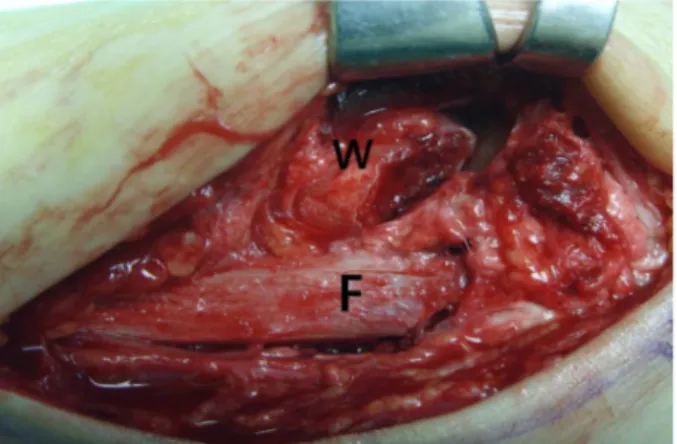
Figure 4. Operative finding of the type III anterior inferior tibiofibular ligament avulsion fracture (F: fibula, W: Wagstaffe fragment).
Figure 5. Anterior inferior tibiofibular ligament avulsion fragment was fixed with mini screw.
Type I Type II Type III Type IV
Figure 6. A modified Wagstaffe classification of anterior inferior tibiofibular ligament avulsion fracture.
중요한 영향을 미친다.4,14) 원위 경비 인대 결합의 손상은 주로 외회전력에 의해 발생되는데, Lauge-Hansen 족관절 분류법 회외-외회전형, 회내-외회전형, 회내-외전형의 골절 에서 원위 경비 인대 결합의 손상이 동반된다고 하였다.5,15,16)
전하 경비 인대 견열 골절은 1875년 Wagstaffe2)가 원위 비골 전방 피질골의 수직 골절에 대하여 최초로 기술함으 로써 알려졌고, 이는 회외-외회전형 족관절 골절과 관계 가 있다고 하였다. 전하 경비 인대 견열 골절에 대한 Pankovich의 분류(변형 Wagstaffe 분류)는 비골측에 견열 골절이 발생하는 I형, 비골 골절에 동반된 견열 골절을 II형, 경골측의 견열 골절을 III형, 경골, 비골 양측에 견열 골절 이 발생하는 IV형으로 분류되며, 주로 II형의 골절이 많이 발생한다(Fig. 6).3,4) Park 등4)에 의하면 전하 경비 인대 견 열 골절은 93예 중 13예로 14%의 발생 빈도를 보였고, 전 예가 Weber B형17,18)의 골절에서 발생하였으며, 변형 Wagstaffe 분류상 모두 2형이었고, Pankovich도 9예의 보고 중 8예가 2형으로 대부분을 차지했다. 본 연구에서 전하 경비 인 대 견열 골절은 전체 족관절 골절의 12%에서 발생하였고,
93%가 족관절 Lauge-Hansen 분류법상 회외-외회전 형에 서 발생하였으며, 변형 Wagstaffe 분류상 II형이 대부분 (76%)이었다.
일반 방사선 검사상 전하 경비 인대 견열 골절의 경우 경 비골의 중첩으로 인해 진단이 어려운 경우가 있으며, Park 등4)은 45도 내회전 방사선 검사가 진단에 유용하다고 하였 다. 본 연구에서는 전후면 또는 측면 사진상 의심이 되는 경우에는 컴퓨터 단층촬영 검사를 통하여 진단하였고, 수술 중 진단이 된 경우도 있었다.
족관절 골절에 동반된 전하 경비 인대 견열 골절의 치료 는 족관절 골절의 해부학적 정복 및 견고한 내고정뿐만 아 니라 전하 경비 인대의 정확한 복원 또한 치료에 중요하
다.4,15,19) 전하 경비 인대의 정확한 복원이 되지 않을 경우
원위 경비 관절의 이개에 의한 원위 경비 관절 부위 만성 적인 통증과 거골의 만성적인 전위로 인한 외상성 관절염, 골절편의 감입에 의한 동통 등이 발생할 수 있다.20,21) Park 등4)은 전한 경비 인대 견열 골절의 불유합으로 인해 1예의 보통의 결과를 보였으나, 전하 경비 인대 견열 골절에 대한 정확한 진단과 정복, 동반된 족관절 골절에 대한 적절한 치 료를 할 경우 좋은 결과를 보였다.
결 론
동반된 족관절 골절의 초기 수상 정도가 심하였던 경 우를 제외하고, 전하 경비 인대 견열 골절의 치료 결과는 대부분 우수한 결과를 보였다. 단순 방사선 검사, 컴퓨터 단층촬영 혹은 수술 중 소견으로 전하 경비 인대 견열 골 절을 발견하여 정확한 내고정을 시행한다면, 이를 간과하 여 발생하는 추가적인 합병증을 예방할 수 있을 것으로 생각된다.
REFERENCES
1. Kannus P, Palvanen M, Niemi S, Parkkari J, Järvinen M.
Increasing number and incidence of low-trauma ankle
fractures in elderly people: Finnish statistics during 1970- 2000 and projections for the future. Bone. 2002;31:430-3.
2. Wagstaffe WW. An unusual form of fracture of the fibula.
Cited by. ST thomas Hosp Rep. 1875;6:43.
3. Pankovich AM. Fractures of the fibula at the distal tibiofibular syndesmosis. Clin Orthop Relat Res. 1979;138-47.
4. Park JW, Kim SK, Hong JS, Park JH. Anterior tibiofibular ligament avulsion fracture in weber type B lateral malleolar fracture. J Trauma. 2002;52:655-9.
5. Lauge-Hansen N. Fractures of the ankle. II. Combined experimental- surgical and experimental-roentgenologic investigations.
Arch Surg. 1950;60:957-85.
6. Pettrone FA, Gail M, Pee D, Fitzpatrick T, Van Herpe LB.
Quantitative criteria for prediction of the results after displaced fracture of the ankle. J Bone Joint Surg Am. 1983;65:667-77.
7. Sarkisian JS, Cody GW. Closed treatment of ankle fractures: a new criterion for evaluation - a review of 250 cases. J Trauma.
1976;16:323-6.
8. Joy G, Patzakis MJ, Harvey JP Jr. Precise evaluation of the reduction of severe ankle fractures. J Bone Joint Surg Am.
1974;56:979-93.
9. Stoffel K, Wysocki D, Baddour E, Nicholls R, Yates P.
Comparison of two intraoperative assessment methods for injuries to the ankle syndesmosis. A cadaveric study. J Bone Joint Surg Am. 2009;91:2646-52.
10. Mizel MS. Technique tip: a revised method of the Cotton test for intra-operative evaluation of syndesmotic injuries. Foot Ankle Int. 2003;24:86-7.
11. Kitaoka HB, Alexander IJ, Adelaar RS, Nunley JA, Myerson MS, Sanders M. Clinical rating systems for the ankle-hindfoot, midfoot, hallux, and lesser toes. Foot Ankle Int. 1994;15:349-53.
12. Grath GB. Widening of the ankle mortise. A clinical and experimental study. Acta Chir Scand Suppl. 1960;Suppl 263:
1-88.
13. Ogilvie-Harris DJ, Reed SC, Hedman TP. Disruption of the ankle syndesmosis: biomechanical study of the ligamentous restraints. Arthroscopy. 1994;10:558-60.
14. Lee HS, Park SS, Kim JW, et al. Diagnostic value of ultrasonography for acute tear of tibiofibular syndesmosis in ankle. J Korean Foot Ankle Soc. 2004;8:1-6.
15. Ramsey PL, Hamilton W. Changes in tibiotalar area of contact caused by lateral talar shift. J Bone Joint Surg Am.
1976;58:356-7.
16. Rasmussen O. Stability of the ankle joint. Analysis of the function and traumatology of the ankle ligaments. Acta Orthop Scand Suppl. 1985;211:1-75.
17. Danis R. Les fratures malleolaires. In: Danis R, ed. Theorie et pratique de osteosynthese. Paris: Masson; 1949. 133-65.
18. Cedell CA. Ankle lesions. Acta Orthop Scand. 1975;46:
425-45.
19. Broos PL, Bisschop AP. Operative treatment of ankle fractures in adults: correlation between types of fracture and final results. Injury. 1991;22:403-6.
20. Akseki D, Pinar H, Bozkurt M, Yaldiz K, Araç S. The distal fascicle of the anterior inferior tibio-fibular ligament as a cause of anterolateral ankle impingement: results of arthroscopic resection. Acta Orthop Scand. 1999;70:478-82.
21. Bassett FH 3rd, Gates HS 3rd, Billys JB, Morris HB, Nikolaou PK. Talar impingement by the anteroinferior tibiofibular ligament. A cause of chronic pain in the ankle after inversion sprain. J Bone Joint Surg Am. 1990;72:55-9.