INTRODUCTION
Ischial sore is frequent in paraplegia patients or quadriplegia patients capable of sitting in a wheelchair or taking a seated posture.1 Despite diverse operative reconstruction methods, its recurrence has been common.2 Recently, as a method of ischial sore reconstruction, a gluteal-region perforator-based flap has most commonly been used. In addition to addressing the diverse variables such as guardians’ care and patients’ self- care efforts, to reduce recurrence after such perforator flap- based reconstruction, we have come to realize that it is very important to lessen the flap tension surgically. Generally, ischial sore reconstruction with a perforator flap is performed in a
prone position. However, because ischial sore occurs in a seated position, it seems that, among the possible operative postures, it seems reasonable that patients be positioned close to the sitting position in order to perform insetting without any tension in the flap.
Therefore, this paper seeks to evaluate the difference in flap tension between the prone position and seated position in healthy subjects and to describe the technique and importance of performing flap insetting in hip flexion.
Hip Flexion during Intraoperative Insetting of a Perforator Flap for Reconstruction of an Ischial Sore
Su Bong Nam, Heung Chan Oh, Jae Woo Lee*, Kyeong Ho Song, Seong Hwan Bae
Department of Plastic and Reconstructive Surgery, Pusan National University School of Medicine, Yangsan, Korea
CC This is an open-access article distributed under the terms of the Creative Commons Attribution Non-Commercial License (http://creativecommons.org/licenses/by-nc/4.0) which permits unrestricted noncommercial use, distribution, and reproduction in any medium, provided the original work is properly cited.
Copyright © 2016 by the Korean Society for Microsurgery. All Rights Reserved.
Received September 21, 2016 Revised November 7, 2016 Accepted November 7, 2016
*Correspondence to: Jae Woo Lee Department of Plastic and Reconstructive Surgery, Pusan National University School of Medicine, 49 Busandaehak-ro, Mulgeum-eup, Yangsan 50612, Korea Tel: +82-55-360-2573
Fax: +82-55-360-2158 E-mail: [email protected]
ORCID: http://orcid.org/0000-0002-0945-6966
Financial support: This work was supported by a 2-year research grant from Pusan National University.
Conflict of interest: None.
Purpose: Perforator flap-using ischial sore reconstruction is performed in a prone position.
But after the surgery, recurrence frequently occurs in a sitting position. In this sense, we introduce modified flap insetting method which closely resembles patient’s sitting position to lessen the flap tension surgically.
Materials and Methods: Authors tried to check a skin tension difference between prone position and sitting position in normal people group and to find out the importance of performing flap insetting in hip flexion position. Healthy volunteers were collected (n=20) and designed the same length of 4 divided sections around the ischium. Lengths of each section were measured when hip joint was flexed to 90 degree and when both hip and knee joints were flexed to 90 degree and the statistical evaluation was performed. Twenty cases with ischial sore underwent reconstructive surgery using perforator flap under hip flexion position and followed-up for any recurrences.
Results: There was a meaningful difference between the joint flexed skin length and that of the neutral position. Flap showed sufficient thickness over 12 months.
Conclusion: It seems that recurrence could be reduced when the reconstructed flap could sufficiently cover in a sitting position regarding its significant length difference in normal people group.
Key Words: Pressure ulcer, Ischium, Perforator flap
ARMS
Archieves of Reconstructive Microsurgery https://doi.org/10.15596/ARMS.2016.25.2.43MATERIALS AND METHODS
Study on ischial-region skin tension differences in prone/hip flexion positions in healthy subjects
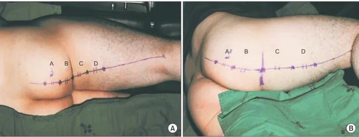
Twenty healthy volunteers were recruited, 10 males and 10 females. The body mass index of all the volunteers ranged from 18.5 to 30.0 kg/m2. All volunteers had 4 divided sections of the same length (5 cm) designed around the ischium (A, B, C, and D). Then the length of each section was measured when the hip joint was flexed to 90 degrees and when both the hip and knee joints were flexed to 90 degrees (Fig. 1), and the statistical evaluation was performed through a t-test. We recognized a gap to be significant if the difference from the neutral position had a p-value less than 0.05.
Perforator flap-based reconstruction
1) Patients and subjectsSince January 2011, we have performed 20 cases of flap insetting in a hip flexion position during reconstruction using the perforator flap-based ischial sore treatment method.
Nineteen cases were male and 1 was female.
2) Operative method
In a prone position, complete debridement is performed to treat the ischial sore. The direction and perforator location of the V-Y advancement flap is designed taking into consideration
the defect size. If the patient has no previous surgical scar, the flap design should be drawn in parallel with gluteal fold for flap movement. The flap width should be designed to be about 2 cm wider than the defect and twice as long. A handheld Doppler probe should be used before the operation to ensure the chosen perforator is located closer to the defect rather than the flap center. If the perforator is 5 cm away from the defect or more, or if cannot be detected well, the flap width and length should be larger and longer to accommodate musculocutaneous flaps in random patterns. If a defect is large in size, the flap should not only be moved in the same direction as V-Y advancement but also rotated about 45~90 degrees according to the location and number of perforators.
A #15 scalpel should be used for incision along the designed border down to the subcutaneous layer. The gluteus maximus muscle fascia should then be incised with monopolar electrocautery. Dissection should then be carefully performed between the fascia and muscle to find a healthy perforator. If possible, 3 to 4 perforators should be chosen rather than a single one, and of them, one or two perforators should be chosen for final use according to the direction and distance of flap motion. Based on the major axis of the flap, ligation should be performed if the perforator is closer to the distal end rather than the central part. All but the healthy perforators found in the proximal portion and part of their surrounding muscles should be exfoliated to make a true island flap.
A B
A B C D
A B C D
Fig. 1. Design of the 4 divided sections around the ischium. (A) A line was drawn on the gluteal fold while in the neutral prone position. The point that corresponds to the position of the ischial tuberosity on the gluteal fold line was marked. An extended line from the center of the popliteal fossa to that marked point was drawn, and 4 divided sections 5 cm in length were taken (A, B, C, and D). The lengths of each line were measured while the hip joint alone was flexed to 90 degrees. (B) Then both the hip and knee joints were flexed to 90 degrees. The averages of each line were used as representative values.
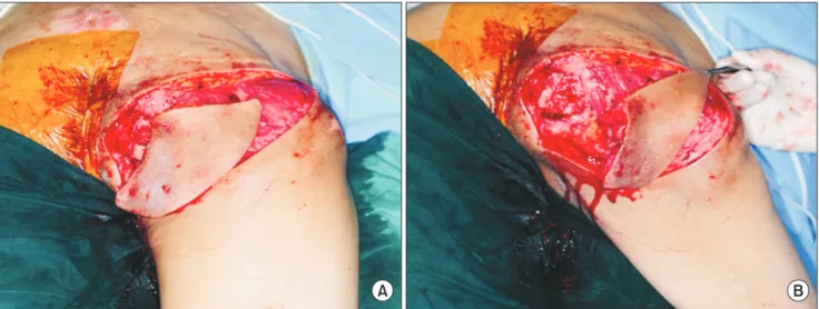
While avoiding damage to the flap, the hips should be flexed at least 90 degrees and then the flap should be checked to determine whether it is mobile enough to pass over the demarcation of the defect without tension. If any tension is noted, dissection should be performed into the muscles along the perforator to increase flap mobility. The flap should move primarily in V-Y advancement form, but if a defect is unusually deep or large, a 45- to 60-degree rotation advancement should be performed for de-epithelialization of the proximal part of the flap to fix it to the defect area and seal the junction. With 90-degree hip flexion (Fig. 2), temporary key suturing should be performed with a skin stapler around the flap. Then position a 400 mL suction drain at the defect area with the patient back in a prone position. The fascia, deep dermis, and skin layer should then be sealed in that order.
After the wound is sealed, cotton rolls should be used to ensure that the flap area is not pressed while in the supine position during the recovery from anesthesia. When moving the patient from the recovery room to the ward, a prone position or opposite-surgery side lateral decubitus position must be maintained. Stitch removal should be performed about 12 to 14 days after the operation, and the suction drain should be removed around the 14th day after the operation. After stitch removal, for about a month, tape should be applied to the suture site to reduce tension on the wound. A supine position is allowed about 2 weeks after the operation. A seated position can be taken increasingly frequently 6 to 8 weeks after surgery.
The first follow-up outpatient visit should take place around 3 months after the operation.
RESULTS
Study on ischial-region skin tension differences in prone/hip flexion positions in healthy subjects
There was a meaningful difference between the length of the designed area for reconstruction with only the hip joint flexed or both the hip and knee joints flexed and the length of the area in the neutral prone position (p=0.00); but there was no significant difference between the length with only the hip joint flexed and with both the hip and knee joints flexed (p=0.76) (Table 1), suggesting that the skin tension was mainly influenced by the hip joint movement, not by the knee joints.
A B
Fig. 2. Increasing size of defect with hip joint movement. (A) Before hip joint flexion. (B) The hip joint was flexed to 90 degrees and the flap was temporarily sutured around the wound margin with a skin stapler. The defect becomes markedly larger with hip joint movement.
Table 1. The average values of each section (n=20) Section Neutral
(cm)
Only hip joint flexed (cm)
Hip and knee joint flexed (cm)
A 5 6.03 (120.6) 6.02 (120.4)
B 5 7.62 (152.4) 7.65 (153.0)
C 5 7.61 (152.2) 7.55 (151.0)
D 5 6.05 (121.0) 6.05 (121.0)
Values are presented as number (%). The lengths of section B and C varied more than section A or D. The length during elongation over the ischial region (sections B and C) was more than 50% in both of the two postures.
Perforator flap-based reconstruction
A total of 20 cases underwent reconstruction with the operative method presented above. They underwent follow-up for at least 12 months. Recurrence was found in 2 cases (10.0%).
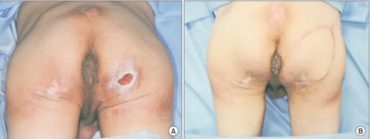
Of them, one showed urinary incontinence caused by ureteral fistula. For this case, the same flap-based reconstruction was performed and thereafter no recurrence has been found. The other case of recurrence continuously showed seroma around the hip joint without a known cause. For this case, a 2-part reconstruction operation was performed, and no recurrence has been spotted. Other than the two cases of recurrence, postoperational complications were found in 3 cases with dehiscence in the triangular suture area of the VY pattern. The cases healed with simple sealing. No infection, flap necrosis, or other issues were found. During observation of postoperative progression, the flap was found to show little reduction in flap thickness and sufficient padding was maintained (Fig. 3). To prevent the recurrence of postoperative bedsores, the patients and their guardians have received education at 6-month intervals.
DISCUSSION
As Foster et al.3 reported an overall 17% reoperation rate after pressure ulcer surgery and dehiscence rate of 42% after ischial reconstructions, 20% of sacral reconstructions and 15%
after trochanteric reconstructions, ischial sores have a higher
tendency to recur than sores located at different positions.
In study of the recurrence rate of 69 pressure sores,4 ischial sores also demonstrated a higher recurrence rate. Particularly when the patient is in a wheelchair, the upper body weight is concentrated in the ischial region, increasing the possibility of pressure sore development or recurrence. The ischial region has no muscle tissue, and the ischial tuberosity is covered by thin hypoderma skin, which is vulnerable to pressure.
As a method of ischial sore reconstruction, the defect area could be supplied with sufficient padding by using a musculocutaneous flap from the posterior thigh or buttock.5 However, this method involves high morbidity of the donor tissue and a large operative area,6 which could limit the potential for additional reconstructive operations in the event of pressure sore recurrence. With recent advances in the understanding of perforators, skin flaps or fasciocutaneous perforator flaps can generally be used to obtain good surgical outcomes.1,2 The method requires a smaller surgical area than the previously used musculocutaneous flap and can be used in diverse directions and forms.7 If a pressure sore recurs, the perforator used previously can be used again to elevate the flap. Or another perforator neighboring the used flap can be used to elevate a new flap. For these reasons, it has been increasingly used as a primary reconstruction method for pressure sores.
In ischial sore reconstruction, not only perforator flaps but also other flaps can be used, just as for sacral sores, for surgical reconstruction in a prone position. However, unlike sacral
Fig. 3. A case: pre- and postoperative figures. (A) Preoperative ischial pressure sore. (B) Postoperative 1 year. Sufficient flap thickness was maintained and no signs of recurrence were found.
A B
sores, ischial sores and trochanteric sores have different levels of flap tension in a sitting position than in a prone position, even though the operation is typically performed in a prone position. For ischial sores especially, pressure sores can develop in a seated position. It is natural that flaps possibly subject to reconstruction should be delineated and fixed in their location in a seated position. Excluding while sleeping, most of the time, people maintain a standing or sitting position. Therefore, in reduction mammoplasty, for instance, producing a surgical design and performing surgery only in a supine position would never produce a good result. Therefore, as for ischial sores, though a preoperative design cannot be drawn in a sitting position exactly, it is important to form 90-degree hip flexion close to what will be the in-operative position and check whether the flap is reconstructed sufficiently without tension.
In the process of this verification during surgery, if flap insetting is performed with 90-degree hip flexion, slight wrinkles will form in the flap in a prone position, showing thickness. In the prognosis evaluation, the central part of the flap was touched in a seated position. In this flap location, the flap’s seal area does not come into direct contact with the seat in a seated position.
It showed little thinning and good results without recurrence.
No matter what measures are taken for ischial sore-related reconstruction, most patients experience recurrence at different points of time after the operation.8 The degree of recurrence may be related to the surgical method, but there are multiple variables including the patients’ temperament, health status, duration of paralysis, occupation, guardian’s care, financial status, and surgeon’s attentiveness. Therefore, in order to assess the effectiveness of a specific surgical method, a control group under similar conditions should be included. However, for pressure sores, unlike cosmetic surgeries, it is extremely difficult to define a control group with similar conditions. Also, the recurrence ratio, computed tomography, and other objective measures are necessary to prove the effective maintenance of postoperative flap thickness. However, for financial reasons, not all of these measures were carried out. This study aimed to demonstrate that in reconstruction of ischial sore using a perforator flap, flap insetting while in 90-degree hip flexion helps reduce the tension of the reconstructed flap as well as the possibility of postoperative recurrence.
Recurrence of ischial sores is common. In study of recurrence rate of pressure sores reconstructed with fasciocutaneous flap,
myocutaneous or muscle flap, ischial sore recurrence rate was 48.9%.4 A 33% recurrence rate was reported by Relander and Palmer,9 Disa et al.10 reported a 69% sore recurrence. In this study, we were able to demonstrate that 10% of all patients had recurrence after ischial sore reconstruction with hip flexion position over 12 months of follow-up period.
CONCLUSION
It seems that recurrence could be reduced if the reconstructed flap were to sufficiently cover the ischial region in a seated position without tension. Therefore, in a prone position, the perforator flap should be serrated, and the flap location should be determined and fixed with the patient in a position as close as possible to a seated position, with 90-degree hip flexion. By doing so, the reconstructed flap can be located closer to the center of a defect in a seated position. Maintaining almost no tension in the flap demarcation is expected to be a significant step toward reducing ischial sore recurrence.
REFERENCES
1. Kim YS, Lew DH, Roh TS, Yoo WM, Lee WJ, Tark KC. Inferior gluteal artery perforator flap: a viable alternative for ischial pressure sores. J Plast Reconstr Aesthet Surg 2009;62:1347-54.
2. Yang CH, Kuo YR, Jeng SF, Lin PY. An ideal method for pressure sore reconstruction: a freestyle perforator-based flap.
Ann Plast Surg 2011;66:179-84.
3. Foster RD, Anthony JP, Mathes SJ, Hoffman WY, Young D, Eshima I. Flap selection as a determinant of success in pressure sore coverage. Arch Surg 1997;132:868-73.
4. Yamamoto Y, Tsutsumida A, Murazumi M, Sugihara T. Long- term outcome of pressure sores treated with flap coverage. Plast Reconstr Surg 1997;100:1212-7.
5. Lee SS, Huang SH, Chen MC, Chang KP, Lai CS, Lin SD.
Management of recurrent ischial pressure sore with gracilis muscle flap and V-Y profunda femoris artery perforator-based flap. J Plast Reconstr Aesthet Surg 2009;62:1339-46.
6. Lin H, Hou C, Xu Z, Chen A. Treatment of ischial pressure sores with double adipofascial turnover flaps. Ann Plast Surg 2010;64:59-61.
7. Pérez de la Fuente T, González González I, Calderón Muñoz F. The IGAP flap for ischial pressure sore reconstruction in tetraplegic patients. Int J Surg 2008;6:e1-3.
8. Unal C, Ozdemir J, Yirmibesoglu O, Yucel E, Agir H. Use of inferior gluteal artery and posterior thigh perforators in management of ischial pressure sores with limited donor sites
for flap coverage. Ann Plast Surg 2012;69:67-72.
9. Relander M, Palmer B. Recurrence of surgically treated pressure sores. Scand J Plast Reconstr Surg Hand Surg 1988;22:89-92.
10. Disa JJ, Carlton JM, Goldberg NH. Efficacy of operative cure in pressure sore patients. Plast Reconstr Surg 1992;89:272-8.