INTRODUCTION
The etiology of sporadic Parkinson’s disease (PD) remains unknown, but there is increasing evidence of a role of inflammation in the brain as a factor in its pathogenesis.1 Microglia, the resident immune cells in the brain, play a role in immune surveillance under normal conditions, and become readily activated in response to injuries in the brain and immunologic
challenges. Activated microglia produce a wide array of cytotoxic factors, including tumor necrosis factor-α (TNF α), interleukin (IL)-1β, eicosanoids, nitric oxide, and reactive oxygen species that impact neurons and induce neurodegeneration.1 Evidence of microglial activation has been derived mostly from postmortem analyses of the substantia nigra of PD patients.2
The active metabolite of vitamin D3, 1,25-dihydroxy- vitamin D3(1,25-(OH)2D3), has been reported to exert profound anti-inflammatory and immunosuppressive
1α,25-Dihydroxyvitamin D
3Protects Dopaminergic Neurons in Rodent Models of Parkinson’s Disease
through Inhibition of Microglial Activation
Joong-Seok Kim, M.D., Sun-Young Ryu, M.D., Injin Yun, M.D., Woo-Jun Kim, M.D., Kwang-Soo Lee, M.D., Jeong-Wook Park, M.D., Yeong-In Kim, M.D.
Department of Neurology, The Catholic University of Korea, Seoul, Korea
Background: Recent studies have demonstrated the molecular basis of the immunomodulatory and anti- inflammatory activities of 1,25-dihydroxyvitamin D3(1,25-(OH)2D3). This hormone improves behavioral deficits and normalizes the nigral dopamine levels in animal models of Parkinson’s disease (PD).
Methods: We studied whether the administration of 1,25-(OH)2D3 would protect against 6-hydroxydopa (6-OHDA)- and 1-methyl-4-phenyl-1,2,3,6-tetrahydropyridine (MPTP)-induced neuronal injury, and its potential regulatory effect on microglia activation.
Results: We found that 1,25-(OH)2D3 pretreatment significantly decreased 6-OHDA- and MPTP-induced dopaminergic neuronal loss in the substantia nigra pars compacta by preventing the activation of microglia. This observed neuroprotective effect in MPTP-treated mice that were given 1,25-(OH)2D3 may be attributable to inhibition of proinflammatory cytokine expression.
Conclusion: These results suggest that 1,25-(OH)2D3 is a potentially valuable neuroprotective agent; it may therefore be considered for the treatment of pathologic conditions of the central nervous system, such as PD, where inflammation-induced neurodegeneration occurs.
J Clin Neurol 2(4):252-257, 2006
Key Words : Vitamin D, Parkinson’s disease, 6-hydroxydopa (6-OHDA), 1-methyl-4-phenyl-1,2,3,6-tetrahydropyridine (MPTP), Cytokine, Inflammation, Rat, Mouse
Received : June 7, 2006 / Accepted : September 13, 2006 / Address for correspondence : Yeong-In Kim, M.D.
Department of Neurology, Kangnam St. Mary’s Hospital, 505, Banpo-dong, Seocho-gu, Seoul 137-701, Korea Tel: +82-2-590-2091, Fax: +82-2-599-9686, E-mail: nuyikim@catholic.ac.kr
* This work was supported by the Korea Research Foundation Grant (grant no. KRF-2003-003-E00188).
effects. This hormone inhibits B- and T-lymphocyte proliferation, downregulates the production of IL-2 and interferon (IFN)γ by T lymphocytes, and prevents immunoglobulin secretion by B lymphocytes.3 More- over, 1,25-(OH)2D3 enhances endogenous glial cell-line- derived neurotrophic factor (GDNF) expression in vitro and in vivo,4,5 which is believed to play an important role in microglial activation.
In the present study, we investigated the potential neuro- protective effect of 1,25-(OH)2D3 against 6-hydroxy- dopa (6-OHDA) and 1-methyl-4-phenyl-1,2,3,6-tetra- hydropyridine (MPTP) toxicity in rodent models of PD.
METHODS
1. Model 1: 6-OHDA in rats
A total of 24 adult male Sprague-Dawley rats (weight 250–300 g) were used. These animals received either a daily dose of 1,25-(OH)2D3 (n=12, 1 μg/ml at 1 ml/kg/day; Bonky, Calcitriol, Yuyu, Anyang, Korea) or a vehicle (n=12, ethanol 0.1%, 1 ml/kg;
Merck, Darmstadt, Germany), injected intraperitoneally for 7 consecutive days. On the 7th day of injection, the animals were anesthetized and injected unilaterally with 6-OHDA (8μg/3μl in normal saline containing 0.2 mg/ml ascorbic acid; Merick, MA, USA) over 5 min using a Hamilton syringe into the medial forebrain bundle (coordinates: 4.3 mm posterior from bregma, 1.4 mm from the midline relative to the bregma, and 8.4 mm below the dural surface).
Locomotor activity was tested with the aid of apomorphine (0.1 mg/kg injected subcutaneously) 4 weeks after inflicting unilateral 6-OHDA lesions. Rats were killed 4 weeks after surgery, and their brains were fixed and processed for immunohistochemical staining as described previously.6 The primary antibodies used in this study were as follows: rabbit polyclonal anti-tyrosine-hydroxylase (TH) antibody (diluted 1:1000; Santa Cruz Biotechnology, CA, USA) and mouse monoclonal anti-rat-CD11b antibody (diluted 1:500; OX-42, Serotec, Oxford, UK). Immunostaining was visualized using the EnvisionTM detection kit
(K5007, Dako, Carpinteria, CA, USA) and 3,3- diaminobenzidine (Sigma, St Louis, MO, USA) containing 0.01% H2O2. Coronal 30-μm-thick sections of the substantia nigra were made (coordinates: bregma -5.30 mm, interaural 3.70 mm), and measurements were made on pairs of sections separated from each other by 300 μm.7 The numbers of TH-immunoreactive neurons and activated microglia (CD11b-immunoreactive cells) were counted in each section from normal and lesioned substantia nigra pars compacta (SNpc), while viewing at a magnification of x100. Average numbers of immuno- reactive cells per section were calculated. The ANOVA with two variables test was used for statistical analysis.
2. Model 2: MPTP in mice
A total of 56 C57BL/6 mice weighing 25-30 g received a daily intraperitoneal injection of either 1,25-(OH)2D3 (n=28, 1μg/ml at 1 ml/kg/day; Bonky, Calcitriol) or vehicle (n=28, ethanol 0.1%, 1 ml/kg Merck) for 7 consecutive days. On the 7th day of injection, the animals also received four intraperitoneal injections of 20 mg/kg MPTP (calculated as free base, dissolved in 0.9% saline solution) at 2-h intervals.
Control mice received saline, which was administered using the same administration schedule (n=16).
The mice were sacrificed, transcardial perfusion fixation was performed at 24 or 48 h after MPTP injection, and the brains were fixed and processed for immunohistochemical staining as described above.
Measurements were made only for those cells that fell clearly within the confines of the substantia nigra (coordinates: bregma -3.16 mm, interaural 0.64 mm) in three coronal sections that were 30-μm thick and separated from each other by 300 μm.8
To investigate the temporal pattern of expression of the key cytokines involved in inflammation and immune responses (IFNγ and TNFα), gene expression was quantified using a modification of a previously described reverse transcriptase (RT)-polymerase chain reaction (PCR) method.9 Animals were killed by spinal cord dislocation and then decapitated at 4, 12, 24, or 48 h after MPTP intoxication (3 animals for each time point). RNA was prepared from the substantia nigra, as
described previously. Briefly, after the RT reaction the cDNA was amplified using the following primers: IFNγ, forward: AGC GGC TGA CTG AAC TCA GAT TGT AG (26-mer), reverse: GGTC ACA GTT TTC AGC TGT ATA GGG (25-mer); TNFα, forward: GGC AGG TCT ACT TTG GAG TCA TTG C (25-mer), reverse:
ACA TTC GAG GCT CCA GTG AAT TCG G (25-mer); and glyceraldehyde-3-phosphate dehydrogenase (GAPDH), forward: TGA AGG TCG GAG TCA ACG GAT TTG GT (26-mer), reverse: CAT GTG GGC CAT GAG GTC CAC CAC (24-mer). As a control for genomic contamination, a sample without RT was added to each PCR analysis. To eliminate sample- to-sample differences in RNA extraction and conversion to cDNA,
we amplified the housekeeping gene GAPDH. The thermal cycling parameters were as follows: 35 cycles of 95℃ for 2 min, 94℃ for 50 s, 60℃ for 50 s, and 72℃
for 1 min; 72℃ for 5 min for TNFα and INFγ and 30 cycles of 94℃ for 10 min, 94℃ for 30 s, 57℃ for 30 s, and 72℃ for 30 s; 72℃ for 10 min for GAPDH. PCR products were separated on 1% agarose gels stained with ethidium bromide and visualized with the aid of UV light and a camera linked to an image analyzer. The results are expressed in relative units, as determined by normalization of the optical density of each band to that of the GAPDH band. Three cytokine PCR assays were performed for each group and all results were analyzed statistically using the Mann-Whitney U test.
(A) (B)
(C) (D)
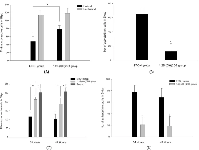
Figure 1. Expression of TH-immunoreactive cells and activated microglia in the SNpc. (A) TH-positive cell loss following 6-OHDA injection. (B) Microglia reaction after 6-OHDA injection. (C) TH-positive cell loss after intraperitoneal MPTP injection. (D) Microglia reaction after MPTP injection. Cells represent numbers of immunoreactive cells per section (mean±SD values). *P<0.05 †P<0.01.
RESULTS
1. 6-OHDA model in rats
The loss of TH-immunoreactive cells in the ipsilateral SNpc reached 58% at 4 weeks after 6-OHDA injection relative to the noninjected side (lesioned substantia nigra, 48.27±12.42 cells, mean
±SD; nonlesioned substantia nigra, 114.12±9.84, P<0.01, n=12). 1,25-(OH)2D3 pretreatment protected against 6-OHDA-mediated damage in the SNpc (lesioned SNpc, 77.93±9.52; nonlesioned, 118.34±
12.90, P<0.05, n=12; Fig. 1A). In 1,25-(OH)2D3- pretreated, 6-OHDA-injected rats, only a few im- munoreactive activated microglia were observed in the ipsilateral SNpc (12.24±11.31). In contrast, vehicle-treated, 6-OHDA-injected rats exhibited a more intense expression of activated microglia along
the lesioned SNpc (65.38±9.82, P<0.01; Fig. 1B).
2. MPTP model in mice
MPTP exposure led to a more than 54% loss of TH-positive neurons (117.3±24.6 vs. 252.6±16.8, P<0.01, 24 h, n=8; and 104.6±25.2 vs. 258.4±22.3, P<0.01, 48 h, n=8 Fig. 1C). Pretreatment with 1,25- (OH)2D3 protected this reduction in nigral TH im- munoreactivity. The mean number of TH-positive neurons was significantly higher in the 1,25-(OH)2D3
-treated groups than in the MPTP- and vehicle- treated groups (214.7±12.6 vs. 117.3±24.6, P<0.01, 24 h, n=8; and 188.6±31.3 vs. 104.6±25.2; P<0.01, 48 h, n=8 Fig. 1C). In 1,25-(OH)2D3-treated mice, there was an approximately 73% inhibition of microglia activation in the number of microglial profiles at 24 h after MPTP injection compared with those MPTP mice that were not given 1,25-(OH)2D3
(A) (B) (C)
(D) (E)
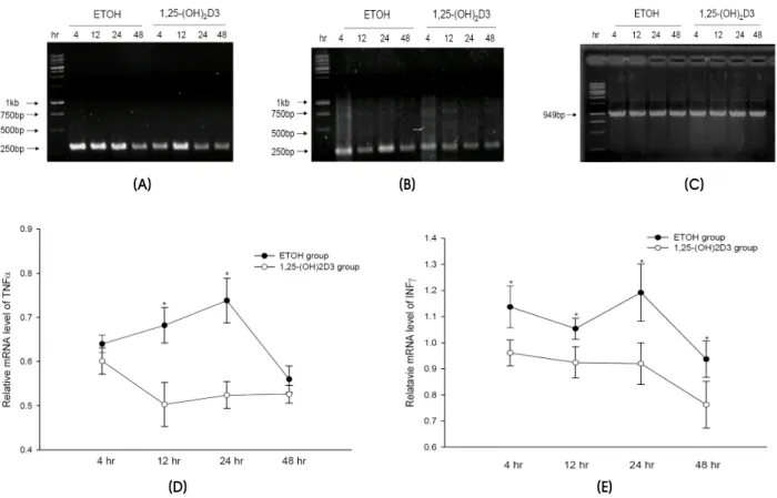
Figure 2. Representative RT-PCR demonstrating the expression of TNFα (A), INFγ (B), and GAPDH (C) mRNAs, and the expression pattern of the TNFα (D) and INFγ (E) mRNA in the SNpc at various times after MPTP intoxication. Data are mean±SEM values (n=3).
*P<0.05, Mann-Whitney U test.
(1,25-(OH)2D3 group, 21.22±13.33; ethanol group, 77.32±12.21, P<0.01; Fig. 1D).
RT-PCR analysis revealed a similar pattern in the kinetics of INFγ and TNFα mRNA expression following MPTP injection (Fig. 2). A rapid increase in the amount of mRNA for INFγ and TNFα was observed between 4 and 24 h after MPTP injection. Comparison with the vehicle-treated group revealed that pretreatment with 1,25-(OH)2D3 protected against the overexpression of proinflammatory cytokines.
DISCUSSION
Our experiments employed two representative pharma- cologically generated rodent models of PD. The 6- OHDA-lesioned rat is a well-validated model for motor alteration related to the advanced phases of PD.
Moreover, the systematic injection of MPTP in mice may be considered a good model of acute-phase PD, which can cause sustained nigral degeneration, bio- chemical changes, and motor abnormalities.10,11
The main finding of this study is that inhibition of microglial activation by pretreatment with 1,25-(OH)2D3
protects against nigrostriatal degeneration in a rodent model of PD. In rats that received 1,25-(OH)2D3, protective effects against 6-OHDA were confirmed by histopathological findings. These effects were also identified in the MPTP-induced acute mouse model of PD.
In mice that received 1,25-(OH)2D3, the neuro- protective effect of 1,25-(OH)2D3 might be related to a decrease in proinflammatory cytokines. The detection of elevated levels of proinflammatory cytokines in post- mortem PD brains provides strong evidence that microglia are involved in the degenerative process.12 The important role of activated microglia in inflammation- mediated neurodegeneration and potentially in the path- ogenesis of PD, prompted us to speculate that the inhibition of microglial activation by 1,25-(OH)2D3 is neuroprotective. The result of this experiment, that 1,25-(OH)2D3 appeared to attenuate the neurotoxin- induced microglial activation and expression of proin- flammatory cytokines, raises the possibility that 1,25-
(OH)2D3 is neuroprotective in experimental models of inflammation-mediated neurodegenerative disease.
Other mechanisms may also be involved in the neuroprotective effects of 1,25-(OH)2D3 in preventing nigral degeneration. First, 1,25-(OH)2D3 treatment might enhance GDNF mRNA and protein expression in the striatum.5 A previous study has shown that the exo- genous administration of GDNF reduces the toxicity of MPTP- and 6-OHDA-induced nigral dopaminergic cell injury.13 Second, the influence on the antioxidant defense mechanisms of 1,25-(OH)2D3 may play a role in the pathogenesis of nigral cell damage.14,15
Our results suggest that 1,25-(OH)2D3 is a potentially valuable neuroprotective agent that may be used for the treatment of pathologic conditions of the central nervous system, such as PD, where inflammation-induced neuro- degeneration occurs. Further study is planned to evaluated the efficacy of 1,25-(OH)2D3 for the treatment of PD and associated neurodegenerative disorders.
REFERENCES
1. Liu B, Hong JS. Role of microglia in inflammation- mediated neurodegenerative diseases: mechanisms and strategies for therapeutic intervention. J Pharmacol Exp Ther 2003;304:1-7.
2. McGeer PL, Itagaki S, Boyes BE, McGeer EG. Reactive microglia are positive for HLA-DR in the substantia nigra of Parkinson’s and Alzheimer’s disease brains. Neurology 1988;38:1285-1291.
3. May E, Asadullah K, Zugel U. Immunoregulation through 1,25-dihydroxyvitamin D3 and its analogs. Curr Drug Targets Inflamm Allergy 2004;3:377-393.
4. Naveilhan P, Neveu I, Wion D, Brachet P. 1,25-Dihydroxy- vitamin D3, an inducer of glial cell line-derived neurotrophic factor. Neuroreport 1996;7:2171-2175.
5. Sanchez B, Lopez-Martin E, Segura C, Labandeira-Garcia JL, Perez-Fernandez R. 1,25-Dihydroxyvitamin D(3) increases striatal GDNF mRNA and protein expression in adult rats. Brain Res Mol Brain Res 2002;108:143-146.
6. Ryu SY, Kim JS, Choi YB, Han SR, Park JW, Park SK, et al. The Effect of 1,25-Dihydroxyvitamin D3 on dopaminergic neurons and microglial activation in Parkinsonian rat model induced by 6-hydroxydopamine. J Korean Neurol Assoc 2005;23:368-373.
7. Paxinos G, Watson C. The Rat Brain in Stereotaxic Coordinates. 2nd ed. Sydney, Australia: Academic Press,
1986.
8. Franklin KBJ, Paxinos G. The Mouse Brain in Stereotaxic Coordinates. 1st ed. San Diego: Academic Press, 1997.
9. Ciesielska A, Joniec I, Przybylkowski A, Gromadzka G, Kurkowska-Jastrzebska I, Czlonkowska A, et al. Dynamics of expression of the mRNA for cytokines and inducible nitric synthase in a murine model of the Parkinson’s disease. Acta Neurobiol Exp (Wars) 2003;63:117-126.
10. Gerlach M, Riederer P. Animal models of Parkinson’s disease: an empirical comparison with the phenomenology of the disease in man. J Neural Transm 1996;103:987- 1041.
11. Kurosaki R, Muramatsu Y, Kato H, Araki T. Biochemical, behavioral and immunohistochemical alterations in MPTP- treated mouse model of Parkinson’s disease. Pharmacol Biochem Behav 2004;78:143-153.
12. Nagatsu T, Sawada M. Inflammatory process in Parkinson’s disease: role for cytokines. Curr Pharm Des 2005;11:999- 1016.
13. Gash DM, Zhang Z, Ovadia A, Cass WA, Yi A, Simmerman L, et al. Functional recovery in parkinsonian monkeys treated with GDNF. Nature 1996;380:252-255.
14. Shinpo K, Kikuchi S, Sasaki H, Moriwaka F, Tashiro K.
Effect of 1,25-dihydroxyvitamin D(3) on cultured mesencephalic dopaminergic neurons to the combined toxicity caused by L-buthionine sulfoximine and 1-methyl-4- phenylpyridine. J Neurosci Res 2000;62:374-382.
15. Garcion E, Sindji L, Montero-Menei C, Andre C, Brachet P, Darcy F. Expression of inducible nitric oxide synthase during rat brain inflammation: regulation by 1,25- dihydroxyvitamin D3. Glia 1998;22:282-294.