94
Introduction
Pulmonary artery (PA) aneurysm is a rare condition and has been associated with structural cardiac and vascular abnormal- ity, pulmonary hypertension, infection, vasculitis, connective tissue disease and trauma.1) Most patients present with non- specific symptoms and are referred with the abnormal mass on chest X-ray.2) The two-thirds of PA aneurysms are associated with pulmonary hypertension. There are no definitive guide- lines about its optimal management. In asymptomatic low pressure PA aneurysm, possibility of rupture is low due to low PA pressure. A conservative treatment may be considered and follows up with echocardiography.
Case
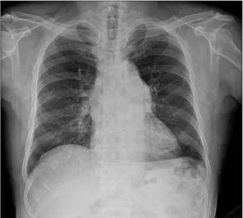
A 69-year-old male was referred to our department for eval- uation of a lung mass which is incidentally found on a routine chest X-ray. He was asymptomatic and physical examination did not reveal cardiac murmurs. There were no finding to doubt infection, vasculitis, collagen vascular disease, and trau- ma in the physical examination. Specific finding was not found in the laboratory finding. Chest X-ray showed left hilar enlargement (Fig. 1). Chest computed tomography showed aneurysmal dilatation of main PA (Fig. 2). The maximum di- ameter of the aneurysm was 56 mm. Transthoracic echocar-
diography showed PA aneurysmal formation with mild pulmo- nary regurgitation (Fig. 3A) and small atrial septal defect (Fig.
3B). The pulmonary to systemic flow ratio (Qp : Qs ratio) was 1.2. The pulmonary transvalvular peak pressure gradient was 8 mmHg. The right heart chambers appeared mildly dilated
pISSN 1975-4612/ eISSN 2005-9655 Copyright © 2013 Korean Society of Echocardiography www.kse-jcu.org http://dx.doi.org/10.4250/jcu.2013.21.2.94
CASE REPORT J Cardiovasc Ultrasound 2013;21(2):94-95
Low Pressure Pulmonary Artery Aneurysm with Atrial Septal Defect
Jae-Kyun Kim, MD1, Sang-Hoon Seol, MD1, Tae-Jin Kim, MD1, Guang-Won Seo, MD1, Bo-Min Park, MD1, Pil-Sang Song, MD1, Dong-Kie Kim, MD1, Ki-Hun Kim, MD1,
Doo-Il Kim, MD1 and Dong-Soo Kim, MD2
1Division of Cardiology, Department of Internal Medicine, Haeundae Paik Hospital, Inje University College of Medicine, Busan, Korea
2Division of Cardiology, Department of Internal Medicine, Busan Paik Hospital, Inje University College of Medicine, Busan, Korea
Pulmonary artery (PA) aneurysm is a rare finding in the thoracic cavity, accompanied by pulmonary hypertension. Clinical presentation of PA aneurysms is usually asymptomatic. The guideline for PA aneurysm treatment is unclear. We report an unusual case of low pressure PA aneurysm associated with atrial septal defect in a 69-year-old man.
KEY WORDS: Low pressure · Pulmonary artery · Aneurysm · Atrial septal defect.
• Received: January 28, 2013 • Revised: May 20, 2013 • Accepted: May 22, 2013
• Address for Correspondence: Sang-Hoon Seol, Division of Cardiology, Department of Internal Medicine, Haeundae Paik Hospital, Inje University College of Medicine, 875 Haeundae-ro, Haeundae-gu, Busan 612-896, Korea Tel: +82-51-797-3070, Fax: +82-51-797-3009, E-mail: [email protected]
• This is an Open Access article distributed under the terms of the Creative Commons Attribution Non-Commercial License (http://creativecommons.org/licenses/by-nc/3.0) which permits unrestricted non-commercial use, distribution, and reproduction in any medium, provided the original work is properly cited.
Fig. 1. Chest X-ray shows dilated main pulmonary artery.
Low Pressure Pulmonary Artery Aneurysm | Jae-Kyun Kim, et al.
95 with pulmonary systolic artery pressure of 26 mmHg. He has
been considered surgery for the PA aneurysm but refused it.
And he was stable at 6 months follow-up.
Discussion
PA aneurysm is a rare disease and its estimated incidence is 1 in 14000 in autopsies.1) The definition is a focal dilatation greater than 4 cm in diameter.3) It has been associated with congenital heart disease, especially patent ductus arteriosus, pulmonary valve stenosis, atrial septal defect, pulmonary hy- pertension, infection, vasculitis, collagen vascular disease, trauma, idiopathic pulmonary aneurysm.1)2)4) The two-thirds of PA aneurysms are associated with pulmonary hyperten- sion.5) The pathophysiology of the PA aneurysm is associated to vessel wall stress that leads to progressive dilatation or even rupture. The law of Laplace dictates that wall stress is directly proportional to the vascular pressure and radius of the vessel and is inversely related to the wall thickness.6) However, in our patient, the natural history of low pressure PA aneurysm was unclear and has been thought to be due to inherent weak- ness of the arterial wall. Most patients with PA aneurysms are asymptomatic or unexplained dyspnea and are referred with the suspicion of a mass or vascular dilatation seen on chest X- ray.2) The confirmative diagnosis may be made with echocar- diography or computed tomography. There are no definite guidelines on the management of PA aneurysm due to its low prevalence. Surgical management may be needed. However, low pressure PA aneurysm is a benign condition with better survival than other etiologies and conservative treatment is may recommended such as our patient.7)
References
1. Deterling RA Jr, Clagett OT. Aneurysm of the pulmonary artery; review of the literature and report of a case. Am Heart J 1947;34:471-99.
2. Bartter T, Irwin RS, Nash G. Aneurysms of the pulmonary arteries.
Chest 1988;94:1065-75.
3. Chetty KG, McGovern J, Mahutte CK. Hilar mass in a patient with chest pain. Chest 1996;109:1643-4.
4. Gould L, Yang DC, Patel C, Patel D, Lee J, Judge D, Taddeo M.
Aneurysms of the pulmonary arteries--a case report. Angiology 1987;38:
474-8.
5. Sakuma M, Demachi J, Suzuki J, Nawata J, Takahashi T, Shirato K.
Proximal pulmonary artery aneurysms in patients with pulmonary artery hypertension: complicated cases. Intern Med 2007;46:1789-93.
6. Butto F, Lucas RV Jr, Edwards JE. Pulmonary arterial aneurysm. A pathologic study of five cases. Chest 1987;91:237-41.
7. Vural AH, Türk T, Ata Y, Göncü T, Ozyazicioglu A. Idiopathic as- ymptomatic main pulmonary artery aneurysm: surgery or conservative man- agement? A case report. Heart Surg Forum 2007;10:E273-5.
Fig. 2. Chest computed tomography demonstrates dilated main pulmon- ary artery and proximal branches.
Fig. 3. Transthoracic echocardiography reveals (A) enlarged main pulmonary trunk and both major pulmonary arteries are seen in the basal short-axis view. (B) There is a small atrial septal defect in the subcostal view.
A
B